

Phospholipase D (PLD) drives cell invasion, tumor growth and metastasis in a human breast cancer xenograph model
- PMID: 23752189
- PMCID: PMC3966651
- DOI: 10.1038/onc.2013.207
Phospholipase D (PLD) drives cell invasion, tumor growth and metastasis in a human breast cancer xenograph model
Abstract
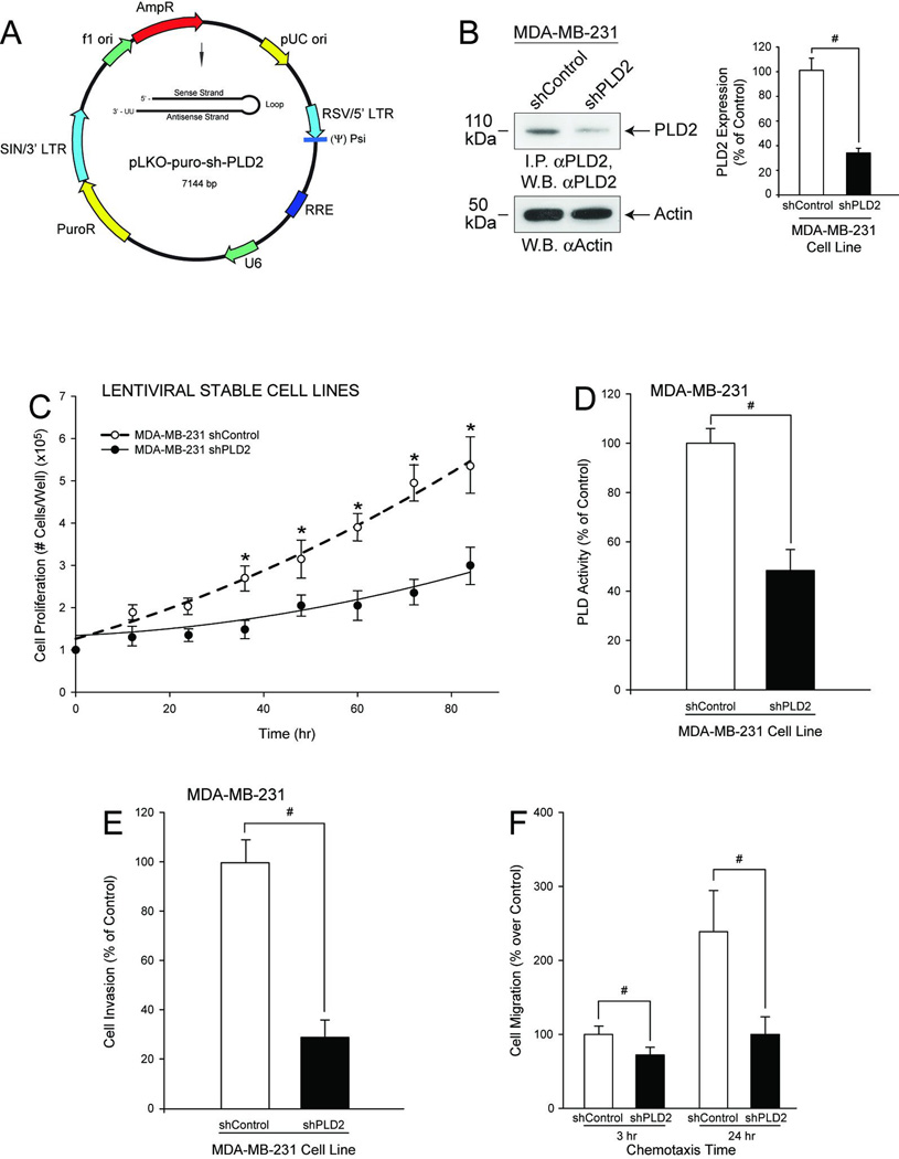
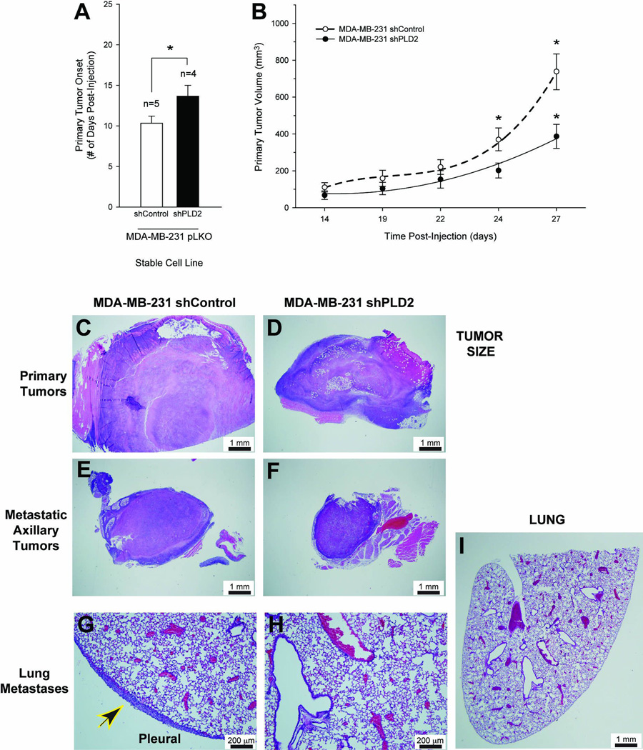
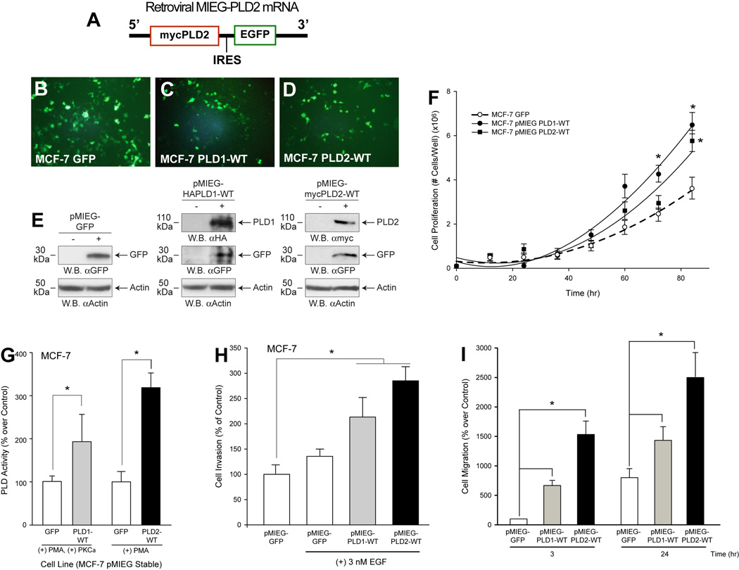
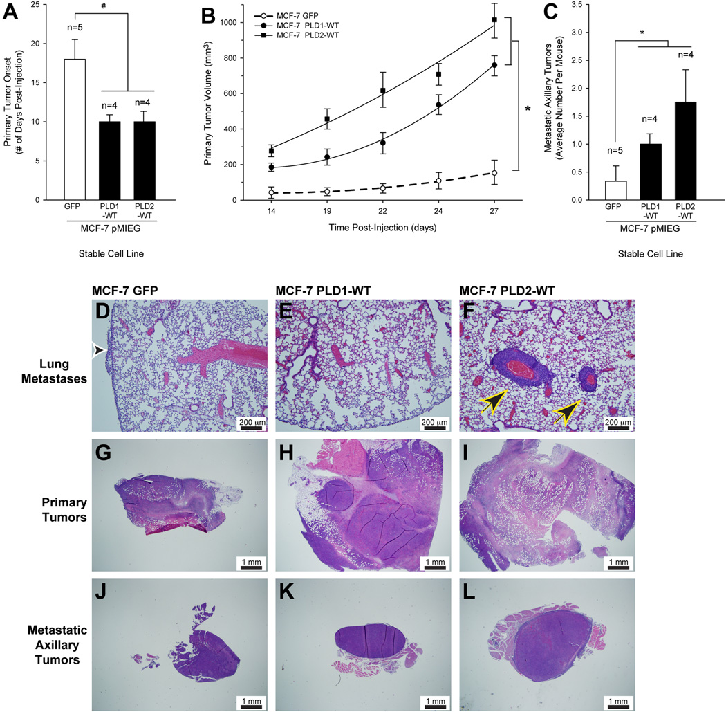
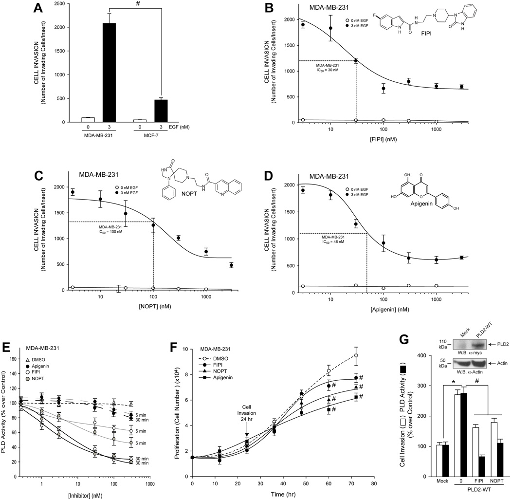
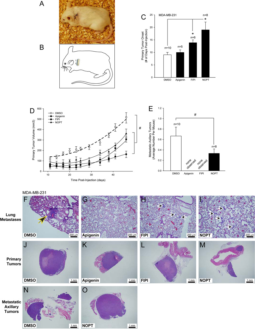
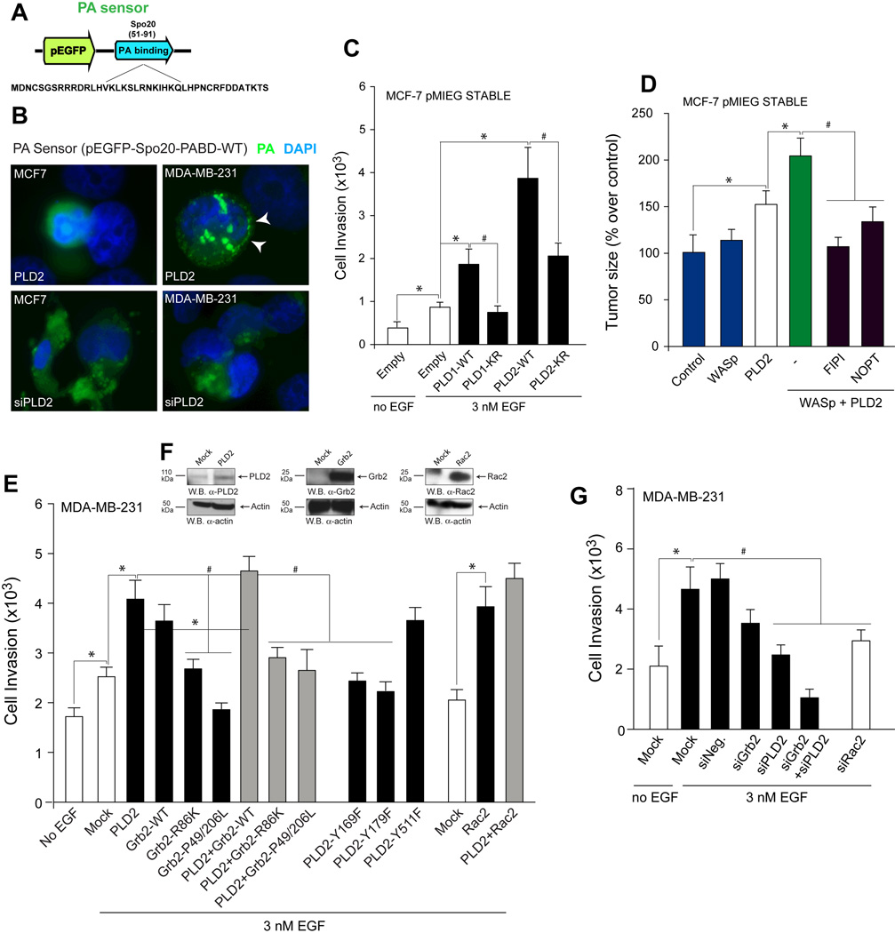
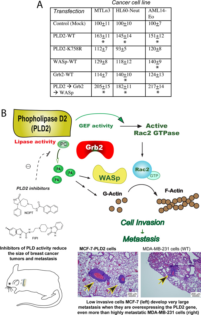
Breast cancer is one of the most common malignancies in human females in the world. One protein that has elevated enzymatic lipase activity in breast cancers in vitro is phospholipase D (PLD), which is also involved in cell migration. We demonstrate that the PLD2 isoform, which was analyzed directly in the tumors, is crucial for cell invasion that contributes critically to the growth and development of breast tumors and lung metastases in vivo. We used three complementary strategies in a SCID mouse model and also addressed the underlying molecular mechanism. First, the PLD2 gene was silenced in highly metastatic, aggressive breast cancer cells (MDA-MB-231) with lentivirus-based short hairpin RNA, which were xenotransplanted in SCID mice. The resulting mouse primary mammary tumors were reduced in size (65%, P<0.05) and their onset delayed when compared with control tumors. Second, we stably overexpressed PLD2 in low-invasive breast cancer cells (MCF-7) with a biscistronic MIEG retroviral vector and observed that these cells were converted into a highly aggressive phenotype, as primary tumors that formed following xenotransplantation were larger, grew faster and developed lung metastases more readily. Third, we implanted osmotic pumps into SCID xenotransplanted mice that delivered two different small-molecule inhibitors of PLD activity (5-fluoro-2-indolyl des-chlorohalopemide and N-[2-(4-oxo-1-phenyl-1,3,8-triazaspiro[4,5]dec-8-yl)ethyl]-2-naphthalenecarboxamide). These inhibitors led to significant (>70%, P<0.05) inhibition of primary tumor growth, metastatic axillary tumors and lung metastases. In order to define the underlying mechanism, we determined that the machinery of PLD-induced cell invasion is mediated by phosphatidic acid, Wiscott-Aldrich Syndrome protein, growth receptor-bound protein 2 and Rac2 signaling events that ultimately affect actin polymerization and cell invasion. In summary, this study shows for the first time that PLD2 has a central role in the development, metastasis and level of aggressiveness of breast cancer, raising the possibility that PLD2 could be used as a new therapeutic target.
Conflict of interest statement
The authors declare no conflict of interest.
Figures
Similar articles
-
The phospholipase D inhibitor FIPI potently blocks EGF-induced calcium signaling in human breast cancer cells.Cell Commun Signal. 2021 Apr 8;19(1):43. doi: 10.1186/s12964-021-00724-z. Cell Commun Signal. 2021. PMID: 33832505 Free PMC article.
-
Serum deprivation confers the MDA-MB-231 breast cancer line with an EGFR/JAK3/PLD2 system that maximizes cancer cell invasion.J Mol Biol. 2013 Feb 22;425(4):755-66. doi: 10.1016/j.jmb.2012.11.035. Epub 2012 Dec 10. J Mol Biol. 2013. PMID: 23238254 Free PMC article.
-
Cell invasion of highly metastatic MTLn3 cancer cells is dependent on phospholipase D2 (PLD2) and Janus kinase 3 (JAK3).J Mol Biol. 2011 May 20;408(5):850-62. doi: 10.1016/j.jmb.2011.03.017. Epub 2011 Mar 22. J Mol Biol. 2011. PMID: 21414324 Free PMC article.
-
Phospholipase D in cell signaling: from a myriad of cell functions to cancer growth and metastasis.J Biol Chem. 2014 Aug 15;289(33):22557-22566. doi: 10.1074/jbc.R114.574152. Epub 2014 Jul 2. J Biol Chem. 2014. PMID: 24990944 Free PMC article. Review.
-
Proliferative and metastatic roles for Phospholipase D in mouse models of cancer.Adv Biol Regul. 2018 Jan;67:134-140. doi: 10.1016/j.jbior.2017.11.004. Epub 2017 Nov 14. Adv Biol Regul. 2018. PMID: 29154090 Free PMC article. Review.
Cited by
-
Development of an Immune-Related Prognostic Signature in Breast Cancer.Front Genet. 2020 Jan 28;10:1390. doi: 10.3389/fgene.2019.01390. eCollection 2019. Front Genet. 2020. PMID: 32047513 Free PMC article.
-
Phospholipase D2 promotes degradation of hypoxia-inducible factor-1α independent of lipase activity.Exp Mol Med. 2015 Nov 27;47(11):e196. doi: 10.1038/emm.2015.87. Exp Mol Med. 2015. PMID: 26611735 Free PMC article.
-
Phospholipase D and Its Essential Role in Cancer.Mol Cells. 2017 Nov 30;40(11):805-813. doi: 10.14348/molcells.2017.0241. Epub 2017 Nov 16. Mol Cells. 2017. PMID: 29145720 Free PMC article. Review.
-
Metabolic fingerprinting by nuclear magnetic resonance of hepatocellular carcinoma cells during p53 reactivation-induced senescence.NMR Biomed. 2024 Sep;37(9):e5157. doi: 10.1002/nbm.5157. Epub 2024 Apr 8. NMR Biomed. 2024. PMID: 38589764 Free PMC article.
-
The phospholipase D inhibitor FIPI potently blocks EGF-induced calcium signaling in human breast cancer cells.Cell Commun Signal. 2021 Apr 8;19(1):43. doi: 10.1186/s12964-021-00724-z. Cell Commun Signal. 2021. PMID: 33832505 Free PMC article.
References
-
- Bray F, Ren JS, Masuyer E, Ferlay J. Global estimates of cancer prevalence for 27 sites in the adult population in 2008. Int J Cancer. 2013;132(5):1133–1145. - PubMed
-
- Benson JR, Jatoi I. The global breast cancer burden. Future Oncol. 2012;8(6):697–702. - PubMed
-
- Scully OJ, Bay BH, Yip G, Yu Y. Breast cancer metastasis. Cancer Genomics Proteomics. 2012;9(5):311–320. - PubMed
-
- Lorusso G, Ruegg C. New insights into the mechanisms of organ-specific breast cancer metastasis. Semin Cancer Biol. 2012;22(3):226–233. - PubMed
-
- Steeg PS. Tumor metastasis: mechanistic insights and clinical challenges. Nat Med. 2006;12(8):895–904. - PubMed
Publication types
MeSH terms
Substances
Grants and funding
LinkOut - more resources
Full Text Sources
Other Literature Sources
Medical
Research Materials
Miscellaneous
