. 2014 Apr;19(4):417-26.
doi: 10.1038/mp.2013.66. Epub 2013 Jun 11.

Reelin, an extracellular matrix protein linked to early onset psychiatric diseases, drives postnatal development of the prefrontal cortex via GluN2B-NMDARs and the mTOR pathway

Affiliations
Free PMC article

Reelin, an extracellular matrix protein linked to early onset psychiatric diseases, drives postnatal development of the prefrontal cortex via GluN2B-NMDARs and the mTOR pathway

J Iafrati et al. Mol Psychiatry. 2014 Apr.
Free PMC article

Erratum in

  • Mol Psychiatry. 2014 Apr;19(4):527. Gonzalez-Campo, C [added]

Abstract

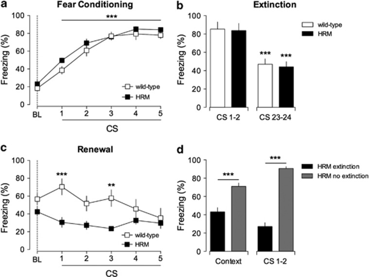
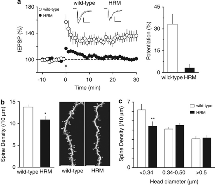
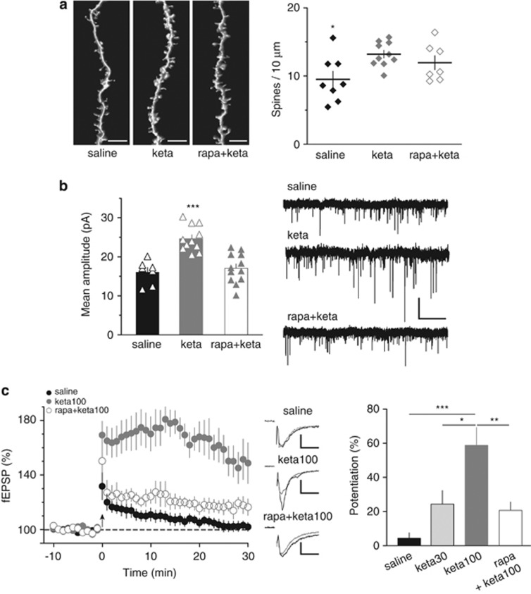
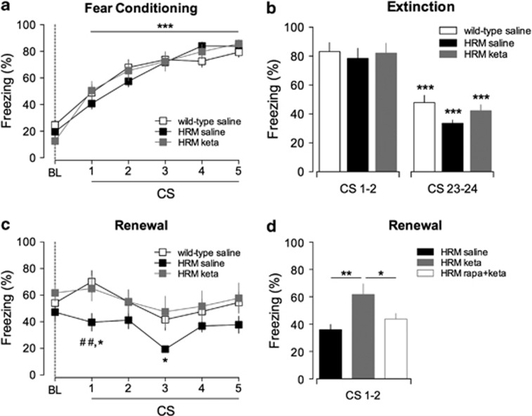
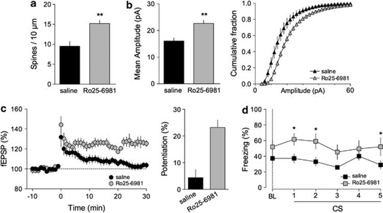
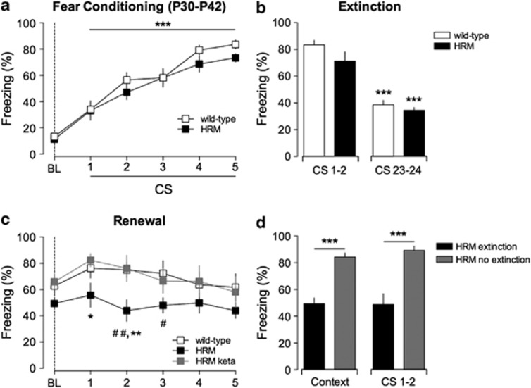
Defective brain extracellular matrix (ECM) is a factor of vulnerability in various psychiatric diseases such as schizophrenia, depression and autism. The glycoprotein reelin is an essential building block of the brain ECM that modulates neuronal development and participates to the functions of adult central synapses. The reelin gene (RELN) is a strong candidate in psychiatric diseases of early onset, but its synaptic and behavioral functions in juvenile brain circuits remain unresolved. Here, we found that in juvenile reelin-haploinsufficient heterozygous reeler mice (HRM), abnormal fear memory erasure is concomitant to reduced dendritic spine density and anomalous long-term potentiation in the prefrontal cortex. In juvenile HRM, a single in vivo injection with ketamine or Ro25-6981 to inhibit GluN2B-N-methyl-D-aspartate receptors (NMDARs) restored normal spine density, synaptic plasticity and converted fear memory to an erasure-resilient state typical of adult rodents. The functional and behavioral rescue by ketamine was prevented by rapamycin, an inhibitor of the mammalian target of rapamycin pathway. Finally, we show that fear memory erasure persists until adolescence in HRM and that a single exposure to ketamine during the juvenile period reinstates normal fear memory in adolescent mice. Our results show that reelin is essential for successful structural, functional and behavioral development of juvenile prefrontal circuits and that this developmental period provides a critical window for therapeutic rehabilitation with GluN2B-NMDAR antagonists.


Figures

Figure 1
Figure 1
Extinction erased fear memories in juvenile heterozygous reeler mice (HRM). (a) Freezing behavior of HRM (n=9; black squares) and wild-type (n=8; white squares) during fear conditioning. Freezing levels after each five pairs of tone-shock presentations were significantly different when compared with baseline (BL) measured before paired conditioned stimuli–unconditioned stimuli (CS–US) presentation (***P<0.001). (b) Freezing behavior evoked by the first (CS 1 and 2) and last pair (CS 23 and 24) of CS presentation during extinction. At the end of extinction training, both groups exhibited a significant auditory fear extinction compared with the initial two trials of extinction session (***P<0.001). (c) CS-evoked freezing showing a significant context-dependent renewal in wild-type (white squares; n=8) but not in HRM (black squares, n=9; ***P<0.001 and **P<0.01, wild-type versus HRM for each CS presentation). (d) In the absence of extinction training (gray bars), HRM exhibited stable fear memories 10 days after conditioning when presented to the conditioning context (context) and the CS (CS 1 and 2). ***P<0.001 extinction HRM (black bars; n=9) versus no extinction HRM (gray bars; n=8; analysis of variance (ANOVA)). Data are expressed as mean±s.e.m. percentage of time spent freezing and n is the number of animals. Statistical analysis is shown in the Supplementary Information.
Figure 2
Figure 2
Functional and structural deficits at the prefrontal cortex synapses of juvenile heterozygous reeler mice (HRM). (a) Left: Theta-burst stimulation (TBS) induces long-term potentiation at layer II/III–V/VI prelimbic area of the prefrontal cortex synapses in juvenile wild-type (white circles), whereas no long-term potentiation was observed in age-matched HRM (black circles). Grouped time courses of field excitatory postsynaptic potential (fEPSP) responses expressed as the percentage of baseline before and after TBS (indicated by arrow). Representative traces averaged from 10 fEPSP responses before (gray) and 30 min after plasticity induction (black) in mice from both genotype. Calibration: 0.1 mV, 10 ms. Right: The percentage of potentiation measured between 20 and 30 min after TBS was 33.2±7.0% in wild-type (n=10) and 1.9±2.7% in HRM (n=9). Error bars represent s.e.m. (b) Left: The average spine density per oblique dendritic length of layer V/VI prelimbic area of the prefrontal cortex pyramidal neurons was reduced in juvenile HRM compared with wild-type (10.9±0.8 spines per 10 μm, n=15 cells from 7 HRM versus 13.8±0.5 spines per 10 μm, n=11 cells from 5 wild-type; *P=0.026, Mann–Whitney t-test). Error bars represent s.e.m. Right: Representative three-dimensional volume rendering of reconstructed spines and shafts from juvenile wild-type mice and HRM. Calibration bars: 5 μm. (c) The classification of spine density by head diameter show a selective decrease in spines with head diameter <0.34 μm in HRM (6.1±0.6 spines per 10 μm, n=11 cells in wild-type versus 4.4±0.5 spines per 10 μm, n=15 cells in HRM). The density of spines with larger head diameter (≥0.34 μm) is not different between both genotypes (F(5,72)=8.72, **P<0.01 analysis of variance (ANOVA)).
Figure 3
Figure 3
Ketamine restored normal spine density and synaptic plasticity in juvenile haploinsufficient reelin mice. (a) Left: Representative images of three-dimensional-reconstructed z-stack projections of the oblique dendrites from neurobiotin-labeled layer V/VI pyramidal neurons. Calibration bars: 5 μm. Right: Average spine density per dendritic length (±s.e.m.) is shown with individual values. In vivo ketamine (keta; 30 or 100 mg kg−1) increased spine density to 13.2±0.6 spines per 10 μm (n=10 cells from nine mice, keta) in juvenile haploinsufficient reelin mice compared with age-matched saline-injected haploinsufficient reelin mice (9.5±1.2 spines per 10 μm, n=8 cells from four mice, saline). This effect of ketamine was not prevented by the mammalian target of rapamycin inhibitor rapamycin (rapa, 3 mg kg−1; 12.0±1.0 spines per 10 μm, n=7 cells from 5 mice, rapa+keta; F(2,22)=4.406, *P<0.05 analysis of variance (ANOVA)). (b) Left: In vivo ketamine increased the mean amplitude of AMPA (2-amino-3-(3-hydroxy-5-methyl-isoxazol-4-yl)propanoic acid) receptor-mediated spontaneous excitatory postsynaptic currents (AMPAR-spEPSCs) of haploinsufficient reelin mice from 16.1±1.2 pA (n=7, saline) to 24.6±1.0 pA (keta, n=11). This effect of ketamine was prevented by rapamycin (rapa+keta, 17.1±1.1 pA, n=12; F(2,27)=18.17, ***P<0.001 ANOVA). Error bars represent s.e.m. and n is the number of cells. Right: Representative recordings of AMPAR-spEPSCs taken from the different conditions. Calibration: 20 pA, 2 s. (c) Left: Average time courses of theta-burst long-term potentiation in haploinsufficient reelin mice injected with saline (saline, n=9), ketamine 100 mg kg−1 (keta100, n=8) and rapamycin before ketamine 100 mg kg−1 (rapa+keta100, n=6). Middle: Representative traces averaged from 10 field excitatory postsynaptic potential (fEPSP) responses before (gray) and 30 min after TBS (black) in the different conditions. Calibration: 0.1 mV, 10 ms. Right: Ketamine restored long-term potentiation in a dose-dependent manner. The percentages of long-term potentiation were 4.4±3.1% in heterozygous reeler mice (HRM) injected with saline (n=10) and increased to 24.4±7.9% (n=5) and to 58.8±10.4% (n=8, ***P<0.001) in HRM after injection with ketamine 30 mg kg−1 (keta30) or 100 mg kg−1 (keta100) respectively. Rapamycin inhibited the action of 100 mg kg−1 ketamine (20.7±5.0% n=6; F(3,24)=11.64, **P<0.01 ANOVA). Error bars represent s.e.m. and n is the number of animals.
Figure 4
Figure 4
Ketamine prevents fear memory erasure by extinction in juvenile heterozygous reeler mice (HRM) via the mammalian target of rapamycin pathway. (a) Similar freezing levels were measured during fear conditioning of saline-injected wild-type (open squares; n=11), saline-injected HRM (black squares, n=9) and ketamine (keta)-injected HRM (gray squares, n=9). All groups showed a significant increase in the percentage of freezing time after each paired conditioned stimulus–unconditioned stimulus (CS–US) presentation compared with their respective baseline (BL; ***P<0.001). (b) All groups exhibited a significant decrease of freezing levels between the first 2 (CS 1 and 2) and the last 2 (CS 23 and 24) trials of extinction sessions (***P<0.001). (c) Ketamine rescued context-dependent renewal in HRM. During context-dependent renewal, the freezing levels of ketamine-injected HRM were significantly different to that of saline-injected HRM and similar to those of saline-injected wild-type littermates (*P<0.05 HRM saline versus HRM keta and ##P<0.01 HRM saline versus wild-type saline). (d) Blockade of mammalian target of rapamycin signaling prevents the behavioral effects of ketamine in juvenile HRM. During context-dependent renewal, pretreatment with rapamycin (rapa) decreased the freezing levels of ketamine-injected HRM to those of saline-injected HRM (**P<0.01 saline versus keta and *P<0.05 rapa+keta versus keta). Data are the average of freezing responses to the first two CS presentations. Data are expressed as mean±s.e.m. percentage of time spent freezing and n is the number of animals. Statistical analysis is shown in the Supplementary Information.
Figure 5
Figure 5
The selective GluN2B antagonist Ro25-6981 rescues functional and behavioral deficits of juvenile heterozygous reeler mice (HRM). (a) The average spine density per dendritic length (±s.e.m.) was increased to 15.2±0.8 spines per 10 μm (n=8 cells from 4 mice) in Ro25-6981-injected HRM compared with age-matched saline-injected HRM (**P=0.002, Mann–Whitney t-test). (b) Left: In vivo injection of Ro25-6981 (10 mg kg−1) increased the mean amplitude of AMPA (2-amino-3-(3-hydroxy-5-methyl-isoxazol-4-yl)propanoic acid) receptor-mediated spontaneous excitatory postsynaptic currents (AMPAR-spEPSCs) of HRM from 16.1±1.2 pA (n=7, saline) to 22.7±1.2 pA (n=14, Ro25-6981; **P=0.0008, Mann-Whitney t-test). Right: Cumulative histograms of the distribution of AMPAR-spEPSCs amplitudes showing that Ro25-6981 caused a significant shift towards larger amplitudes compared with saline-injected HRM. Error bars represent s.e.m. and n is the number of cells. (c) Left: Average time courses of theta burst long-term potentiation showing that injection of Ro25-6981 (10 mg kg−1), 6 h before experiments, restored long-term potentiation in juvenile HRM. Right: Potentiation was 4.4±3.1% (n=10) in saline-injected HRM and 23.2±2.8% (n=7) after Ro25-6981 injection (n=7). Error bars represent s.e.m. and n is the number of animals. (d) HRM exposed to Ro25-6981 (10 mg kg−1) showed enhanced freezing (gray squares; n=8) during context-dependent renewal in contrast to saline-treated HRM littermates (black squares; n=9; *P<0.05 HRM injected with Ro25-6981 versus saline-injected HRM). Data are expressed as mean±s.e.m. percentage of time spent freezing and n is the number of animals. Statistical analysis is shown in the Supplementary Information.
Figure 6
Figure 6
Injection of ketamine at juvenile stages suppresses fear memory erasure in older heterozygous reeler mice (HRM). (a) Adolescent wild-type (n=8) and HRM (n=8) aged between P30 and P42 showed comparable freezing levels during fear conditioning. Freezing progressively increases in both groups after each paired conditioned stimulus–unconditioned stimulus (CS–US) presentation compared with their respective baseline (BL; ***P<0.001). (b) During extinction, both groups showed significant decrease in freezing percentage in the last pair of CS presentation when compared with the initial two trials of extinction session (***P<0.001). (c) CS-evoked freezing showing a significant context-dependent renewal in wild-type (n=8) but not in HRM (n=8; #P<0.05 and ##P<0.01 HRM versus wild-type for each CS presentation). A single injection of ketamine (keta), during the juvenile period, rescued context-dependent renewal in adolescent HRM. The freezing levels of ketamine-injected HRM (gray squares; n=7) were significantly different to that of age-matched HRM and similar to those of age-matched wild-type mice (*P<0.05 and **P<0.01 HRM versus HRM keta for each CS presentation). (d) In the absence of extinction training (gray bars), adolescent HRM exhibited stable fear memories 10 days after conditioning when presented to the conditioning context (context) and the CS (CS 1 and 2). ***P<0.001 HRM extinction (black bars; n=8) versus HRM no extinction (gray bars; n=9). Data are expressed as mean±s.e.m. percentage of time spent freezing and n is the number of animals. Statistical analysis is shown in the Supplementary Information.

References

    1. Dityatev A, Schachner M, Sonderegger P. The dual role of the extracellular matrix in synaptic plasticity and homeostasis. Nat Rev Neurosci. 2010;11:735–746. - PubMed
    1. Knuesel I. Reelin-mediated signaling in neuropsychiatric and neurodegenerative diseases. Prog Neurobiol. 2010;91:257–274. - PubMed
    1. Berretta S. Extracellular matrix abnormalities in schizophrenia. Neuropharmacology. 2011;62:1584–1597. - PMC - PubMed
    1. D'Arcangelo G, Miao GG, Chen SC, Soares HD, Morgan JI, Curran T. A protein related to extracellular matrix proteins deleted in the mouse mutant reeler. Nature. 1995;374:719–723. - PubMed
    1. Herz J, Y Chen. Reelin lipoprotein receptors and synaptic plasticity. Nat Rev Neurosci. 2006;7:850–859. - PubMed

Publication types

MeSH terms