

NADH-dehydrogenase type-2 suppresses irreversible visual loss and neurodegeneration in the EAE animal model of MS
- PMID: 23752309
- PMCID: PMC3808146
- DOI: 10.1038/mt.2013.104
NADH-dehydrogenase type-2 suppresses irreversible visual loss and neurodegeneration in the EAE animal model of MS
Abstract
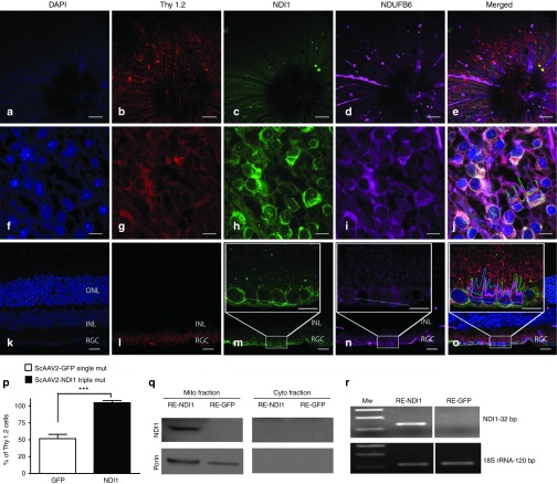
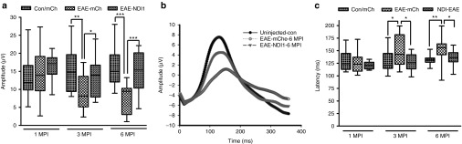
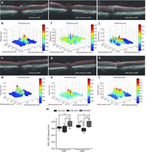
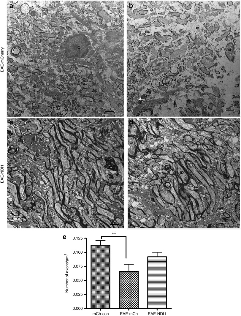
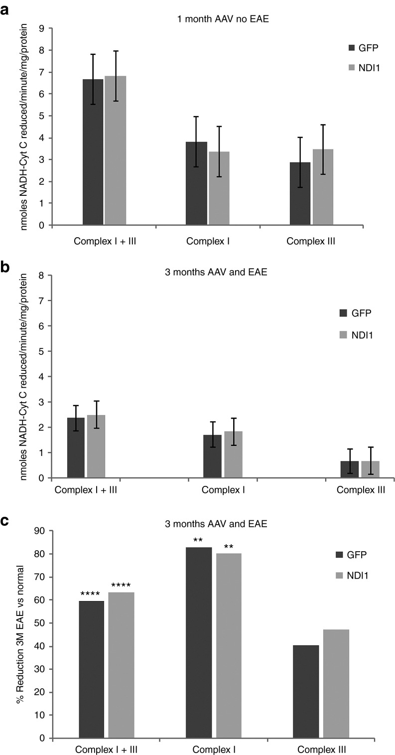
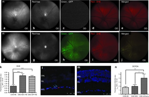
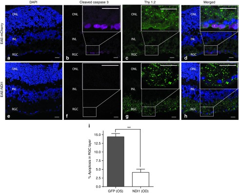
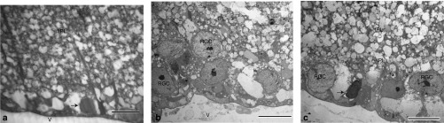
To address mitochondrial dysfunction that mediates irreversible visual loss and neurodegeneration of the optic nerve in the experimental autoimmune encephalomyelitis (EAE) animal model of multiple sclerosis (MS), mice sensitized for EAE were vitreally injected with self-complementary adenoassociated virus (scAAV) containing the NADH-dehydrogenase type-2 (NDI1) complex I gene that quickly expressed in mitochondria of almost all retinal ganglion cells (RGCs). Visual function assessed by pattern electroretinograms (PERGs) reduced by half in EAE showed no significant reductions with NDI1. Serial optical coherence tomography (OCT) revealed significant inner retinal thinning with EAE that was suppressed by NDI1. Although complex I activity reduced 80% in EAE was not improved by NDI1, in vivo fluorescent probes indicated mitochondrial oxidative stress and apoptosis of the EAE retina were reduced by NDI1. Finally, the 42% loss of axons in the EAE optic nerve was ameliorated by NDI1. Targeting the dysfunctional complex I of EAE responsible for loss of respiration, mitochondrial oxidative stress and apoptosis may be a novel approach to address neuronal and axonal loss responsible for permanent disability that is unaltered by current disease modifying drugs for MS that target inflammation.
Figures
Similar articles
-
Gene therapy with mitochondrial heat shock protein 70 suppresses visual loss and optic atrophy in experimental autoimmune encephalomyelitis.Invest Ophthalmol Vis Sci. 2014 Jul 11;55(8):5214-26. doi: 10.1167/iovs.14-14688. Invest Ophthalmol Vis Sci. 2014. PMID: 25015358 Free PMC article.
-
Gene Therapy with Single-Subunit Yeast NADH-Ubiquinone Oxidoreductase (NDI1) Improves the Visual Function in Experimental Autoimmune Encephalomyelitis (EAE) Mice Model of Multiple Sclerosis (MS).Mol Neurobiol. 2020 Apr;57(4):1952-1965. doi: 10.1007/s12035-019-01857-6. Epub 2020 Jan 3. Mol Neurobiol. 2020. PMID: 31900864
-
Mutant NADH dehydrogenase subunit 4 gene delivery to mitochondria by targeting sequence-modified adeno-associated virus induces visual loss and optic atrophy in mice.Mol Vis. 2012;18:1668-83. Epub 2012 Jun 20. Mol Vis. 2012. PMID: 22773905 Free PMC article.
-
Optical Coherence Tomography and Magnetic Resonance Imaging in Multiple Sclerosis and Neuromyelitis Optica Spectrum Disorder.Int J Mol Sci. 2016 Nov 15;17(11):1894. doi: 10.3390/ijms17111894. Int J Mol Sci. 2016. PMID: 27854301 Free PMC article. Review.
-
Psychophysical testing in rodent models of glaucomatous optic neuropathy.Exp Eye Res. 2015 Dec;141:154-63. doi: 10.1016/j.exer.2015.06.025. Epub 2015 Jul 2. Exp Eye Res. 2015. PMID: 26144667 Free PMC article. Review.
Cited by
-
Deletion of Arginase 2 Ameliorates Retinal Neurodegeneration in a Mouse Model of Multiple Sclerosis.Mol Neurobiol. 2019 Dec;56(12):8589-8602. doi: 10.1007/s12035-019-01691-w. Epub 2019 Jul 6. Mol Neurobiol. 2019. PMID: 31280447 Free PMC article.
-
Gene therapy with mitochondrial heat shock protein 70 suppresses visual loss and optic atrophy in experimental autoimmune encephalomyelitis.Invest Ophthalmol Vis Sci. 2014 Jul 11;55(8):5214-26. doi: 10.1167/iovs.14-14688. Invest Ophthalmol Vis Sci. 2014. PMID: 25015358 Free PMC article.
-
Male-specific association between MT-ND4 11719 A/G polymorphism and ulcerative colitis: a mitochondria-wide genetic association study.BMC Gastroenterol. 2016 Oct 3;16(1):118. doi: 10.1186/s12876-016-0509-1. BMC Gastroenterol. 2016. PMID: 27716073 Free PMC article.
-
Delimiting MOGAD as a disease entity using translational imaging.Front Neurol. 2023 Dec 7;14:1216477. doi: 10.3389/fneur.2023.1216477. eCollection 2023. Front Neurol. 2023. PMID: 38333186 Free PMC article. Review.
-
Mitochondrial Dysfunction and Multiple Sclerosis.Biology (Basel). 2019 May 11;8(2):37. doi: 10.3390/biology8020037. Biology (Basel). 2019. PMID: 31083577 Free PMC article. Review.
References
-
- Guy J. Optic nerve degeneration in experimental autoimmune encephalomyelitis. Ophthalmic Res. 2008;40:212–216. - PubMed
-
- Bjartmar C, Trapp BD. Axonal and neuronal degeneration in multiple sclerosis: mechanisms and functional consequences. Curr Opin Neurol. 2001;14:271–278. - PubMed
-
- Shirani A, Zhao Y, Karim ME, Evans C, Kingwell E, van der Kop ML, et al. Association between use of interferon beta and progression of disability in patients with relapsing-remitting multiple sclerosis. JAMA. 2012;308:247–256. - PubMed
-
- Nikic I, Merkler D, Sorbara C, Brinkoetter M, Kreutzfeldt M, Bareyre FM, et al. A reversible form of axon damage in experimental autoimmune encephalomyelitis and multiple sclerosis. Nat Med. 2011;17:495–499. - PubMed
-
- Stys PK. General mechanisms of axonal damage and its prevention. J Neurol Sci. 2005;233:3–13. - PubMed
Publication types
MeSH terms
Substances
Grants and funding
LinkOut - more resources
Full Text Sources
Other Literature Sources
Medical
