. 2013 Aug;21(8):1507-16.
doi: 10.1038/mt.2013.116. Epub 2013 Jun 11.

Functional cloning of recurrence-specific antigens identifies molecular targets to treat tumor relapse

Affiliations

Functional cloning of recurrence-specific antigens identifies molecular targets to treat tumor relapse

Nicolas Boisgerault et al. Mol Ther. 2013 Aug.

Abstract

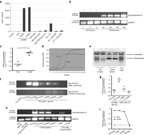
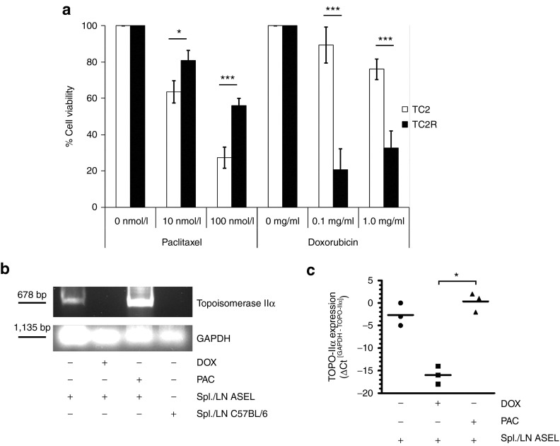
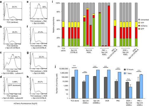
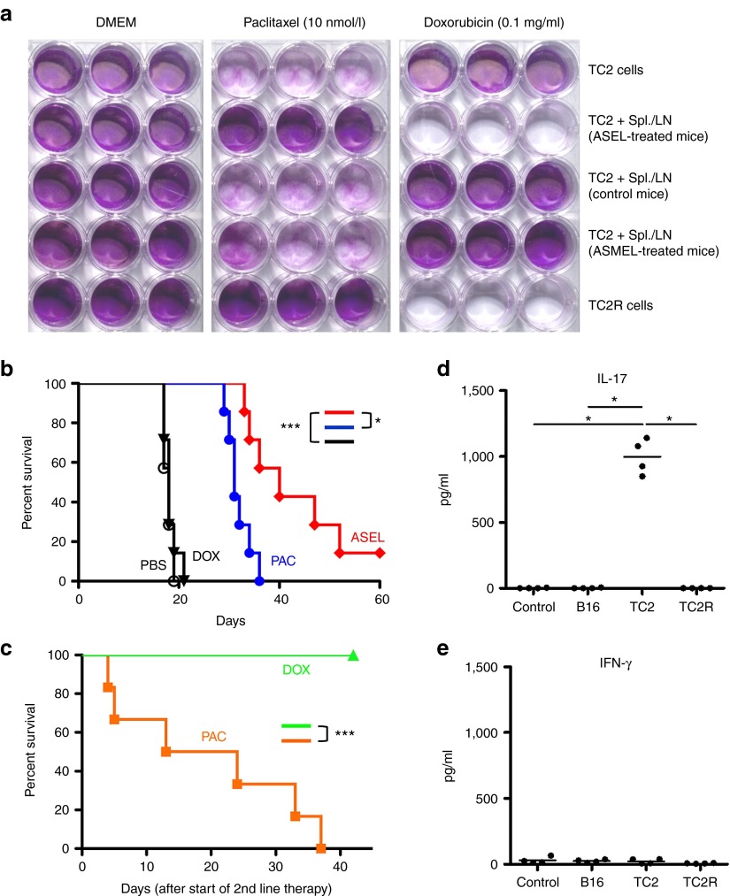
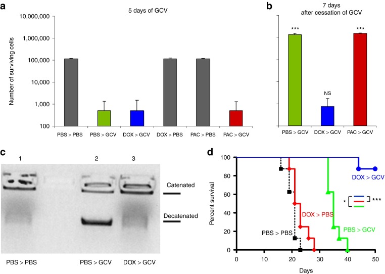
Aggressive regrowth of recurrent tumors following treatment-induced dormancy represents a major clinical challenge for treatment of malignant disease. We reported previously that recurrent prostate tumors, which underwent complete macroscopic regression followed by aggressive regrowth, could be cured with a vesicular stomatitis virus (VSV)-expressed cDNA library derived from recurrent tumor cells. By screening the protective, recurrence-derived VSV-cDNA library, here we identify topoisomerase-IIα (TOPO-IIα) as a recurrence-specific tumor antigen against which tolerance can be broken. Tumor recurrences, in two different types of tumor (prostate and melanoma), which had evaded two different frontline treatments (immunotherapy or chemotherapy), significantly overexpressed TOPO-IIα compared with their primary tumor counterparts, which conferred a novel sensitivity to doxorubicin (DOX) chemotherapy upon the recurrent tumors. This was exploited in vivo using combination therapies to cure mice, which would otherwise have relapsed, after suboptimal primary therapy in both models. Our data show that recurrent tumors-across histologies and primary treatments-express distinct antigens compared with the primary tumor which can be identified using the VSV-cDNA library technology. These results suggest that it may be possible to design a few common second-line therapies against a variety of tumor recurrences, in some cases using agents with no obvious activity against the primary tumor.


Figures

Figure 1
Figure 1
Selective pressure drives TC2R recurrent phenotype evolution. (ac) Mice bearing 7-day–established TC2 tumors were injected intravenously with either (a) VSV-GFP (5 × 108 pfu, days 7, 9, 11, 14, and 16) or (b) with ASEL (107 pfu) six times (days 7, 9, 11, 14, 16, and 18) or (c) nine times (days 7, 9, 11, 14, 16, 18, 21, 23, and 25). Three representative animals for each group, from which tumor cell lines were derived and used in following experiments, are shown. (d) Parental TC2 cells were cocultured (E:T ratio of 10:1) with pooled splenocytes/LN from (e) untreated C57BL/6 mice or from (f) 6×ASEL-treated mice which had been restimulated as described in Materials and Methods. Five days after the last addition of splenocytes/LN, cultures were examined by microscopy and cDNA was screened by PCR for expression of E-cadherin and N-cadherin. (g) Early passage culture (5 days post-explant) of a tumor from a 6×ASEL-treated mouse. Scale bar = 50 μm. (h) Functional cloning of viruses encoding tumor rejection antigens for recurrent prostate tumors. LN/splenocyte cultures (104/well) from mice cured of TC2 and TC2R tumors by vaccination with a combination of ASEL and IEEL were screened for secretion of IFN-γ induced by infection with aliquots of ~104 pfu of the parental IEEL virus stock in the presence of recombinant hsp70. Aliquots which contained virus competent for inducing the IFN-γ recall response were pooled and expanded in BHK cells (24–36 hours). New LN/splenocyte cultures from IEEL-vaccinated mice were infected with serial dilutions of this expanded stock in the presence of recombinant hsp70, and assayed for IFN-γ production. The highest dilution of the virus stock which induced IFN-γ at levels significantly above background was amplified by passaging through BHK cells for 24–36 hours. Serial dilutions of this expanded stock were screened for their ability to induce IFN-γ. A 10 µl aliquots of the highest dilution of the virus which induced IFN-γ were used as the starting point for limiting dilution cloning on BHK cells to identify the dilution at which a single virus particle generated cytopathic effect (+). ASEL, Altered-Self Epitope Library; IEEL, Immune Escape Epitope Library; LN, lymph node; S, syncytia; VSV, vesicular stomatitis virus.
Figure 2
Figure 2
Topoisomerase-IIα is a tumor antigen for recurrent prostate tumors. (a) Splenocyte/LN cultures from IEEL-vaccinated mice were screened for IFN-γ secretion following no infection, stimulation with lysates (TC2, TC2R1, and TC2R2 cells), infection with VSV-GFP (107 pfu), or infection/restimulation with VSV-cDNA#2 (CD44), VSV-cDNA#17 (TOPO-IIα) or a combination (5 × 106 pfu/each) of VSV-cDNA#2 and #17, VSV-cDNA#2 and VSV-GFP, or VSV-cDNA#17 and VSV-GFP. (b) cDNA from freshly explanted TC2.1–3, TC2R1-3 (Figure 1b), or TC2R4 tumor from a separate experiment were screened by PCR (15 cycles), or (c,d) quantitative real-time PCR for expression of murine TOPO-IIα. *Samples positive for TOPO-IIα after 30 cycles. Relative quantities of TOPO-IIα mRNA were determined using GAPDH as calibrator gene (*P < 0.05). (d) Amplification curves from a representative quantitative real-time PCR experiment using cDNA (1:100) from TC2.1 and TC2R1 are shown. (e) Nuclear extracts (107 cells, 1:100) from two freshly explanted TC2 or TC2R tumors, or from TC2-cultured cells were incubated with kDNA to test TOPO-IIα activity. (f,g) TC2 cells (105/well) were either left untreated or cocultured with pooled LN/splenocytes (106/well) from untreated mice, or from 6×ASEL-treated mice, all of which previously restimulated as described in Materials and Methods. cDNA were screened by (f) PCR (15 cycles) or (g) three independent quantitative real-time PCR experiments for expression of TOPO-IIα at different time points following the coculture initiation. (h) cDNA from a freshly explanted TC2 tumor, TC2 cells cocultured with ASEL-restimulated LN/splenocytes from a C57BL/6 mouse, a TC2R explanted tumor at different time points post-explant, or TC2 cells cocultured for 1 week or 1 month with ASEL-restimulated LN/splenocytes were screened for expression of TOPO-IIα. (i) cDNA were prepared from freshly explanted TC2 or TC2R tumors at different time points following explant and screened by quantitative real-time PCR for expression of TOPO-IIα. ASEL, Altered-Self Epitope Library; IEEL, Immune Escape Epitope Library; LN, lymph node; VSV, vesicular stomatitis virus.
Figure 3
Figure 3
Immune escape variants acquire a novel chemosensitivity. (a) TC2 parental cells or freshly explanted TC2R tumor cells were treated in vitro with different concentrations of PAC or DOX as indicated. After 48 hours, cell viability was assessed using an MTT assay (mean ± SD). *P < 0.05; ***P < 0.001. (b,c) TC2 cells were left untreated or were treated for 48 hours with DOX (0.1 mg/ml) or PAC (10 nmol/l). Cells were then cocultured with ASEL-restimulated LN/splenocytes as described in Materials and Methods or with LN/splenocytes from an untreated C57BL/6 mouse. Twenty-four hours following washing, cDNA were screened for expression of TOPO-IIα by (b) PCR (15 cycles) or (c) quantitative real-time PCR. mRNA relative quantities were determined using GAPDH as calibrator gene (*P < 0.05). ASEL, Altered-Self Epitope Library; LN, lymph node; PAC, paclitaxel.
Figure 4
Figure 4
A doxorubicin (DOX)-sensitive subpopulation senses immune selective pressure. (ad) TC2 cells were separately infected with three lentiviral vectors encoding fluorescent marker genes (GFP/mCherry/YFP) and mixed with unmarked TC2 cells at approximately 1:1:1:1. The resulting TC2 Rainbow population contained a stable level of cells of each lineage over long-term (>2 weeks) culture. TC2 Rainbow cells (105/well) were either (a) left alone or (bd) cocultured with (b) pooled LN/splenocytes (106/well) from untreated or (c,d) 6×ASEL-treated mice, which had been restimulated as described above. Populations were analyzed by flow cytometry for fluorescent markers expression. Percentages of mCherry+ cells are shown. (ef) The experiment was repeated as above and TC2 Rainbow cultures were treated (48 hours) with either (e,f) PAC (10 nmol/l) or (g,h) DOX (0.1 mg/ml) before being washed and cocultured with ASEL-restimulated LN/splenocytes from (e,g) ASEL-treated or (f,h) untreated mice. (i) The percentages of cells expressing the fluorescent markers following treatment (described in ah) are shown. (j) TC2 cells were left untreated, or were cocultured with LN/splenocytes from prostate-specific ASEL-, or melanoma-specific ASMEL-treated mice. Alternatively, TC2 cells were treated with DOX or PAC with or without subsequent coculture with LN/splenocytes from ASEL-treated mice as described in eh. MTT assays were carried out on surviving cell populations 72 hours or 1 week after coculture. ASEL, Altered-Self Epitope Library; LN, lymph node; PAC, paclitaxel.
Figure 5
Figure 5
TC2R recurrences are cured by early treatment with doxorubicin (DOX). (a) TC2 and TC2R cells were either left alone or cocultured with pooled LN/splenocytes from 6×ASEL-treated, untreated C57BL/6, or 6×ASMEL-treated mice. All LN/splenocyte cultures had been restimulated as described in Materials and Methods. Two days after the last addition of LN/splenocytes, cocultures were washed three times and then cultured in medium containing no drug (Dulbecco's Modified Eagle's Medium, DMEM), PAC (10 nmol/l), or DOX (0.1 mg/ml) for 5 days. Surviving cells were visualized by crystal violet staining. (b) C57BL/6 mice (n = 7/group) bearing 4-day–established TC2 tumors were treated with i.v. PBS, PAC (10 mg/kg), DOX (10 mg/kg), or ASEL (107 pfu) on days 4, 6, 8, 11, 13, and 15. Survival with time is shown. (c) C57BL/6 mice (n = 6/group) bearing 4-day–established TC2 tumors were treated with i.v. ASEL (107 pfu) on days −14, −12, −10, −7, −5, and −3. Mice were then treated with either PAC or DOX on days 0, 2, 4, 7, 9, and 11 (six mice/group). (d,e) Splenocytes/LN from four of the mice which survived the sequential treatment of ASEL followed by DOX in c above were cocultured with either nothing (control) or with lysates of B16 murine melanoma cells, TC2 murine prostate cells or TC2R cells every 24 hours for 3 days. After 48 hours, supernatants were assayed for (d) IL-17 or (e) IFN-γ secretion. *P < 0.05; ***P < 0.001. ASEL, Altered-Self Epitope Library; LN, lymph node; PBS, phosphate-buffered saline; PAC, paclitaxel.
Figure 6
Figure 6
Doxorubicin (DOX)-sensitive, TOPO-IIαHi melanoma cells drive escape from chemotherapy. (a,b) B16tk cells were treated with PBS, DOX (0.1 mg/ml), or PAC (10 nmol/l) for 48 hours before being treated with ganciclovir (GCV, 5 µg/ml) for 5 days. Surviving cells were (a) counted (mean ± SD) at the end of GCV treatment or were (b) harvested and replated at 500 cells/well and grown in normal medium for 7 days before being counted. Statistical significance is shown for comparisons of the same treatments between a and b. (c) B16tk cells were treated with PBS or DOX for 48 hours before being treated with GCV for 3 days. After 24 hours, cells were harvested and TOPO-IIα activity was assessed. (d) C57Bl/6 mice (n = 8/group) bearing 5-day–established B16tk tumors were treated daily with PBS or DOX (10 mg/kg) on days 6–10 and then daily with PBS or GCV (50 mg/kg) on days 13–17 and 20–24. Tumor survival with time is shown. GCV, ganciclovir; NS, not significant; PBS, phosphate-buffered saline; PAC, paclitaxel.

Comment in

References

    1. Aguirre-Ghiso JA. Models, mechanisms and clinical evidence for cancer dormancy. Nat Rev Cancer. 2007;7:834–846. - PMC - PubMed
    1. Goss PE, Chambers AF. Does tumour dormancy offer a therapeutic target. Nat Rev Cancer. 2010;10:871–877. - PubMed
    1. Hensel JA, Flaig TW, Theodorescu D. Clinical opportunities and challenges in targeting tumour dormancy. Nat Rev Clin Oncol. 2013;10:41–51. - PubMed
    1. McGowan PM, Kirstein JM, Chambers AF. Micrometastatic disease and metastatic outgrowth: clinical issues and experimental approaches. Future Oncol. 2009;5:1083–1098. - PubMed
    1. Gerlinger M, Rowan AJ, Horswell S, Larkin J, Endesfelder D, Gronroos E, et al. Intratumor heterogeneity and branched evolution revealed by multiregion sequencing. N Engl J Med. 2012;366:883–892. - PMC - PubMed

Publication types

MeSH terms