Review
. 2013 Sep;23(9):1026-37.
doi: 10.1093/glycob/cwt045. Epub 2013 Jun 10.

Mucin-type O-glycans and their roles in intestinal homeostasis

Affiliations
Review

Mucin-type O-glycans and their roles in intestinal homeostasis

Kirk S B Bergstrom et al. Glycobiology. 2013 Sep.

Abstract

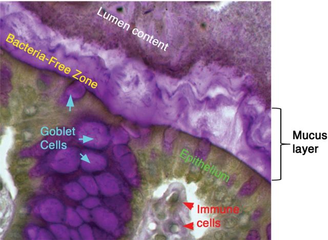
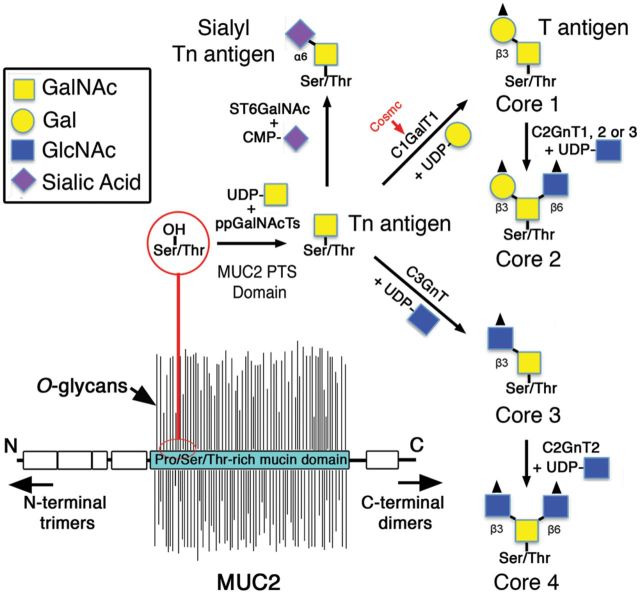
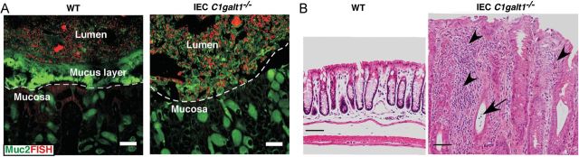
Mucin-type O-glycans are the primary constituents of mucins that are expressed on various mucosal sites of the body, especially the bacteria-laden intestinal tract. Mucins are the main components of mucus, which is secreted by goblet cells and forms a protective homeostatic barrier between the resident microbiota and the underlying immune cells in the colon. However, the specific role of mucin-type O-glycans in mucus barrier function has been uncertain. Recent studies utilizing mice deficient in key glycosyltransferases involved in O-glycan biosynthesis on intestinal mucins have underscored the importance of mucin-type O-glycosylation in mucus barrier function. This review will highlight recent advances in our understanding of mucin-type O-glycan function in the mucus barrier and how they promote mutualism with our resident microbiota.

Keywords: colitis; intestinal homeostasis; microbiota; mucin; mucin-type O-glycans.


Figures

Fig. 1.
Fig. 1.
The intestinal mucus layer and host–microbiota interactions. Periodic acid-Schiff staining of the murine colon revealing the stratified mucus layer creating the bacteria-free zone between the bacteria-laden intestinal lumen and the underlying tissue. The mucus layer is primarily composed of Muc2 (human, MUC2), the major secreted gel-forming mucin of the intestinal tract. Muc2 is synthesized by goblet cells and is secreted as a polymer into the intestinal lumen where it coats the epithelial surface. In the colon, this layer is critical to separating the dense luminal content, including bacteria, from the underlying epithelial and immune cells to limit unwanted stimulation of the mucosal immune system. As described in the text, the Muc2-mucus layer is composed primarily of O-glycans.
Fig. 2.
Fig. 2.
The biosynthesis of mucin-type O-glycans. MUC2 is densely O-glycosylated within its PTS domains, giving the molecule its “bottle-brush” appearance. MUC2 O-glycosylation begins with the addition of GalNAc by the action of ppGalNAcTs, creating the Tn antigen. Tn antigen is modified by C1GalT1 via the help of the chaperone Cosmc to create the core 1 structure, or by C3GnT to create the core 3 structure, or else by ST6GalNAc (α2,6 sialytransferase) to form the sialyl Tn antigen. Core 1 and 3 structures can be extended to form the core 2 and 4 structures, respectively; however, sialyl Tn cannot be extended further. Moreover, each core structure can be further modified and/or terminated by fucosyltransferases, sulfotransferases or sialotransferases (not shown).
Fig. 3.
Fig. 3.
Loss of intestinal core 1-derived O-glycans impairs mucus barrier function and leads to spontaneous colitis. (A) Fluorescence in situ hybridization (FISH, bacterial 16S rDNA probe) revealed that WT colon epithelium was covered by an intact inner layer of mucus (FITC-labeled glycosylation-independent polyclonal antibody to Muc2) that was devoid of bacteria. In contrast, IEC C1galt1–/– epithelium had no mucus layer and directly contacted commensal bacteria. Dashed lines mark epithelial surfaces. Scale bar, 20 mm. (B) H&E staining showing distal colon sections from 16-week-old WT and IEC C1galt1−/− mice. Severe inflammatory disease that resembles human UC occurs in IEC C1galt1−/− mice, as shown by numerous infiltrating inflammatory cells (arrowheads), and a crypt abscess (arrow).

Similar articles

Cited by

References

    1. Allen A, Hutton DA, Pearson JP. The MUC2 gene product: A human intestinal mucin. Int J Biochem Cell B. 1998;30:797–801. - PubMed
    1. An G, Wei B, Xia B, McDaniel JM, Ju T, Cummings RD, Braun J, Xia L. Increased susceptibility to colitis and colorectal tumors in mice lacking core 3-derived O-glycans. J Exp Med. 2007;204:1417–1429. - PMC - PubMed
    1. Arumugam M, Raes J, Pelletier E, Le Paslier D, Yamada T, Mende D, Fernandes G, Tap J, Bruls T, Batto J, et al. Enterotypes of the human gut microbiome. Nature. 2011;473:174–180. - PMC - PubMed
    1. Asker N, Axelsson MA, Olofsson SO, Hansson GC. Dimerization of the human MUC2 mucin in the endoplasmic reticulum is followed by a N-glycosylation-dependent transfer of the mono- and dimers to the Golgi apparatus. J Biol Chem. 1998;273:18857–18863. - PubMed
    1. Atuma C, Strugala V, Allen A, Holm L. The adherent gastrointestinal mucus gel layer: Thickness and physical state in vivo. Am J Physiol Gastrointest Liver Physiol. 2001;280:G922–G929. - PubMed

Publication types