. 2013 Aug;81(8):2962-71.
doi: 10.1128/IAI.00161-13. Epub 2013 Jun 10.

Contribution of interleukin-12 p35 (IL-12p35) and IL-12p40 to protective immunity and pathology in mice infected with Chlamydia muridarum

Affiliations

Contribution of interleukin-12 p35 (IL-12p35) and IL-12p40 to protective immunity and pathology in mice infected with Chlamydia muridarum

Lili Chen et al. Infect Immun. 2013 Aug.

Abstract

The p35 molecule is unique to interleukin-12 (IL-12), while p40 is shared by both IL-12 and IL-23. IL-12 promotes Th1 T cell responses, while IL-23 promotes Th17 T cell responses. The roles of IL-12p35- and IL-12p40-mediated responses in chlamydial infection were compared in mice following an intravaginal infection with Chlamydia muridarum. Mice deficient in either IL-12p35 or p40 both developed similar but prolonged infection time courses, confirming the roles of IL-12-mediated immune responses in clearing primary infection. However, all mice, regardless of genotype, cleared reinfection within 2 weeks, suggesting that an IL-12- or IL-23-independent adaptive immunity is protective against chlamydial infection. All infected mice developed severe oviduct hydrosalpinx despite the increased Th2 responses in IL-12p35- or IL-12p40-deficient mice, suggesting that Th2-dominant responses can contribute to Chlamydia-induced inflammatory pathology. Compared to IL-12p35 knockout mice, the IL-12p40-deficient mice exhibited more extensive spreading of chlamydial organisms into kidney tissues, leading to significantly increased incidence of pyelonephritis, which both confirms the role of IL-12 or IL-23-independent host responses in Chlamydia-induced pathologies and suggests that in the absence of IL-12/IFN-γ-mediated Th1 immunity, an IL-23-mediated response may play an important role in restricting chlamydial organisms from spreading into distal organs. These observations together provide important information for both understanding chlamydial pathogenesis and developing anti-Chlamydia vaccines.


Figures

Fig 1
Fig 1
Effect of IL-12p35 or p40 deficiency on live organism shedding following chlamydial infection. (A) Wild-type mice or mice deficient in IL-12p35 (IL-12p35 KO) or IL-12p40 (IL-12p40 KO) were infected intravaginally with C. muridarum organisms, and vaginal swabs were taken on the days indicated on the x axis for measuring the number of live organisms (inclusion forming units [IFUs]). The number of IFUs from each swab was converted to a log10 value, and the log10 IFUs were used to calculate the mean and standard deviation for each mouse group at each time point. The wild-type group started with 15 mice, the IL-12p35KO group with 13, and the IL-12p40KO with 16. On day 114 after the primary infection, 7 (both KO groups) or 8 (wild-type group) mice from each group were reinfected with C. muridarum organisms. Mice that were negative for shedding 2 consecutive times were no longer swabbed. The number of mice that showed positive live organism shedding at each time point from each group were used to calculate the percentage of mice positive for shedding (B). All mice with primary infection only were sacrificed on day 114, while mice with both primary and secondary infection on day 143 after primary infection. The log10 IFUs and percent shedding-positive mice were compared between each two groups (**, P < 0.01). Note that the wild-type mice resolved their infections within 30 days after primary infection, while mice deficient in IL-12p35 or IL-12p40 maintained statistically significantly higher levels of live organism shedding starting on day 21, and the infection lasted up to 87 days. However, all mice cleared reinfection within 2 weeks.
Fig 2
Fig 2
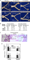
Effect of IL-12p35 or p40 deficiency on mouse hydrosalpinx development following chlamydial infection. (A) When urogenital tract tissues from wild-type (panel a, n = 7 with primary infection; panel d, n = 8 with secondary infection), IL-12p35 KO (b, n = 6; e, n = 7), and IL-12p40 KO (c, n = 9; f, n = 7) mice were examined for gross appearance, obvious hydrosalpinx (OH) was noted; representative images from all groups are shown. The hydrosalpinx severity was semiquantitatively graded from 0 to 4, and scores are given in parentheses. The total score from both oviducts was assigned as the score for a given mouse. Both the incidence (percentage of mice with either unilateral or bilateral OH) and severity (mean ± standard deviation) of hydrosalpinx (but the medians were used for statistical analyses) are listed (B). There were no significant differences in either the number of mice positive for OH or the severity of hydrosalpinx between different groups. After images were taken and gross pathology was evaluated, the oviduct tissues were further examined microscopically (C). Both inflammatory infiltration (gray arrows) and dilated lumen were noted and further semiquantitated according to the criteria listed in the Materials and Methods section; data are expressed as medians plus standard deviations (D). As with gross pathology, there was no significant difference in oviduct histopathology between different groups of mice.
Fig 3
Fig 3
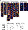
Effect of IL-12p35 or p40 deficiency on renal gross pathology following chlamydial infection. (A) Kidneys from wild-type (images 1 to 7 are from mice with primary infection and 23 to 30 are from those with secondary infection), IL-12p35 KO (images 8 to 13 and 31 to 37), and IL-12p40 KO (images 14 to 22 and 38 to 44) mice were examined for gross appearance, and enlarged, edematous, or necrotic kidneys were noted. Most of these kidneys exhibited typical gross pathology of pyelonephritis. All kidneys identified as abnormal are marked with green stars. Abnormal kidneys were found in most mice deficient in IL-12p40, while no obvious kidney abnormality was noted in any wild-type (WT) mice. The abnormal kidneys were also found in some IL-12p35−/− mice. The numbers of mice with abnormal kidneys from different groups are summarized in panel B (*, P < 0.05; **, P < 0.01; ***, P < 0.001; NS, not significant). The number of mice with abnormal kidneys was the highest in the IL-12p40 KO group. The age-matched KO mice with mock infection (images 45 to 48 for IL-12p35 KO and images 49 to 52 for IL-12p40 KO) were sacrificed on day 114 after primary mock infection for kidney gross pathology evaluation. Vaginal swabs were also taken from these mock-infected mice. No abnormal kidneys were found in these mice.
Fig 4
Fig 4
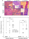
Effect of IL-12p35 or p40 deficiency on renal inflammatory pathology following chlamydial infection. (A) Kidneys harvested from wild-type (representative images in panels a and d), IL-12p35 KO (panels b and e), and IL-12p40 KO (c and f) mice as described in the legend to Fig. 3 were further examined for inflammatory pathologies under a microscope. The inflammation was semiquantitatively scored based on the criteria listed in Materials and Methods, and the scores are given in each image as examples. Inflammatory cells or infiltrates are marked with white arrows. The inset in the lower left corner of each panel shows how the polymorphic nuclear cells (PMNs; green arrows) and mononuclear cells (MCs; red arrows) in the corresponding inflammatory infiltration loci were differentiated under a 40× objective lens. Renal tubules (white star) and glomeruli (white arrowheads) are marked. (B) Inflammatory scores for mouse groups, including wild type (n = 7 for primary and n = 8 for secondary infection groups), IL-12p35 KO (n = 6 for primary and n = 7 for secondary infection), and IL-12p40 KO (n = 9 for primary and n = 7 for secondary infection). Each symbol indicates the score from two kidneys of one mouse. Note that the most severe inflammation was in kidney sections from IL-12p40 KO mice (*, P < 0.05; **, P < 0.01; NS, not significant).
Fig 5
Fig 5
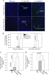
Effect of IL-12p35 or p40 deficiency on chlamydial organism spreading into kidneys. (A) Kidney sections from wild-type (panels a and d), IL-12p35 KO (b and e), and IL-12p40 KO (c and f) mice were subjected to immunolabeling with anti-chlamydial antibodies (green, indicated with white arrows) and a DNA dye (blue). The intact chlamydial inclusions were counted from each section, and 3 sections were used from each kidney. To ensure that the inclusions counted from the 3 sections were not the same inclusions, each of the 3 sections was selected by skipping 5 sections in between. The total number of chlamydial inclusions counted from the 6 sections (from both kidneys) of the same mouse was used to calculate the average number of inclusions per section that was assigned to each mouse. The number of inclusions per section for all mice was summarized (B) and expressed as the mean and standard deviation. Note that chlamydial inclusions were detected only in enlarged and edematous kidneys from the IL-12 gene KO mice, not in kidneys from the wild-type mice. Kruskal-Wallis analysis revealed that the IL-12p40 KO kidney sections contained significantly more chlamydial inclusions than IL-12p35 KO kidney sections. No chlamydial inclusions were detected in wild-type mouse kidney sections using this method. (C) To test whether infectious live organisms could be recovered from the kidney tissues, a parallel experiment with 3 to 5 mice in each group listed on the x axis was carried out. Both kidneys were harvested from each mouse 100 days after C. muridarum infection for making homogenates. The live chlamydial organisms from each renal homogenate were titrated, and the numbers of live organisms per kidney were calculated for each mouse (values are means and standard deviations). Note that the IL-12p40 KO mice had the highest number of live chlamydial organisms in their kidneys. (D) Aliquots of the same kidney homogenates were used for titrating live bacteria on sheep blood agar plates. The number of bacterial colonies counted from each plate was used to calculate the total number of bacteria recovered from each kidney, and the data are expressed as total number of bacteria per mouse. Similar numbers of bacteria were recovered from IL-12p35 KO and IL-12p40 KO mice.
Fig 6
Fig 6
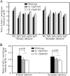
Effect of IL-12p35 or p40 deficiency on mouse antibody responses after chlamydial infection. (A) The 3 groups of mice infected with chlamydial organisms as described in the legend to Fig. 1 were bled on day 114 (after primary infection; n = 4 for wild-type and 6 for IL-12p35 and p40 KO groups) or day 143 (after secondary infection; n = 8 for wild-type and 7 for IL-12p35 and p40 KO groups) for titrating C. muridarum-specific IgG antibodies, including both total IgG and IgG isotypes. The highest dilution at which a given mouse serum still positively stained the C. muridarum inclusions was determined as the titer of that serum. The serum dilutions were converted into log10 titers for calculating means and standard deviations. (B) The ratios of IgG2a titer to IgG1 titer were calculated for each group of mice. Note that the wild-type mice displayed the highest IgG2/IgG1 ratio during both primary and secondary infections (ratios are expressed as means and standard deviations).
Fig 7
Fig 7
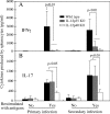
Effect of IL-12p35 or p40 deficiency on mouse T cell responses following chlamydial infection. Splenocytes were harvested from IL-12p35 KO, IL-12p40 KO, and wild-type mice on day 114 (after primary infection) or day 143 (after secondary infection). These mice were infected with C. muridarum as described in the legend to Fig. 1, and numbers of mice in each group are the same as in Fig. 6. The splenocytes were restimulated in vitro with UV-inactivated C. muridarum organisms for 72 h. IFN-γ (A) and IL-17 (B) in culture supernatants were measured using ELISA, and the results are expressed in picograms per ml (data are means and standard deviations). Note that levels of IFN-γ produced by splenocytes from IL-12p35 or IL-12p40 KO mice and IL-17 from IL-12p40 KO mice were significantly lower than those from wild-type mice.

Similar articles

Cited by

References

    1. Sherman KJ, Daling JR, Stergachis A, Weiss NS, Foy HM, Wang SP, Grayston JT. 1990. Sexually transmitted diseases and tubal pregnancy. Sex Transm Dis. 17:115–121 - PubMed
    1. Morrison RP, Feilzer K, Tumas DB. 1995. Gene knockout mice establish a primary protective role for major histocompatibility complex class II-restricted responses in Chlamydia trachomatis genital tract infection. Infect. Immun. 63:4661–4668 - PMC - PubMed
    1. Watford WT, Moriguchi M, Morinobu A, O'Shea JJ. 2003. The biology of IL-12: coordinating innate and adaptive immune responses. Cytokine Growth Factor Rev. 14:361–368 - PubMed
    1. Hunter CA. 2005. New IL-12-family members: IL-23 and IL-27, cytokines with divergent functions. Nat. Rev. Immunol. 5:521–531 - PubMed
    1. Happel KI, Dubin PJ, Zheng M, Ghilardi N, Lockhart C, Quinton LJ, Odden AR, Shellito JE, Bagby GJ, Nelson S, Kolls JK. 2005. Divergent roles of IL-23 and IL-12 in host defense against Klebsiella pneumoniae. J. Exp. Med. 202:761–769 - PMC - PubMed

Publication types

Substances