

Using shape effects to target antibody-coated nanoparticles to lung and brain endothelium
- PMID: 23754411
- PMCID: PMC3696781
- DOI: 10.1073/pnas.1308345110
Using shape effects to target antibody-coated nanoparticles to lung and brain endothelium
Abstract
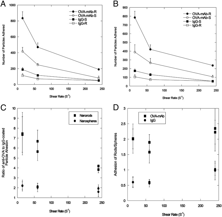
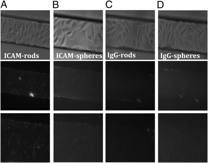
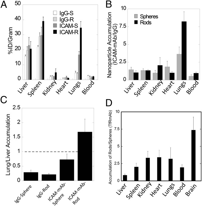
Vascular endothelium offers a variety of therapeutic targets for the treatment of cancer, cardiovascular diseases, inflammation, and oxidative stress. Significant research has been focused on developing agents to target the endothelium in diseased tissues. This includes identification of antibodies against adhesion molecules and neovascular expression markers or peptides discovered using phage display. Such targeting molecules also have been used to deliver nanoparticles to the endothelium of the diseased tissue. Here we report, based on in vitro and in vivo studies, that the specificity of endothelial targeting can be enhanced further by engineering the shape of ligand-displaying nanoparticles. In vitro studies performed using microfluidic systems that mimic the vasculature (synthetic microvascular networks) showed that rod-shaped nanoparticles exhibit higher specific and lower nonspecific accumulation under flow at the target compared with their spherical counterparts. Mathematical modeling of particle-surface interactions suggests that the higher avidity and specificity of nanorods originate from the balance of polyvalent interactions that favor adhesion and entropic losses as well as shear-induced detachment that reduce binding. In vivo experiments in mice confirmed that shape-induced enhancement of vascular targeting is also observed under physiological conditions in lungs and brain for nanoparticles displaying anti-intracellular adhesion molecule 1 and anti-transferrin receptor antibodies.
Keywords: SMN; biodistribution; cylinder; drug delivery; morphology.
Conflict of interest statement
The authors declare no conflict of interest.
Figures
References
-
- Brekken RA, Thorpe PE. Vascular endothelial growth factor and vascular targeting of solid tumors. Anticancer Res. 2001;21(6B):4221–4229. - PubMed
-
- Muzykantov VR. Targeting of superoxide dismutase and catalase to vascular endothelium. J Control Release. 2001;71(1):1–21. - PubMed
-
- Tousoulis D, et al. Novel therapies targeting vascular endothelium. Endothelium. 2006;13(6):411–421. - PubMed
-
- Ruoslahti E. Specialization of tumour vasculature. Nat Rev Cancer. 2002;2(2):83–90. - PubMed
-
- Hanahan D, Folkman J. Patterns and emerging mechanisms of the angiogenic switch during tumorigenesis. Cell. 1996;86(3):353–364. - PubMed
Publication types
MeSH terms
Substances
LinkOut - more resources
Full Text Sources
Other Literature Sources
