. 2013 Aug;33(16):3113-24.
doi: 10.1128/MCB.01597-12. Epub 2013 Jun 10.

ATM mediates pRB function to control DNMT1 protein stability and DNA methylation

Affiliations

ATM mediates pRB function to control DNMT1 protein stability and DNA methylation

Awad Shamma et al. Mol Cell Biol. 2013 Aug.

Abstract

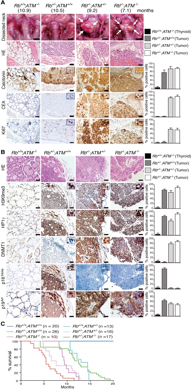
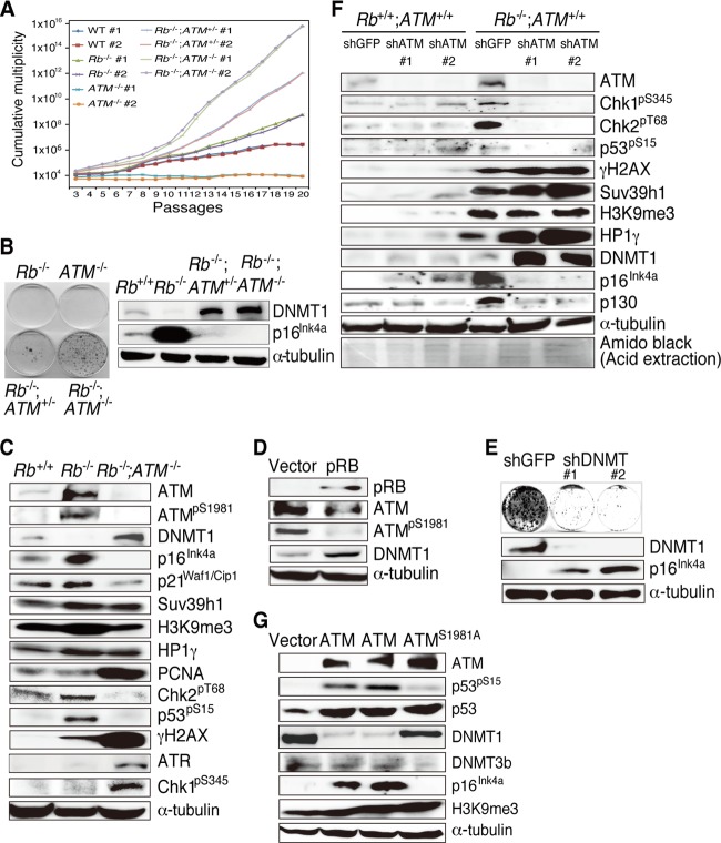
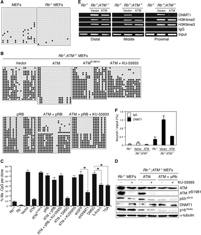
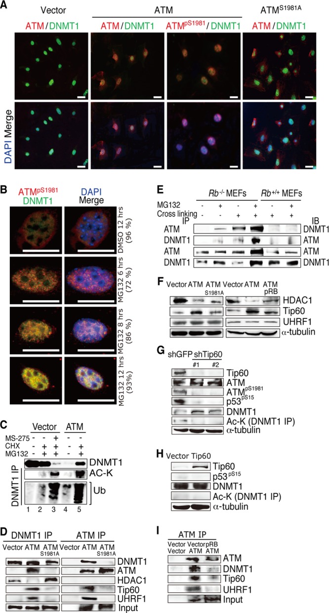
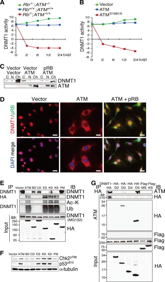
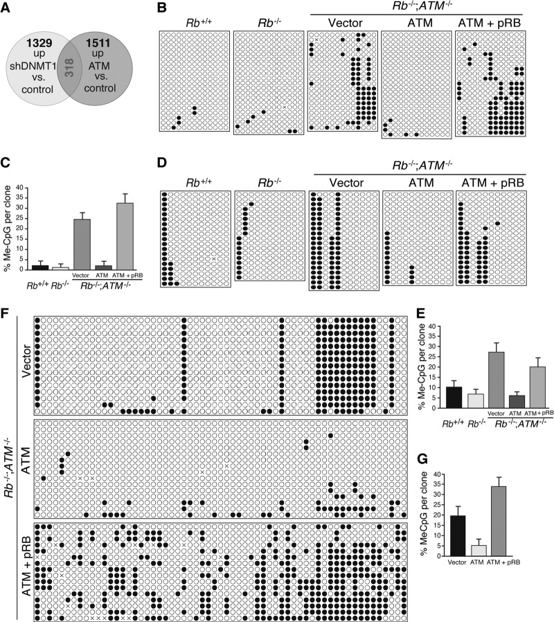
The retinoblastoma tumor suppressor gene (RB) product has been implicated in epigenetic control of gene expression owing to its ability to physically bind to many chromatin modifiers. However, the biological and clinical significance of this activity was not well elucidated. To address this, we performed genetic and epigenetic analyses in an Rb-deficient mouse thyroid C cell tumor model. Here we report that the genetic interaction of Rb and ATM regulates DNMT1 protein stability and hence controls the DNA methylation status in the promoters of at least the Ink4a, Shc2, FoxO6, and Noggin genes. Furthermore, we demonstrate that inactivation of pRB promotes Tip60 (acetyltransferase)-dependent ATM activation; allows activated ATM to physically bind to DNMT1, forming a complex with Tip60 and UHRF1 (E3 ligase); and consequently accelerates DNMT1 ubiquitination driven by Tip60-dependent acetylation. Our results indicate that inactivation of the pRB pathway in coordination with aberration in the DNA damage response deregulates DNMT1 stability, leading to an abnormal DNA methylation pattern and malignant progression.


Figures

Fig 1
Fig 1
Analysis of Rb-deficient C cell tumors with various ATM genotypes. (A) Thyroid tumors developed in mice of the indicated genotypes at the indicated months of age were photographed, and the tissues were then analyzed by hematoxylin-eosin (HE) staining and immunostaining with antibodies to the indicated proteins. The frequency of immunopositive cells per 200 normal thyroid or tumor cells was quantified. Columns show means and SEM. Scale bars, 1 mm in macroscopic and 100 μm in microscopic pictures. (B) Serial sections of the same tumors were immunostained with antibodies to the indicated proteins and analyzed as described above for panel A. (C) Survival rate in mice of the indicated genotypes. Data are expressed as percent survival.
Fig 2
Fig 2
Analysis of Rb−/− MEFs of various ATM genotypes. (A) 3T3 protocol assay of MEFs with the indicated genotypes. MEFs were freshly prepared from embryos of timed pregnant inbred mice and cultivated for 3 passages before the start of the 3T3 assay. The cumulative multiplicity in the representative cultures at the indicated passage number is illustrated in the graph. WT, wild type. (B) Representative results from colony formation assays with MEFs of the indicated genotypes plated at 1 × 10³ cells per 60-mm dish and cultured for 14 days. (C) Immunoblotting analysis of MEFs of the indicated genotypes with antibodies to the indicated proteins. (D) Rb−/− MEFs infected with retroviruses expressing the indicated proteins were selected and analyzed by immunoblotting. (E) Rb−/−; ATM−/− MEFs infected with lentiviruses expressing shRNAs that target the indicated genes were analyzed by a colony formation assay and immunoblotting with antibodies to the indicated proteins. #1 and #2 indicate independently designed shRNAs targeting DNMT1. (F) Rb+/+ or Rb−/− MEFs infected with lentiviruses expressing the shRNAs that target the indicated genes were selected and analyzed by immunoblotting. GFP, green fluorescent protein. (G) Rb−/−; ATM−/− MEFs transduced with vectors expressing the indicated proteins were selected and analyzed by immunoblotting.
Fig 3
Fig 3
DNA methylation status of the Ink4a promoter region in MEFs of various Rb and ATM genotypes. (A) Results of bisulfite sequencing of the Ink4a promoter region in Rb+/+ or Rb−/− MEFs. (B) Rb−/−; ATM−/− MEFs were transfected with the vectors expressing the indicated proteins, selected, cultured for an additional 10 days, and thereafter treated with the indicated reagent (KU-55933 at 10 μM for 7 days) and analyzed. (C) Statistical analysis of the results of bisulfite sequencing of the Ink4a promoter region in MEFs of the indicated genotypes that were transfected with the expression vectors or infected with lentiviruses expressing shRNAs that target the indicated genes, selected for 72 h, and treated with the indicated reagents (trichostatin A [TSA] at 2 μM for 3 days, KU-55933 at 10 μM for 5 days, and 5-AzadC at 2 μM for 3 days). *, P < 0.01 by Mann-Whitney test. (D) Immunoblotting analysis of MEFs of the indicated genotypes with antibodies to the indicated proteins. MEFs were transfected with the vectors expressing the indicated proteins, selected, cultured for an additional 10 days, and thereafter treated with the indicated reagent (KU-55933 at 10 μM for 5 days) and analyzed. (E) ChIP assay of the Ink4a promoter region in MEFs of the indicated genotypes. Rb−/−; ATM−/− MEFs were transduced with vectors expressing the indicated proteins, selected, and analyzed for binding of the indicated proteins by PCR with specific primers (see Fig. S6B and Table S5 in the supplemental material). Proximal, middle, and distal regions are relative to the transcription start site (see Fig. S6B in the supplemental material). (F) Quantitative ChIP assay of the Ink4a promoter region (positions ∼−44 to −149) in MEFs of the indicated genotypes. Rb−/−; ATM−/− MEFs were transduced with vectors expressing the indicated proteins, selected, and analyzed for DNMT1 binding by qPCR with specific primers (see Fig. S6B and Table S5 in the supplemental material).
Fig 4
Fig 4
Effects of pRB and ATM on the DNMT1 degradation complex. (A) IF analysis of the indicated proteins in Rb−/−; ATM−/− MEFs transduced with vectors expressing the indicated proteins. (B) IF analysis of Rb−/−; ATM−/− MEFs transduced with vectors expressing the indicated proteins and treated with vehicle (DMSO) or 5 μM MG132 for the indicated periods of time. Numbers in parentheses indicate percentages of the representative phenotype that appeared in all analyzed cells. (C) Lysates from Rb−/−; ATM−/− MEFs transduced with vectors expressing the indicated proteins and treated with the indicated reagents were immunoprecipitated with DNMT1-specific antibody and analyzed by immunoblotting. Concentrations and durations of treatments are as follows: MS275 at 5 μM for 48 h, cycloheximide (CHX) at 2.5 μg/ml for 12 h, and MG132 at 5 μM for 12 h. Ub, ubiquitin (D) Lysates from Rb−/−; ATM−/− MEFs transduced with vectors expressing the indicated proteins were treated with 5 μM MG132 for 12 h, immunoprecipitated with antibodies to DNMT1 or ATM, and analyzed by immunoblotting. (E) Lysates from MEFs of the indicated genotypes arrested in S phase and treated with the indicated reagents were immunoprecipitated (IP) with antibodies to DNMT1 or ATM and analyzed by immunoblotting (IB). Cells were arrested in S phase by a double-thymidine block at 2 mM for 18 h (first block), released for 9 h, and blocked for another 17 h (second block); MG132 was used at 5 μM for 12 h; and cross-linking was performed with a 1% formaldehyde solution for 10 min at room temperature and terminated with 0.125 M glycine. (F) Rb−/−; ATM−/− MEFs were transduced with vectors expressing the indicated proteins, selected with 4 μg/ml puromycin and 8 μg/ml blasticidin S for 5 days, and analyzed by immunoblotting using antibodies specific to the indicated proteins. (G) Rb−/−; ATM−/− MEFs transduced with vectors expressing ATM were infected with lentiviruses expressing shRNAs that target the indicated genes, selected, and analyzed by immunoblotting. (H) Rb−/−; ATM−/− MEFs transduced with vectors expressing the indicated proteins were analyzed by immunoblotting. Ac-K, acetylated lysine. (I) Lysates from Rb−/−; ATM−/− MEFs transduced with vectors expressing the indicated proteins were treated with 5 μM MG132 for 12 h, immunoprecipitated with ATM-specific antibody, and analyzed by immunoblotting.
Fig 5
Fig 5
Physical interaction between ATM and DNMT1. (A) Measurement of the dose-dependent effects of the lysates from MEFs of the indicated genotypes on DNMT1 enzyme activity. (B) Measurement of the dose-dependent effects of the lysates from Rb−/−; ATM−/− MEFs transduced with vectors expressing the indicated proteins on DNMT1 enzyme activity. (C) Cytosolic (C), nuclear (N), or chromatin (Ch) protein fractions from Rb−/−; ATM−/− MEFs transduced with vectors expressing the indicated proteins and treated with 5 μM MG132 for 12 h were analyzed by immunoblotting. (D) IF analysis with antibodies specific to the indicated proteins in Rb−/−; ATM−/− MEFs transduced with vectors expressing the indicated proteins. Scale bars, 20 μm. (E) Lysates from Rb−/−; ATM−/− MEFs transduced with vectors expressing the indicated proteins (see Fig. S8A in the supplemental material) were untreated or treated with 5 μM MG132 for 12 h (indicated in parentheses), immunoprecipitated, and analyzed by immunoblotting. (F) Lysates from cells prepared as described above for panel E were analyzed by immunoblotting with antibodies to the indicated proteins. (G) Lysates from FLAG-ATM-reconstituted Rb−/−; ATM−/− MEFs transduced with vectors expressing the indicated proteins (see Fig. S8B in the supplemental material) were treated with 5 μM MG132 for 12 h, immunoprecipitated with antibodies specific to the indicated proteins, and analyzed by immunoblotting.
Fig 6
Fig 6
Genome-wide survey for genes epigenetically regulated by the pRB-ATM-DNMT1 nexus. (A) Venn diagram of microarray analysis. Rb−/−; ATM−/− MEFs were transduced with vectors expressing the indicated proteins or infected with lentiviruses expressing the shRNAs that target the indicated genes, selected with 4 μg/ml puromycin or 8 μg/ml blasticidin S for 72 h, cultured for an additional 4 days under normal culture conditions, and analyzed by microarray analysis. Commonly upregulated genes in the 2 combinations of experiments are shown in red. A list of genes and their ontology are indicated in Table S6 in the supplemental material. (B and C) Results of bisulfite sequencing of the Shc2 promoter region in MEFs of the indicated genotypes transduced with vectors expressing the indicated proteins and their statistical analysis. (D and E) Results of bisulfite sequencing of the FoxO6 promoter region in MEFs of the indicated genotypes transduced with vectors expressing the indicated proteins and their statistical analysis. (F and G) Results of bisulfite sequencing of the Noggin promoter region in Rb−/−; ATM−/− MEFs transduced with vectors expressing the indicated proteins and their statistical analysis. A detailed statistical analysis of the effects of pRB and ATM on the frequency and location of methylated CpG (Me-CpG) nucleotides in the promoters of these three representative genes is shown in Fig. S9 in the supplemental material.
Fig 7
Fig 7
Model for the functional interaction of ATM and DNMT1 in the absence of pRB. The molecular species participating in the DNMT1 protein stability regulation complex are dynamically altered depending on the status of pRB and ATM, and these changes actively modulate the DNA methylation status of target gene promoters. DKO, double knockout; AC, acetylation.

References

    1. Burkhart DL, Sage J. 2008. Cellular mechanisms of tumour suppression by the retinoblastoma gene. Nat. Rev. Cancer 8:671–682 - PMC - PubMed
    1. Viatour P, Sage J. 2011. Newly identified aspects of tumor suppression by RB. Dis. Model. Mech. 4:581–585 - PMC - PubMed
    1. Takahashi C, Sasaki N, Kitajima S. 2012. Twists in views on RB functions in cellular signaling, metabolism and stem cells. Cancer Sci. 103:1182–1188 - PMC - PubMed
    1. Robertson KD, Ait-Si-Ali S, Yokochi T, Wade PA, Jones PL, Wolffe AP. 2000. DNMT1 forms a complex with Rb, E2F1 and HDAC1 and represses transcription from E2F-responsive promoters. Nat. Genet. 25:338–342 - PubMed
    1. Dick FA. 2007. Structure-function analysis of the retinoblastoma tumor suppressor protein—is the whole a sum of its parts? Cell Div. 2:26.10.1186/1747-1028-2-26 - DOI - PMC - PubMed

Publication types

MeSH terms

Substances