. 2013 Aug;33(16):3099-112.
doi: 10.1128/MCB.00524-13. Epub 2013 Jun 10.

A network of transcription factors operates during early tooth morphogenesis

Affiliations

A network of transcription factors operates during early tooth morphogenesis

Minglian Zhao et al. Mol Cell Biol. 2013 Aug.

Abstract

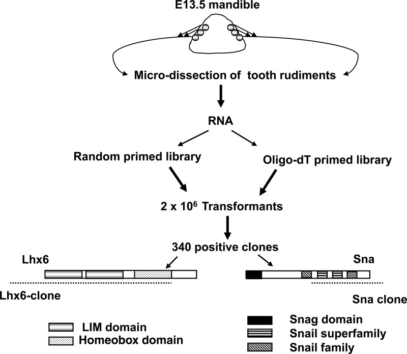
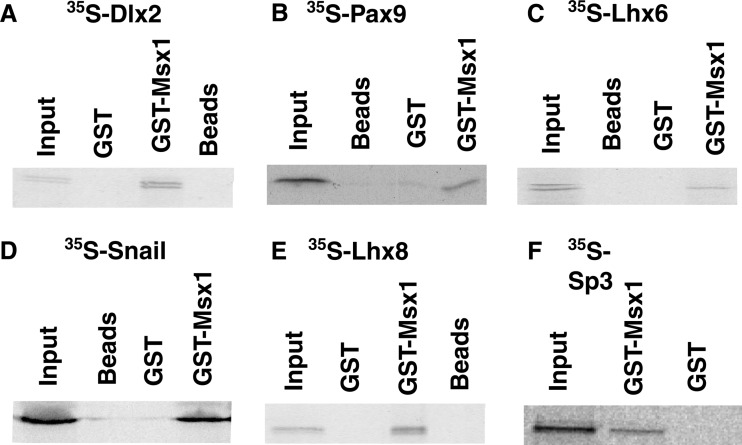
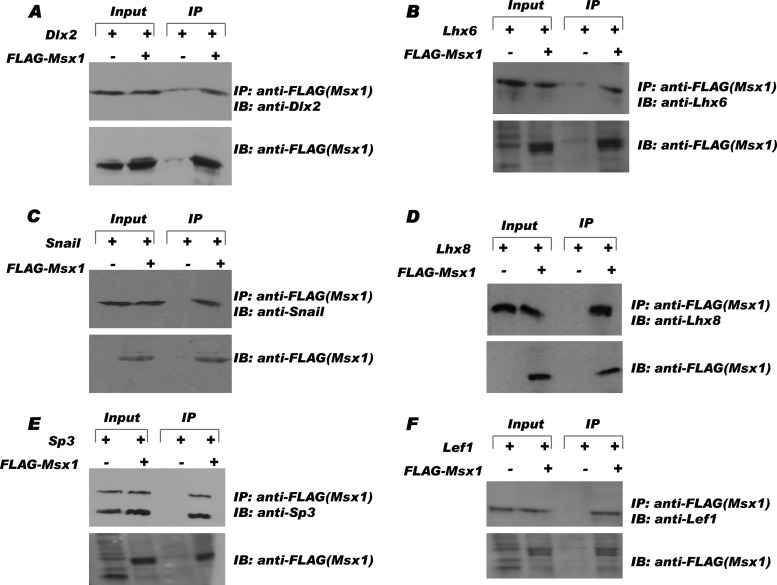
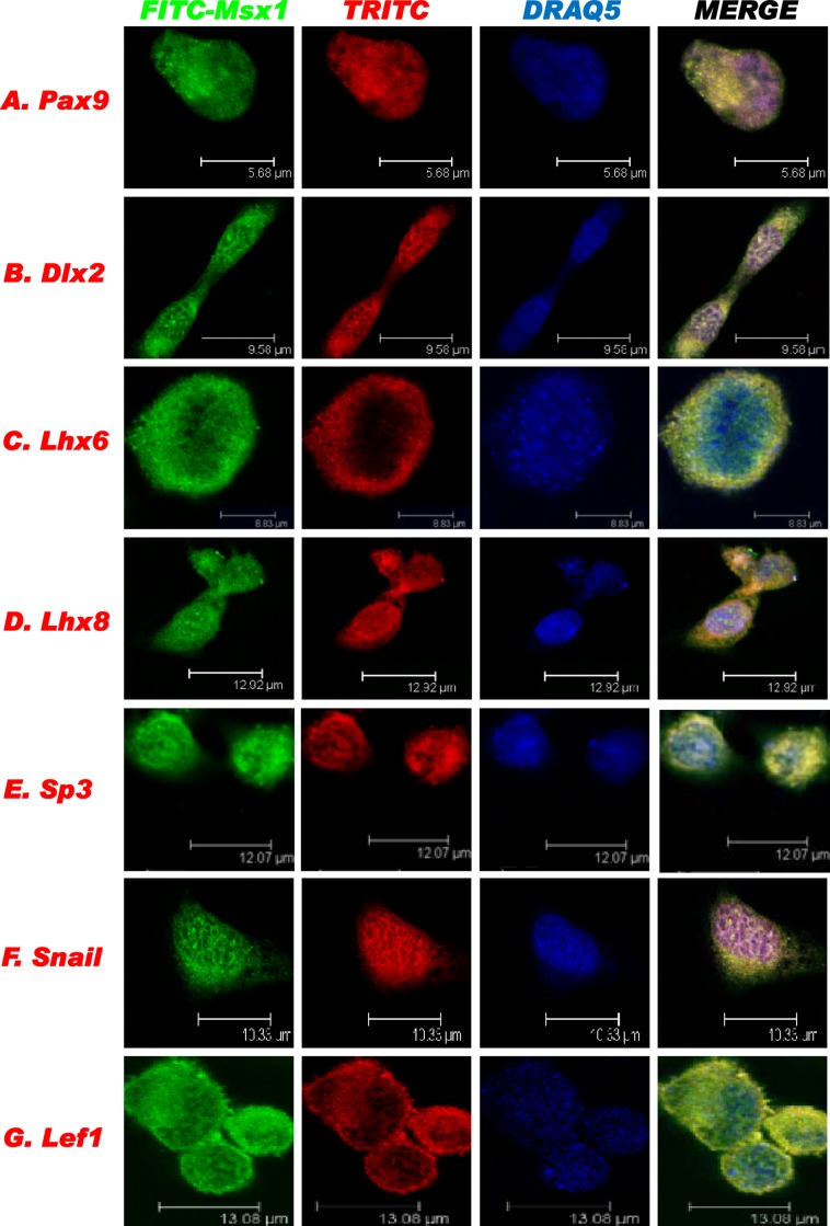
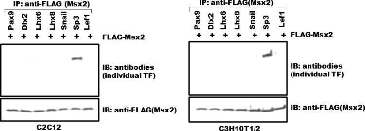
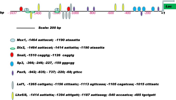
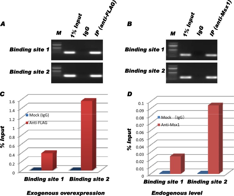
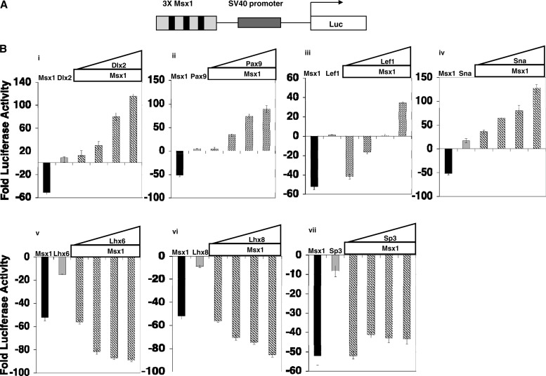
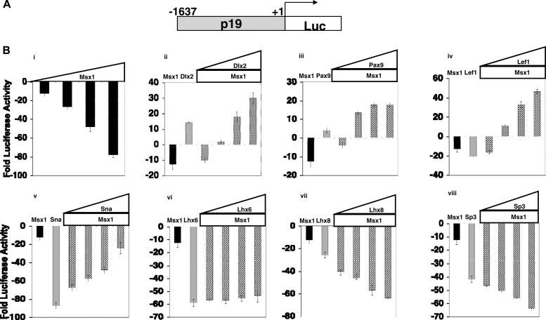
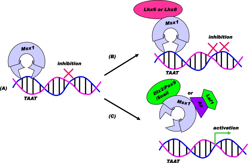
Improving the knowledge of disease-causing genes is a unique challenge in human health. Although it is known that genes causing similar diseases tend to lie close to one another in a network of protein-protein or functional interactions, the identification of these protein-protein networks is difficult to unravel. Here, we show that Msx1, Snail, Lhx6, Lhx8, Sp3, and Lef1 interact in vitro and in vivo, revealing the existence of a novel context-specific protein network. These proteins are all expressed in the neural crest-derived dental mesenchyme and cause tooth agenesis disorder when mutated in mouse and/or human. We also identified an in vivo direct target for Msx1 function, the cyclin D-dependent kinase (CDK) inhibitor p19(ink4d), whose transcription is differentially modulated by the protein network. Considering the important role of p19(ink4d) as a cell cycle regulator, these results provide evidence for the first time of the unique plasticity of the Msx1-dependent network of proteins in conferring differential transcriptional output and in controlling the cell cycle through the regulation of a cyclin D-dependent kinase inhibitor. Collectively, these data reveal a novel protein network operating in the neural crest-derived dental mesenchyme that is relevant for many other areas of developmental and evolutionary biology.


Figures

Fig 1
Fig 1
A schematic representation of the yeast two-hybrid screen using E13.5 tooth cDNA. Microdissected tooth rudiments from E13.5 embryos were used to make two cDNA sources, one oligo(dT) primed and the other random primed. The cDNAs were transformed into the yeast library vector by homologous recombination. From screening 2 × 10⁶ transformants and analyzing 340 clones, two clones that corresponded to the ORFs of Lhx6 and Sna were obtained. The Lhx6 clone contained the LIM domains and the homeodomain shown, in comparison with the full-length ORF. The Sna clone, obtained from the oligo(dT)-primed library, included the 3′ untranslated region and the C-terminal zinc fingers.
Fig 2
Fig 2
Msx1 interacts directly with tooth-specific transcription factors in vitro. GST pulldown assays with in vitro-translated, 35S-labeled Dlx2 (A), Pax9 (B), Lhx6 (C), Snail (D), Lhx8 (E), and Sp3 (F) were performed using the Msx1-GST fusion protein. GST-bound beads and unbound beads were used as controls. Immobilized proteins were eluted in the SDS buffer, resolved by SDS-PAGE, and visualized by autoradiography.
Fig 3
Fig 3
Msx1 interacts with tooth-specific transcription factors in vivo. pCMV-FLAG-Msx1 or control vector (pCMV-FLAG-Tag2B) was coexpressed with transcription factors Dlx2 (A), Lhx6(B), Snail (C), Lhx8 (D), Sp3 (E), and Lef1 (F) in C3H10T1/2 cells. After 36 h, cell lysate (20%) of each was taken as the input sample to verify the level of expression (first and second lanes). All the transcription factors tested were detected only in the presence of FLAG-Msx1 in the immunoprecipitation samples (fourth lanes) and not in the control samples (third lanes). IP, immunoprecipitation; IB, immunoblotting.
Fig 4
Fig 4
Endogenous colocalization of Msx1 and interacting transcription factors in living cells. Msx1 was labeled with FITC-conjugated antibody and the other transcription factors, at their endogenous levels in C3H10T1/2 cells, were labeled with TRITC for confocal imaging analysis. DRAQ5 was used to stain DNA.
Fig 5
Fig 5
Msx2 interacts with Sp3 only. FLAG-Msx2 was cotransfected with transcription factors (TF) Pax9, Dlx2, Lhx6, Lhx8, Snail, Sp3, and Lef1 into C2C12 and C3H10T1/2 cells. Only Sp3 and not the other Msx1 interactors tested was coimmunoprecipitated with Msx2 both in C2C12 and C3H10T1/2 cells.
Fig 6
Fig 6
Schematic representation of the transcription factor binding sites in the murine p19ink4d proximal promoter region. All the binding loci are based on computer analysis and UniProbe database profiling (http://thebrain.bwh.harvard.edu/uniprobe/). Msx1 possesses two binding clusters, labeled 1 (nt 1464) and 2 (nt 1190) in pale blue ovals.
Fig 7
Fig 7
The p19ink4d proximal promoter region is a direct target of Msx1. (A) After chromatin immunoprecipitation, samples from C3H10T1/2 cells transfected with pCMV-Msx1-FLAG were PCR amplified; the binding region was directly amplified prior to immunoprecipitation (1% Input) and specifically amplified in the immunoprecipitated sample (anti-FLAG). No amplification was detected in the nonspecific IgG (normal mouse serum IgG)-immunoprecipitated sample (IgG) due to low background under the exogenous overexpression of Msx1 in cells. (B) After chromatin immunoprecipitation, samples from untransfected C3H10T1/2 cells were PCR amplified; the binding region was directly amplified prior to immunoprecipitation (1% Input) and specifically amplified in the immunoprecipitated sample (anti-Msx1). (C and D) Quantitative analysis of Msx1 binding preference from transfected cells (exogenous expression) (C) and untransfected cells (endogenous expression) (D). In both situations, there was a 3-fold-higher preference of Msx1 for binding site 2 (nt 1190) over binding site 1 (nt 1464) in vivo. M, DNA marker.
Fig 8
Fig 8
Functional consequences of the interaction of Msx1 with tooth-specific transcription factors for the transcriptional regulation of an artificial Msx1-dependent promoter. (A) Schematic representation of reporter plasmid containing 3 Msx1 binding sites (3× Msx1). (B) C3H10T1/2 cells were cotransfected with 1 μg of reporter plasmid containing 3× Msx1-Luc, along with increasing amounts of expression plasmid Msx1-FLAG (0.25 μg, 0.50 μg, 0.75 μg, and 1.0 μg). Cells were harvested 24 h after transfection for reporter gene assays. Transcription efficiencies were determined using Renilla luciferase plasmid. For this and subsequent experiments, the levels of luciferase activity were normalized to Renilla luciferase activity and expressed as fold luciferase activity relative to the level of luciferase activity from cells transfected with the reporter construct and empty expression plasmid. Effects of Dlx2 (i), Pax9 (ii), Lef1 (iii), Sna (iv), Lhx6 (v), Lhx8 (vi), and Sp3 (vii) on Msx1-mediated transcription are shown. One microgram of the 3× Msx1-Luc reporter plasmid was cotransfected with 0.25 μg of Msx1-FLAG or 0.25 μg of the indicated transcription factor, or increasing amounts of transcription factors were added (0.25 μg, 0.50 μg, 0.75 μg, and 1.0 μg) along with 0.25 μg of Msx1-FLAG. All the transfection experiments were performed three times, and results are shown as means ± standard deviations. Pax9, Dlx2, Sna, and Lef1 acted as activators of transcription at the 3× Msx1 promoter in conjugation with Msx1, whereas Lhx8 and Lhx6 with Msx1 resulted in repression. The repression by Sp3 at the 3× Msx1 promoter did not change in the presence of Msx1.
Fig 9
Fig 9
Functional consequences of the interaction of Msx1 with tooth-specific transcription factors for the transcriptional regulation of the p19 promoter. C3H10T1/2 cells were cotransfected with 1 μg of p19-Luc reporter plasmid along with increasing amounts of expression plasmid Msx1-FLAG (0.25 μg, 0.50 μg, 0.75 μg, and 1.0 μg). Cells were harvested 24 h after the transfections for reporter gene assays. All the transfection experiments were performed three times, and the bars indicate the means ± standard deviations. Msx1 used alone repressed the p19 promoter. Pax9, Dlx2, Lef1, and Snail in conjugation with Msx1 acted as activators of transcription at the p19 promoter, whereas Lhx8 and Sp3 used with Msx1 resulted in repression of p19. The repression by Lhx6 at the p19 promoter did not change in the presence of Msx1.
Fig 10
Fig 10
Model for transcriptional consequences of Msx1-dependent combinatorial interactions. (A) Direct repression model: repressor Msx1 may bind a target promoter region containing the core motif TAAT via the homeodomain to exert repression. (B) Synergistic repression model: corepressor Sp3, Lhx8, or Lhx6 can interact with Msx1 to enhance repression via functional synergism. (C) Functional antagonism model: activator Dlx2, Pax9, or Snail or Lef1 via an adaptor protein can interact with Msx1 to relieve repression, leading to downstream gene activation via functional antagonism.

Similar articles

Cited by

References

    1. Thesleff I, Vaahtokari A, Partanen AM. 1995. Regulation of organogenesis. Common molecular mechanisms regulating the development of teeth and other organs. Int. J. Dev. Biol. 39:35–50 - PubMed
    1. Bei M, Peters H, Maas RL. 2002. The role of PAX and MSX genes in craniofacial development, p 101–112 In Lin KY, Ogle RC, Jane JA. (ed), Craniofacial surgery: science & surgical technique. W. B. Saunders Company, Philadelphia, PA
    1. Bei M. 2009. Molecular genetics of ameloblast cell lineage. J. Exp. Zool. B Mol. Dev. Evol. 312B:437–444 - PMC - PubMed
    1. Bei M. 2009. Molecular genetics of tooth development. Curr. Opin. Genet. Dev. 19:504–510 - PMC - PubMed
    1. Hayashi S, Scott MP. 1990. What determines the specificity of action of Drosophila homeodomain proteins? Cell 63:883–894 - PubMed

Publication types

MeSH terms

LinkOut - more resources