Review
. 2013 Jun 10;368(1622):20130088.
doi: 10.1098/rstb.2013.0088. Print 2013 Jul 19.

Early bioenergetic evolution

Affiliations
Review

Early bioenergetic evolution

Filipa L Sousa et al. Philos Trans R Soc Lond B Biol Sci. .

Abstract

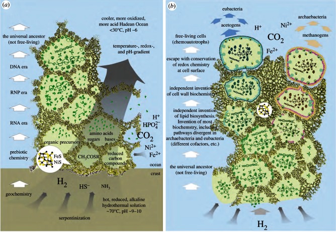
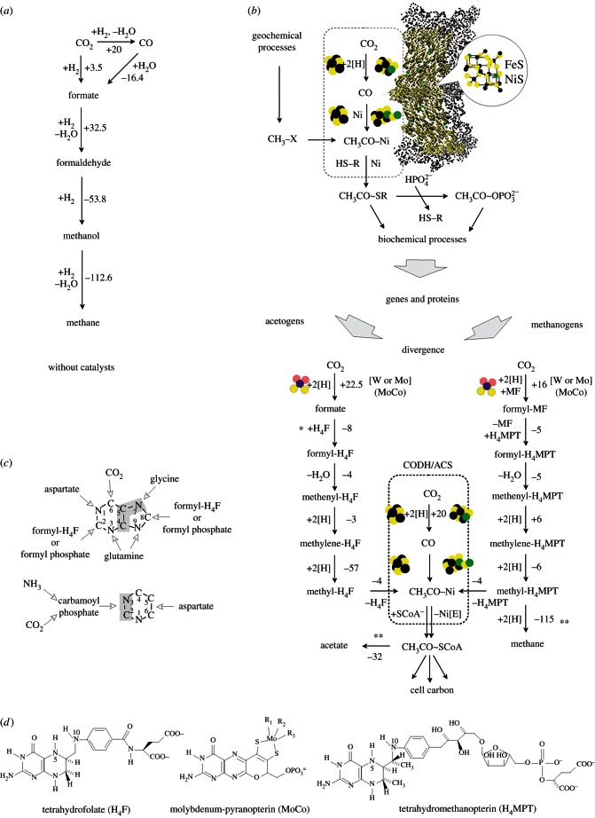
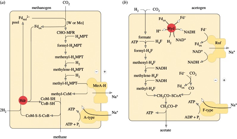
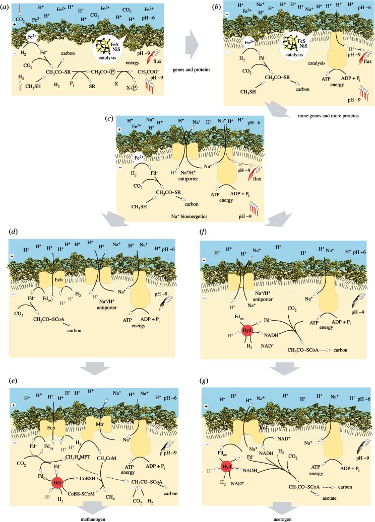
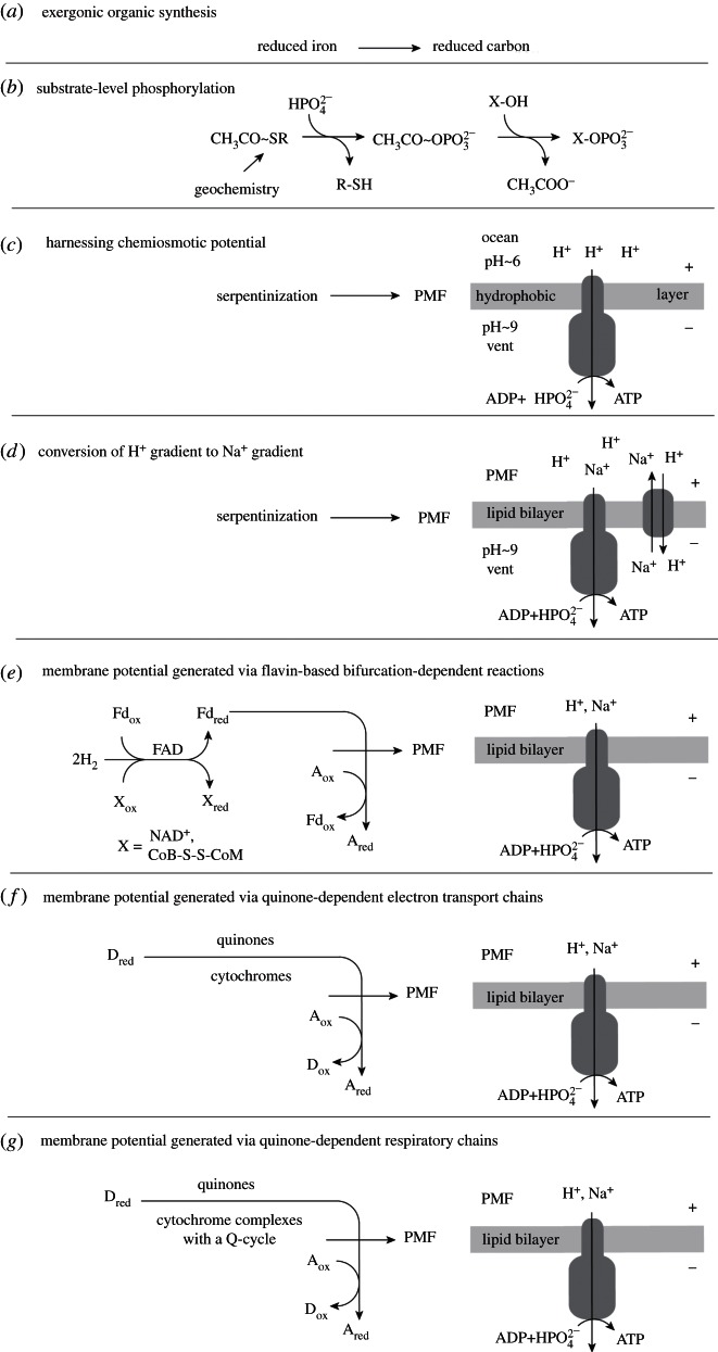
Life is the harnessing of chemical energy in such a way that the energy-harnessing device makes a copy of itself. This paper outlines an energetically feasible path from a particular inorganic setting for the origin of life to the first free-living cells. The sources of energy available to early organic synthesis, early evolving systems and early cells stand in the foreground, as do the possible mechanisms of their conversion into harnessable chemical energy for synthetic reactions. With regard to the possible temporal sequence of events, we focus on: (i) alkaline hydrothermal vents as the far-from-equilibrium setting, (ii) the Wood-Ljungdahl (acetyl-CoA) pathway as the route that could have underpinned carbon assimilation for these processes, (iii) biochemical divergence, within the naturally formed inorganic compartments at a hydrothermal mound, of geochemically confined replicating entities with a complexity below that of free-living prokaryotes, and (iv) acetogenesis and methanogenesis as the ancestral forms of carbon and energy metabolism in the first free-living ancestors of the eubacteria and archaebacteria, respectively. In terms of the main evolutionary transitions in early bioenergetic evolution, we focus on: (i) thioester-dependent substrate-level phosphorylations, (ii) harnessing of naturally existing proton gradients at the vent-ocean interface via the ATP synthase, (iii) harnessing of Na(+) gradients generated by H(+)/Na(+) antiporters, (iv) flavin-based bifurcation-dependent gradient generation, and finally (v) quinone-based (and Q-cycle-dependent) proton gradient generation. Of those five transitions, the first four are posited to have taken place at the vent. Ultimately, all of these bioenergetic processes depend, even today, upon CO2 reduction with low-potential ferredoxin (Fd), generated either chemosynthetically or photosynthetically, suggesting a reaction of the type 'reduced iron → reduced carbon' at the beginning of bioenergetic evolution.

Keywords: acetogens; hydrothermal vents; methanogens; origin of life; sulfate reducers; transition metals.


Figures

Figure 1.
Figure 1.
A scheme for the origin of cells [42,46].
Figure 2.
Figure 2.
(Opposite.) Illustration of some concepts relevant to this paper. (a) Abiotic methane production. Summary representation of the H2-dependent conversions of CO2 to methane without catalysts, adapted from [82]. Numbers next to arrows indicate the approximate change in free energy, ΔG°′, for the step indicated at physiological conditions (25°C and pH 7) in kJ mol−1, conditions which do not generally exist in geochemical environments. Note, however, that Lost City does produce methane of abiotic origin [20]. The thermodynamic values are taken from Maden [83] and Rother & Metcalf [84]. Regarding the equilibria between the different C1 species in the absence of catalysts, see Seewald et al. [82]. (b) Scheme suggesting homology between the acetyl-CoA pathway in modern acetogens and methanogens to geochemical processes in the hydrothermal vent of a serpentinizing system in the crust of the ancient Earth. See text. Numbers next to arrows indicate the approximate change in free energy, ΔG°′, for the step indicated at physiological conditions (25°C and pH 7) in kJ mol−1 as reported in [73]. An asterisk indicates ATP investment, a double asterisk indicates ATP return. Note that net ATP return in acetogens and methanogens requires chemisosmotic coupling. FeS and FeNiS clusters are symbolized. Fuchs [73] gives the free energy change for the CODH/ACS reaction as ΔG°′ = 0 kJ mol−1. The thermodynamic value for the methane-producing step is from [85]. H4F, tetrahydrofolate; MF, methanofuran; H4MPT, tetrahydromethanopterin; Ni(E), an Fe–Ni–S cluster in CODH/ACS; HSCoA, coenzyme A. For the acetyl-CoA pathway, see also Bender et al. [60], Ragsdale [56] and Fuchs [73]. The formate to formyl-H4F conversion in acetogens entails ATP hydrolysis (not shown), which lowers ΔG°′ for the reaction to –10 kJ mol−1. Though all reactions shown are reversible, arrows are shown in one direction only for convenience. (c) The source of carbon and nitrogen atoms in the purine and pyrimidine backbone. Data from Stryer [86] and from Ownby et al. [87]. (d) Pterin cofactors involved in the WL-pathway: MoCo [59] folate and methanopterin [85].
Figure 3.
Figure 3.
Energy metabolism of (a) methanogens and (b) acetogens without cytochromes. Redrawn for Methanothermobacter marburgensis from Thauer et al. [40], Kaster et al. [145] and Thauer & Buckel [143], redrawn for Acetobacterium woodii from Pohlein et al. [146] and Thauer & Buckel [143]. When we refer to acetogens and methanogens that lack cytochromes, we are referring to the physiology in those organisms, as the examples. The use of the symbol Fd2– indicates that the ferredoxin in question has two FeS centres, both of which become reduced. MtrA-H, methyl transferase complex [40]. Rnf: Fd:NADH oxidoreductase, originally named for Rhodobacter nitrogen fixation [147]. Other abbreviations as in figure 2. Enzymes known to perform electron bifurcation are indicated in red: heterodisulfide reductase (Hdr) [145] and hydrogenase (Hyd) [148].
Figure 4.
Figure 4.
A hypothetical path for the events linking figures 2 and 3, redrawn from [43]. Ech: Energy converting hydrogenase [159]. Other abbreviations as in figures 2 and 3. See text.
Figure 5.
Figure 5.
Occurrence of 4Fe–4S cluster motifs among 1606 prokaryotic genomes. The upper part of the figure represents the results of a search for the general form of the 4Fe–4S cluster-forming protein motif CX2CX2CX3C. The proteins of 1606 prokaryotic genomes from the RefSeq database (v03.2012) [166] were under examination for this. The prokaryotes are represented by single bars and are ordered by their taxonomical classification. The height of each bar indicates the proportion of proteins within a single genome containing the motif. The lower part of the figure gives the absolute number of proteins containing one of the four additional motifs, which have different numbers of bridging amino acids. The order of the columns is the same as the corresponding bars in the upper part. The following prokaryotes having a high abundance of CX2CX2CX3C motif containing proteins were marked: archaeoglobi, methanogens (methanobacteria, methanococci, methanomicrobia), coriobacteriales (actinobacteria), dehalococcoidetes (chloroflexi), deferribacteres, clostridia, fusobacteria, deltaproteobacteria and thermodesulfobacteria. All 1606 taxa were also checked for the presence of heterodisulfide reductase subunits (HdrABC) or its relative, the quinone-interacting membrane-bound oxidoreductase subunits of sulfate reducers (QmoABC), as these might hint at the presence of electron bifurcation involving these proteins; black bars indicate taxa where at least one of them was present. Note that several other enzymes known to be involved in electron bifurcation [143] are not indicated.
Figure 6.
Figure 6.
The ‘amazing disappearing tree’ of 48 universal genes for a 100 species set. A tree generated from a concatenated alignment of 48 universal genes, compared with its underlying single gene trees. The species sample comprises 50 archaebacteria and 50 eubacteria. To estimate the inconsistency between single gene trees and the concatenated tree, the frequency of each node in the concatenated tree was compared with its frequency within the single gene trees. The transparency of the branches reflects how often the associated node was present within the single gene trees. The 48 universal genes consist of the 31 genes that were previously identified as universal [226], and later used in phylogenetic analysis [217] namely (ArgRS, RNApol(a), LeuRS, metal-dependent protease, PheRS, GTPase, SecY, Rpl1, Rpl11, Rpl13, Rpl14, Rpl15, Rpl16/L10E, Rpl18, Rpl22, Rpl3, Rpl5, Rpl6, Rps11, Rps12, Rps13, Rps15/13E, Rps17, Rps2, Rps3, Rps4, Rps5, Rps7, Rps8, Rps9, Rps, SerRS), plus 17 additional genes (PRPP, AlaRS, PCNA homologue, RNApol(b), HisRS, Met-aminopeptidase, MetRS, PheRS beta subunit, ProRS, RecA, Rpl4, ThrRS, EfG, translation release factor, eIF5A, TyrRS, ValRS) that are present in this prokaryote sample, which contains no members with highly reduced genomes. The taxa were chosen for broad sampling. For this, proteomes of 1606 prokaryotes were retrieved from the RefSeq database (v03.2012) [166]. Pairwise sequence comparisons were run for the ribosomal protein L3. Based on these results, all prokaryotes were clustered by a hierarchical clustering algorithm. From each cluster 100 sample taxa (50 archaeabacteria and 50 eubacteria) were chosen. A complete list of genomes sampled is available in the electronic supplementary material.
Figure 7.
Figure 7.
Distribution of quinone and haem biosynthetic pathways among 1606 prokaryotic genomes. The left part of the figure represents the organization of the selected taxonomic groups from the 1606 completed sequenced genomes (117 archaeal and 1489 eubacterial). The right part of the figure represents the proportion of genomes within a taxa where at least 70% of the genes involved in the pathway are present. Each column represents a different pathway. Homologous proteins involved in the several steps of ubiquinone (ubiC, ubiA, ubiD/ubiX, ubiB, ubiH, ubiE, ubiF and ubiG), menaquinone (MK) alternative (MqnA, MqnB, MqnC and MqnD) [235], menaquinone or phylloquinone (PQQ) (MenF, MenD, MenH, MenC, MenE, MenB, MenA and UbiE/MenG) [236], haem (HemE, HemF/HemN, HemY/HemG and HemH) and haem alternative (AhbA, AhbB, AhbC and AhbD) biosynthesis pathway were identified by BLAST [237]. The BLAST results were filtered for E values better than 10−10 and amino acid identities greater than or equal to 25 per cent. Owing to the high similarity between genes involved in haem d1 biosynthesis with genes from the haem alternative pathway [131], BLAST searches for the presence of cd1 nitrite reductase (the only enzyme containing haem d1) were also performed. In the genomes where both haem alternative pathway genes and cd1 nitrite reductase were present, the former were considered to be involved in haem d1 biosynthesis. Quinone biosynthesis distribution: ubiquinone is only present in the Eubacteria domain, mainly in proteobacteria (beta, alpha and gamma classes) and a few actinobacteria. It is an oxygen-dependent pathway being confined to aerobic organisms. The cyanobacterial ubiquinone hits reflect the presence of genes probably involved in plastoquinone biosynthesis instead. The two MK biosynthesis pathways are present in both prokaryotic domains although the MK alternative pathway has a broader distribution. The MK alternative pathway is the main pathway in both anaerobic and aerobic organisms such as archaeoglobi, thermoproteales, chrysiogenetes, deferribacteres, aquifecales, gemmatimonadetes, chlamydiae, fibrobacteres, acidobacteria, deinococcus-thermus, epsilonproteobacteria and deltaproteobacteria. The MK (and phylloquinone) ‘classical’ pathway is present in halobacteria, but it was acquired in their common ancestor by lateral gene transfer [231]. The classical pathway is also present in actinobacteria, gammaproteobacteria, cyanobacteria (PQQ and MK), chlorobia and bacteroidetes. Sulfolobales have benzothiophene quinone derivatives instead of typical quinones. Haem biosynthesis distribution: with very few exceptions (five out of 117 archaeal organisms surveyed here), the classical haem pathway is only present in the eubacteria domain. On the contrary, the alternative haem pathway is mostly confined to archaeal haem containing taxa and a few mostly anaerobic eubacteria (thermodesulfobacteria, gemmatimonadetes, clostridia, fibrobacterales and deltaproteobacteria). Interestingly, in some methanomicrobiales (organisms that do not contain cytochromes) genes coding for enzymes involved in the alternative haem pathway are present (Methanoculleus marisnigri JR1, Methanoplanus petrolearius DSM11571 and Methanosphaerula palustris). The role of the genes in these organisms is not clear and they might be involved in F430 synthesis instead.
Figure 8.
Figure 8.
A summary diagram outlining a possible sequence of events in early bioenergetic evolution starting with (a) as the most ancient and ending with (g) as the most recent. Of course, respiratory chains are, generally speaking, ancient, just not as ancient as the bioenergetic processes in acetogens and methanogens that lack cytochromes or those in sulfate reducers, in our view (see text). The scheme in (f) could correspond to the situation in sulfate reducers; the scheme in (g) could correspond to the situation in Paracoccus [241] or Rhodobacter [242].

References

    1. Stüeken EE, Anderson RE, Bowman JS, Brazelton WJ, Colangelo-Lillis J, Goldman AD, Som SM, Baross JA. 2013. Did life originate from a global chemical reactor? Geobiology 11, 101–12610.1111/gbi.12025 (doi:10.1111/gbi.12025) - DOI - DOI - PubMed
    1. Haldane JBS. 1929. The origin of life. Rationalist Ann. 3, 3–10
    1. de Duve C. 1994. Vital dust: life as a cosmic imperative. New York, NY: Harper Collins
    1. Ricardo A, Carrigan MA, Olcott AN, Benner SA. 2004. Borate minerals stabilize ribose. Science 303, 196.10.1126/science.1092464 (doi:10.1126/science.1092464) - DOI - DOI - PubMed
    1. Mulkidjanian AY, Bychkov AY, Dibrova DV, Galperin MY, Koonin EV. 2012. Origin of first cells at terrestrial, anoxic geothermal fields. Proc. Natl Acad. Sci. USA 109, E821–E83010.1073/pnas.1117774109 (doi:10.1073/pnas.1117774109) - DOI - DOI - PMC - PubMed

Publication types

MeSH terms

Substances