. 2013;9(6):e1003409.
doi: 10.1371/journal.ppat.1003409. Epub 2013 Jun 6.

Shigella IpaH0722 E3 ubiquitin ligase effector targets TRAF2 to inhibit PKC-NF-κB activity in invaded epithelial cells

Affiliations

Shigella IpaH0722 E3 ubiquitin ligase effector targets TRAF2 to inhibit PKC-NF-κB activity in invaded epithelial cells

Hiroshi Ashida et al. PLoS Pathog. 2013.

Abstract

NF-κB plays a central role in modulating innate immune responses to bacterial infections. Therefore, many bacterial pathogens deploy multiple mechanisms to counteract NF-κB activation. The invasion of and subsequent replication of Shigella within epithelial cells is recognized by various pathogen recognition receptors as pathogen-associated molecular patterns. These receptors trigger innate defense mechanisms via the activation of the NF-κB signaling pathway. Here, we show the inhibition of the NF-κB activation by the delivery of the IpaH E3 ubiquitin ligase family member IpaH0722 using Shigella's type III secretion system. IpaH0722 dampens the acute inflammatory response by preferentially inhibiting the PKC-mediated activation of NF-κB by ubiquitinating TRAF2, a molecule downstream of PKC, and by promoting its proteasome-dependent degradation.


Conflict of interest statement

The authors have declared that no competing interests exist.

Figures
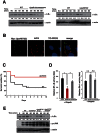
Figure 1
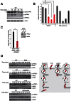
Figure 1. IpaH0722 inhibits Shigella-induced NF-κB activation.
(A) HeLa cells were infected with Shigella WT, ΔipaH-chromosome (left), or ΔipaH0722 (right). The cell lysates were prepared at the indicated time points and subjected to immunoblottings with anti-IκBα. Anti-actin antibody was used as a loading control. The values indicated below the images are the relative intensities of the bands. (B) IpaH0722 localizes to the host cell plasma membrane. Cos-7 cells were transfected with a Myc-IpaH0722 expression vector and immunostained with anti-Myc, actin, and TO-PRO3. (C) The murine pulmonary model of Shigella infection. Mice were intranasally inoculated with Shigella WT or ΔipaH0722 (n = 13) at 5×10⁷ cfu and survival was recorded for 10 days post-infection. *P<0.05. (D) NF-κB-luciferase and Elk-1-luciferase reporter assays were performed after Shigella infection of 293T cells that were transiently transfected with empty vector, IpaH0722, or IpaH0722CA expressing plasmids. Results are presented as ‘fold’ relative to the activity of uninfected or unstimulated cells. *P<0.01, n.s., not significant. (E) HeLa cells were infected with Shigella WT, ΔipaH0722, or ΔipaH0722 harboring ipaH0722 or ipaH0722CA. The cell lysates prepared at the indicated time points were subjected to immunoblotting. Anti-actin antibody served as the loading control.
Figure 2
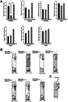
Figure 2. IpaH0722 selectively inhibits PMA–induced activation of the NF-κB pathway.
(A) Luciferase reporter assays of 293T cells transiently transfected with an NF-κB reporter plasmid and empty vector, IpaH0722, or IpaH0722CA expressing plasmids. After 24 h, cells were treated with TNF-α, PMA, LPS, or IL-1β for 3 h and luciferase activity was measured. Results are presented as fold change relative to the activity of uninfected or unstimulated cells. *P<0.01 (B) IL-8 expression in 293T cells. Cells expressing empty vector, IpaH0722, or IpaH0722CA expressing plasmids were treated with TNF-α, PMA, or IL-1β for 3 h. Measurement of IL-8 reporter activity after PMA, TNF-α, and IL-1β stimulation. Results are presented as fold change relative to the activity of uninfected or unstimulated cells. *P<0.01. (C) The levels of IL-8 production were measured by ELISA. Cells expressing empty vector, IpaH0722, or IpaH0722CA expressing plasmids were treated with PMA and TNF-α, then supernatants were harvested for ELISA analysis. *P<0.0. (D)Luciferase reporter assays of 293T cells transiently transfected with NF-κB, AP-1, or Elk-1 reporter plasmids plus empty vector, IpaH0722, or IpaH0722CA expressing plasmids. After 24 h, cells were treated with PMA for 3 h and luciferase activity was measured. Results are presented as fold change relative to the activity of uninfected or unstimulated cells.*P<0.01, n.s., not significant.
Figure 3
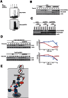
Figure 3. Shigella triggers PKC–NF-κB activation.
(A) HeLa cells were infected with Shigella. Cell lysates were prepared at the indicated time points and subjected to immunoblotting with anti-phospho-PKC antibody. *non-specific bands. (B) NF-κB luciferase assays of 293T cells transiently transfected with an NF-κB reporter plasmid and empty vector or dominant-negative forms of PKC. After 24 h, cells were infected with Shigella or treated with PMA for 3 h, and luciferase activity was measured. Results are presented as fold change relative to the activity of uninfected or unstimulated cells. *P<0.01. (C) Cells were treated with PKCδ siRNA and transiently transfected with an NF-κB reporter plasmid. The cells were infected with Shigella and NF-κB reporter activity was measured. (D) left; HeLa cells were infected with Shigella WT, invasin expressing ΔipaB mutant, ΔvirG mutant, or Salmonella. The cell lysates were harvested at the indicated time points and subjected to immunoblot. right: A model of Shigella invasion into the epithelial cells.
Figure 4
Figure 4. IpaH0722 targets PKC–NF-κB activation but not via the CBM complex.
(A) IpaH0722 inhibits PKC-induced NF-κB activation. Luciferase assays of 293T cells that were transiently transfected with dominant-active forms of PKC and empty vector, IpaH0722, or IpaH0722CA expressing plasmids. Results are presented as fold change relative to the activity of uninfected or unstimulated cells. *P<0.01. (B) HeLa cells were infected with Shigella WT or the ΔipaH0722 mutant. The cell lysates were harvested at the indicated time points and subjected to immunoblotting. (C) Cells that transiently expressed empty vector, IpaH0722, or IpaH0722CA were treated with PMA+ionomycin for the indicated time points. Cell lysates were subjected to immunoblotting. (D) Luciferase reporter assays of 293T cells were transiently transfected with NF-κB and empty vector, IpaH0722, or IpaH0722CA expressing plasmids. Results are presented as fold change relative to the activity of uninfected or unstimulated cells. (E) 293T cells were transfected with FLAG-CARMA1 with or without PKCδ-DA, Myc-IpaH0722, or Myc-IpaH0722CA. After 24 h, cells were harvested and subjected to immunoprecipitation (IP).
Figure 5
Figure 5. IpaH0722 inhibits TRAF2-mediated NF-κB activation.
(A) Luciferase reporter assays of 293T cells transiently transfected with NF-κB signaling factors and empty vector, IpaH0722, or IpaH0722CA expressing plasmids. Results are presented as fold change relative to the activity of uninfected or unstimulated cells. *P<0.01. (B) 293T cells were transfected with FLAG or GFP-tagged NF-κB signaling factor with or without Myc6-IpaH0722CA. After 24 h, cells were harvested and subjected to immunoprecipitation (IP) and immunoblotting. (C) Pull down assays using GST or GST-IpaH0722 were performed with HeLa whole cell lysates. Samples were subjected to immunoblotting with anti-TRAF2 and -TRAF6.
Figure 6
Figure 6. IpaH0722 targets TRAF2 for ubiquitination.
(A) In vitro ubiquitination assay using TRAF2 and a mixture of E1, UbcH5b, ATP, and ubiquitin in the presence or absence of IpaH0722 or IpaH0722CA. (B) 293T cells were transfected with FLAG-TRAF2 with and without Myc-IpaH0722 or Myc-IpaH0722CA. After 24 h, the cells were treated with CHX (50 mg/ml) and cell lysates were prepared at the indicated time points. (C) 293T cells were transfected with FLAG-TRAF2 with and without FLAG-IpaH0722 or FLAG-IpaH0722CA. After 24 h, the cells were treated with DMSO or CHX (50 mg/ml) plus MG132 or E64D+pepstatin A. Cell lysates were prepared after 3 h and samples were subjected to immunoblotting. (D) Traf2/Traf5 −/− MEFs and Traf2/Traf5 −/− MEFs stably expressing Traf2 were infected with Shigella WT or ΔipaH0722. Cell lysates were harvested at the indicated time point and subjected to immunoblotting with anti- IκBα Anti-actin antibody was used as a loading control. (E) A model of this study. Shigella invasion induced phagosome membrane rupture of epithelial cells, which stimulates DAG–PKC–NF-κB activation. However, Shigella deliver IpaH0722 to inhibit PKC–NF-κB activation by ubiquitinating TRAF2 and promoting its proteasome degradation.

References

    1. Chen GY, Nuñez G (2010) Sterile inflammation: sensing and reacting to damage. Nat Rev Immunol 10: 826–837. - PMC - PubMed
    1. Davis BK, Wen H, Ting JP (2011) The inflammasome NLRs in immunity, inflammation, and associated diseases. Annu Rev Immunol 29: 707–735. - PMC - PubMed
    1. Ashida H, Ogawa M, Kim M, Mimuro H, Sasakawa C (2012) Bacteria and host interactions in the gut epithelial barrier. Nat Chem Biol 8: 36–45. - PubMed
    1. Galán JE (2009) Common themes in the design and function of bacterial effectors. Cell Host Microbe 5: 571–579. - PMC - PubMed
    1. Rahman MM, McFadden G (2011) Modulation of NF-κB signalling by microbial pathogens. Nat Rev Microbiol 9: 291–306. - PMC - PubMed

Publication types

MeSH terms

LinkOut - more resources