. 2013 Jun;9(6):e1003536.
doi: 10.1371/journal.pgen.1003536. Epub 2013 Jun 6.

Mutations in CERS3 cause autosomal recessive congenital ichthyosis in humans

Affiliations

Mutations in CERS3 cause autosomal recessive congenital ichthyosis in humans

Franz P W Radner et al. PLoS Genet. 2013 Jun.

Erratum in

  • PLoS Genet. 2013 Jun;9(6): doi/10.1371/annotation/df5af830-8e1d-495a-a206-f881ed85e7fe

Abstract

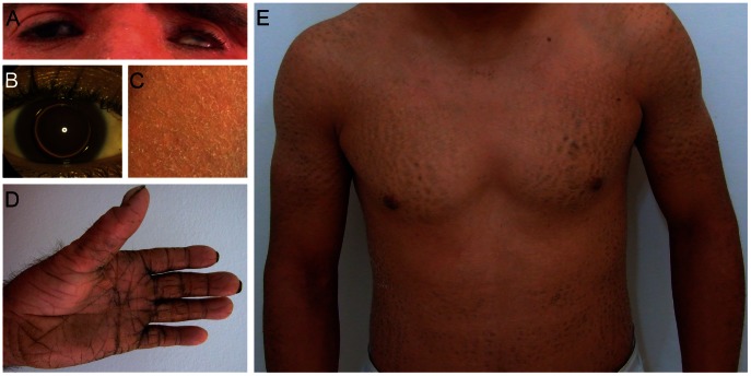
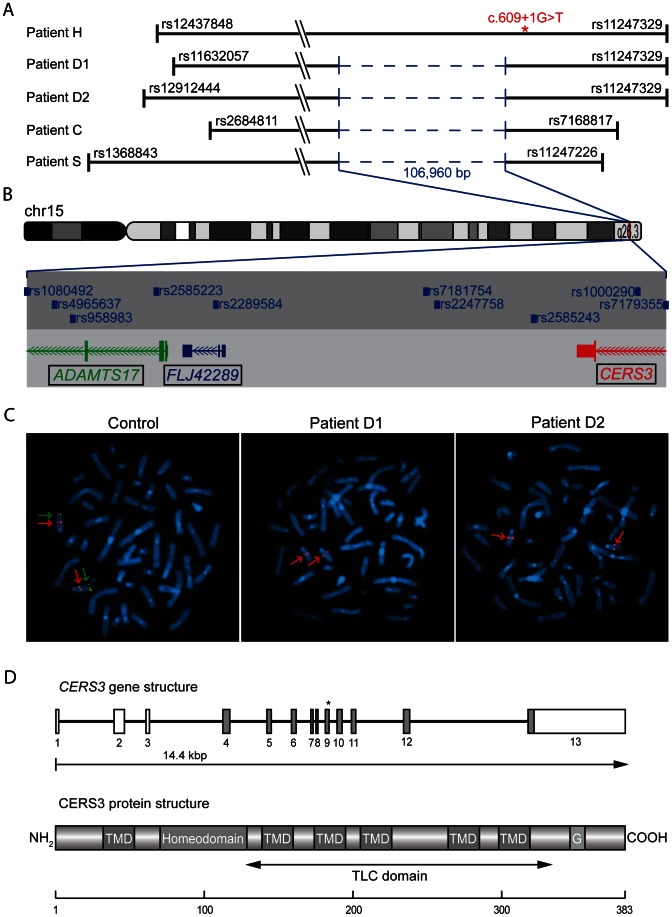
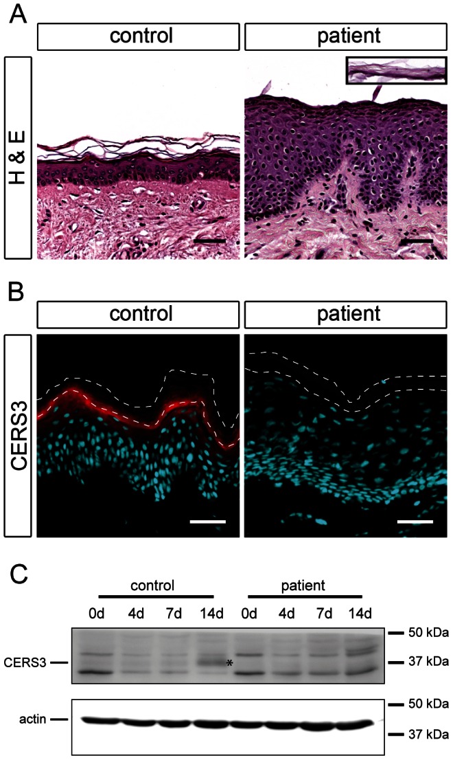
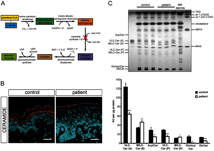
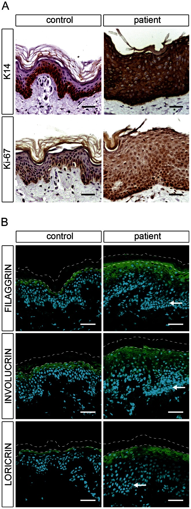
Autosomal recessive congenital ichthyosis (ARCI) is a rare genetic disorder of the skin characterized by abnormal desquamation over the whole body. In this study we report four patients from three consanguineous Tunisian families with skin, eye, heart, and skeletal anomalies, who harbor a homozygous contiguous gene deletion syndrome on chromosome 15q26.3. Genome-wide SNP-genotyping revealed a homozygous region in all affected individuals, including the same microdeletion that partially affects two coding genes (ADAMTS17, CERS3) and abolishes a sequence for a long non-coding RNA (FLJ42289). Whereas mutations in ADAMTS17 have recently been identified in autosomal recessive Weill-Marchesani-like syndrome in humans and dogs presenting with ophthalmologic, cardiac, and skeletal abnormalities, no disease associations have been described for CERS3 (ceramide synthase 3) and FLJ42289 so far. However, analysis of additional patients with non-syndromic ARCI revealed a splice site mutation in CERS3 indicating that a defect in ceramide synthesis is causative for the present skin phenotype of our patients. Functional analysis of patient skin and in vitro differentiated keratinocytes demonstrated that mutations in CERS3 lead to a disturbed sphingolipid profile with reduced levels of epidermis-specific very long-chain ceramides that interferes with epidermal differentiation. Taken together, these data present a novel pathway involved in ARCI development and, moreover, provide the first evidence that CERS3 plays an essential role in human sphingolipid metabolism for the maintenance of epidermal lipid homeostasis.


Conflict of interest statement

The authors have declared that no competing interests exist.

Figures

Figure 1
Figure 1. Clinical features of patients.
(A) Patient D1 with erythema of the face and eye symptoms; (B) Patient S with isolated bilateral microspherophakia without ectopia or hypertonia; (C) Skin of the upper leg of patient S showing ichthyosiform erythroderma with large, white scales; (D) Palmoplantar hyperlinearity, hyperkeratosis, and brachydactyly in patient D1; (E) Upper part of the body in patient D2; large brownish scales on the limbs and trunk.
Figure 2
Figure 2. Deletion and CERS3 mutation scheme.
(A) Homozygous regions on chromosome 15q26.3 in patients. The smallest common interval is defined by rs2684811 in patient C and rs11247226 in patient S. The genomic deletion characterized by breakpoint spanning PCR (in blue) encompasses 106,960 bp with the borders 100,856,031 to 100,962,985 on chromosome 15 (UCSC hg19, February 2009) in patient D1, D2, C, and S. The splice site mutation in patient H is marked by an asterisk. The diagram shows the deletion and the limiting SNPs of the homozygous regions in patient H, D1, D2, C, and S (not to scale). (B) The scale illustration of the deleted region shows the missing SNPs and genes. The internal limit of the deletion is between SNP rs1080429 and rs7179355. (C) FISH signal pattern in a healthy individual shows the control signal on 15q21.2 in red (digoxigenin-labeled BAC RP11-562A8) and the signal corresponding to 15q26.3 in green (arrow). In patients D1 and D2 the green FISH signal is missing confirming the microdeletion in 15q26.3. (D) Diagram depicting the structure of the human CERS3 gene and the site of mutation in exon 9 (c.609+1G>T) of patient H indicated by an asterisk. Below is illustrated the predicted structure of human CERS3 protein including transmembrane domains (TMD), homeobox, polyglutamic acid region (G), and the TLC (TRAM, LAG1 and CLN8 homology) domain.
Figure 3
Figure 3. Histological analysis and CERS3 protein expression in skin biopsies and cultured keratinocytes from healthy controls and patient H.
(A) Hematoxylin and eosin staining (H&E) shows acanthosis with a thickening of the granular layer and psoriasiform epidermal hyperplasia in the patient compared to a healthy control. The inset (same scale) shows the detached stratum corneum of the patient. Scale bars, 50 µm (B) Immunofluorescence staining using a specific antibody for CERS3 (red) and DAPI (blue) as nuclear counterstaining. CERS3 staining is present at the interface between the stratum granulosum and the stratum corneum in control skin but not detectable in the patient skin. The thin dashed lines indicate the interface between the stratum granulosum and the stratum corneum as well as the upper edge of the stratum corneum. Scale bars, 50 µm (C) Western blot analysis of CERS3 in control and patient keratinocytes before differentiation (0 d) and at day 4, 7, and 14 after induction of differentiation. CERS3 was detected at ∼37 kDa (indicated by an asterisk) using an antibody targeting the C-terminus of the protein. An antibody that recognizes actin was used as a loading control.
Figure 4
Figure 4. In situ ceramide localization and sphingolipid profile of cultured keratinocytes.
(A) Diagram depicting the de novo synthesis pathway of (glucosyl)ceramides. Loss-of-function of ceramide synthase 1–6 results in an impaired N-acylation of dihydrosphingosine as indicated by a cross. (B) Confocal microscopy images of healthy control and patient H skin biopsies immunostained with an antibody targeting ceramides (red) with DAPI (blue) as nuclear counterstaining. Ceramides localize to the stratum granulosum and the stratum corneum in healthy control skin. Note the loss of ceramide staining in patient's skin. We observed immunostaining of ceramides in the uppermost layer of the mutant stratum corneum, which results from unspecific binding of the secondary antibody. The thin dashed lines indicate the interface between the stratum granulosum and the stratum corneum as well as the upper edge of the stratum corneum. Scale bars, 50 µm (C) Upper panel: TLC of lipid extracts from healthy control and patient H keratinocytes 14 days after induction of differentiation. Lipids corresponding to 400 µg of cellular protein were extracted from cultures, separated twice by TLC using the solvent system chloroform/methanol/glacial acetic acid (190/9/1 v/v/v), and quantified after carbonization. An epidermal lipid extract of a healthy control individual was used as a reference. Lower panel: Data are presented as mean values +S.D. of triplicate samples and are representative for three independent experiments. Statistical significance was determined by unpaired two-tailed Student's t-test (* p<0.05, ** p<0.01, *** p<0.001). Ceramide species are classified according to the sphingoid base (S, sphingosine or P, phytosphingosine). Abbreviations: AcylCer, acylceramides; DAG, diacylglycerols; GlcAcylCer, glucosylacylceramides; GlcCer, glucosylceramides; MAG, monoacylglycerols; M/LC-Cer, middle and long-chain ceramides; NEFA, non-esterified fatty acids; TAG, triacylglycerols; VLC-Cer, very long-chain ceramides.
Figure 5
Figure 5. Epidermal differentiation in healthy control and patient H.
(A) Light microscopy images of skin biopsies from control and patient H immunolabeled with antibodies specific for keratin 14 (K14) and Ki-67 with hematoxylin as nuclear counterstaining reveal an abnormal differentiation process in patient skin. (B) Confocal microscopy images of the same control and patient skin biopsies immunostained with antibodies specific for filaggrin, involucrin, and loricrin with DAPI (blue) as nuclear counterstaining. The patient skin biopsy shows a thickening of the stratum granulosum compared to the healthy control. Arrows indicate hyperplastic basal cells in the patient skin. The thin dashed lines indicate the interface between the stratum granulosum and the stratum corneum as well as the upper edge of the stratum corneum. Scale bars, 50 µm.

References

    1. Lefévre C, Audebert S, Jobard F, Bouadjar B, Lakhdar H, et al. (2003) Mutations in the transporter ABCA12 are associated with lamellar ichthyosis type 2. Hum Mol Genet 12: 2369–2378. - PubMed
    1. Jobard F, Lefèvre C, Karaduman A, Blanchet-Bardon C, Emre S, et al. (2002) Lipoxygenase-3 (ALOXE3) and 12(R)-lipoxygenase (ALOX12B) are mutated in non-bullous congenital ichthyosiform erythroderma (NCIE) linked to chromosome 17p13.1. Hum Mol Genet 11: 107–113. - PubMed
    1. Lefèvre C, Bouadjar B, Ferrand V, Tadini G, Mégarbané A, et al. (2006) Mutations in a new cytochrome P450 gene in lamellar ichthyosis type 3. Hum Mol Genet 15: 767–776. - PubMed
    1. Lefèvre C, Bouadjar B, Karaduman A, Jobard F, Saker S, et al. (2004) Mutations in ichthyin a new gene on chromosome 5q33 in a new form of autosomal recessive congenital ichthyosis. Hum Mol Genet 13: 2473–2482. - PubMed
    1. Grall A, Guaguère E, Planchais S, Grond S, Bourrat E, et al. (2012) PNPLA1 mutations cause autosomal recessive congenital ichthyosis in golden retriever dogs and humans. Nat Genet 44: 140–147 doi:10.1038/ng.1056 - DOI - PubMed

Publication types