. 2013 May 28:7:100.
doi: 10.3389/fncir.2013.00100. eCollection 2013.

Cellular dissection of the spinal cord motor column by BAC transgenesis and gene trapping in zebrafish

Affiliations

Cellular dissection of the spinal cord motor column by BAC transgenesis and gene trapping in zebrafish

Kazuhide Asakawa et al. Front Neural Circuits. .

Abstract

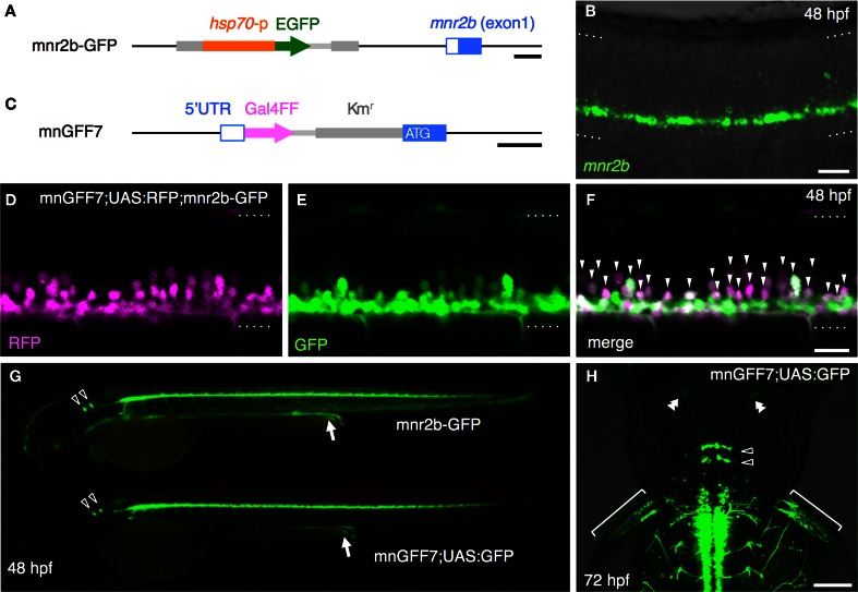
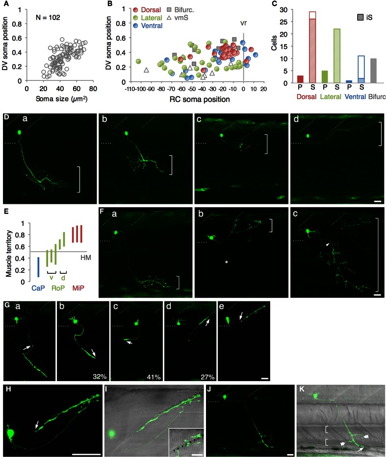
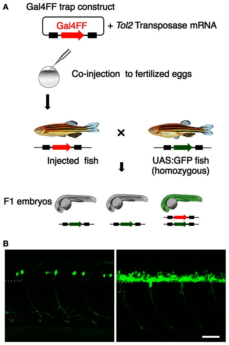
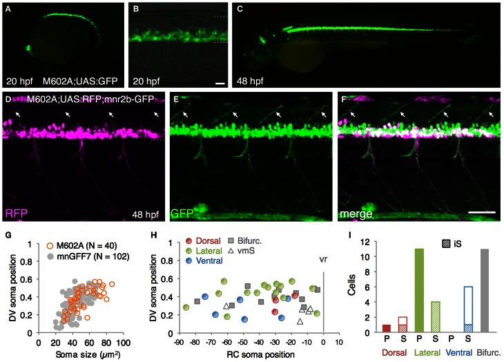
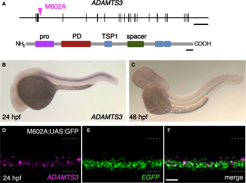
Bacterial artificial chromosome (BAC) transgenesis and gene/enhancer trapping are effective approaches for identification of genetically defined neuronal populations in the central nervous system (CNS). Here, we applied these techniques to zebrafish (Danio rerio) in order to obtain insights into the cellular architecture of the axial motor column in vertebrates. First, by using the BAC for the Mnx class homeodomain protein gene mnr2b/mnx2b, we established the mnGFF7 transgenic line expressing the Gal4FF transcriptional activator in a large part of the motor column. Single cell labeling of Gal4FF-expressing cells in the mnGFF7 line enabled a detailed investigation of the morphological characteristics of individual spinal motoneurons, as well as the overall organization of the motor column in a spinal segment. Secondly, from a large-scale gene trap screen, we identified transgenic lines that marked discrete subpopulations of spinal motoneurons with Gal4FF. Molecular characterization of these lines led to the identification of the ADAMTS3 gene, which encodes an evolutionarily conserved ADAMTS family of peptidases and is dynamically expressed in the ventral spinal cord. The transgenic fish established here, along with the identified gene, should facilitate an understanding of the cellular and molecular architecture of the spinal cord motor column and its connection to muscles in vertebrates.

Keywords: ADAMTS3; BAC; Gal4; Mnx; gene trapping; motor column; zebrafish.


Figures

Figure 1
Figure 1
Construction and characterization of the BAC transgenic line, mnGFF7. (A) The mnr2b genomic locus in the mnr2b-GFP enhancer trap line. The hsp70p-EGFP enhancer trap construct was integrated 1.9 kb upstream of the mnr2b gene. (B) Expression of mnr2b at 48 hpf detected by fluorescent in situ hybridization. A confocal section of the spinal cord above the yolk extension in a wild type embryo is shown. The rostral is to the left. (C) The mnr2b locus on the mnGFF BAC DNA. The PCR-amplified Gal4FF-polyA-Kmr cassette is inserted downstream of the 5′UTR of the mnr2b (open blue box) by homologous recombination. The coding sequence of exon1 of mnr2b (filled blue box) is left intact after the recombination. (D–F) The lateral view of the spinal cord of the mnGFF7;UAS:RFP; mnr2b-GFP triple transgenic embryo at 48 hpf. The images shown are the RFP (left), GFP (middle), and merged (right) images of a single confocal slice. The RFP-positive cells whose somata are identifiable are indicated by the arrowheads in (F). The dotted lines demarcate the dorsal and ventral limit of the spinal cord. The rostral is to the left. (G) GFP expression in mnr2b-GFP (top) and mnGFF7;UAS:GFP (bottom) embryos at 48 hpf. The arrows indicate the GFP expression in the gut. The open arrowheads indicate the abducens motoneurons. (H) The dorsal view of the mnGFF7;UAS:GFP larvae at 72 hpf. GFP is expressed in the abducens and pectoral fin motoneurons. The open and double arrowheads indicate the soma positions and the axon terminals of the abducens motoneurons, respectively. The brackets indicate the axon terminals of the pectoral fin motoneurons. The rostral is to the top. The dotted lines demarcate the dorsal (top) and ventral limit of the spinal cord in (B,D–F). The bars indicate 500 base-pair (bp) in (A) and (C), 20 μm in (B,D,F), and 100 μm in (H).
Figure 2
Figure 2
Single cell labeling of Gal4FF expressing cells in mnGFF7. (A) The plot of the single GFP-labeled motoneurons (open gray circles) with respect to the soma size on the horizontal axis and to the position of the soma along with the DV axis on the vertical axis. The DV soma position is normalized to the dorsal (y = 1) and the ventral (y = 0) edge of the spinal cord. (B) Distribution of the GFP-labeled cells plotted with respect to the soma position along the RC axis on the horizontal axis and to the DV soma position on the vertical axis. The exit point of the ventral root from the spinal cord is indicated on the RC axis by the gray vertical line marked as vr (x = 0). The dorsally, laterally and ventrally projecting cells are shown as red, green, and blue circles, respectively. The cells with bifurcating main axon and the vmS motoneurons are shown as gray squares and open triangles, respectively. (C) Cell counts of the neurons innervating the axial muscles based on the axon trajectory and muscle innervation pattern. P and S indicate primary and secondary motoneurons, respectively. Diagonally-striped bars indicate iS-type neurons. “Bifurc” indicates the motoneurons with a bifurcating main axon. (D) Primary motoneurons innervating ventrally (a), ventrolaterally (b), dorsolaterally (c), or dorsally (d). (E) Muscle innervation territory of the primary motoneurons. The position of the horizontal myoseptum (y = 0.5) is indicated as a gray horizontal bar. (F) Secondary motoneurons innervating ventrally (a, vS), dorsally (b, dS), or dorsoventrally (c, dvS). The arrowhead in (c) indicates the bifurcation in the main axon. The asterisk in (b) is the axon of another motoneuron in the opposite hemi-segment. (G) The secondary motoneuron innervating the ventral (a), ventrolateral (b), horizontal (c), dorsolateral (d), or dorsal (e) myoseptal region. The arrows indicate the axon terminals and roughly indicate their direction of extension. The percentiles show the ratio among the laterally projecting cells. (H) A cell innervating the dorsal myotseptum. The arrow indicates the axon terminal. This cell is identical to the one shown in (Ge) and the image was taken 6 h after the image (Ge) was taken. Note that the morphology of the soma changed dramatically in 6 h. (I) The superficial myoseptal region of (H) merged with the DIC image. The inset shows a stack of a few confocal sections indicated in the dotted square in (I). The axon collaterals cross the myoseptal boundary marked by the black dotted line. (J,K) A vmS-type neuron. The arrow and arrowheads indicate the axon terminal and the collaterals extending along the blood vessels, respectively. All images in (D–I) were taken during 72–100 hpf and the rostral is to the left. In (D,F,G,J), the oblique and horizontal dotted lines demarcate the myoseptal boundaries and the ventral edge of the spinal cord, respectively. The brackets roughly indicate the innervation territory along the dorsoventral axis in (D,F), and the axial blood vessels in (K). The bars indicate 20 μm, except in (I), where the bars indicate 10 μm.
Figure 3
Figure 3
Gene trapping using the Gal4FF-UAS system. (A) Scheme for the Gal4FF gene trapping. A plasmid DNA containing a Gal4FF construct and the transposase mRNA were co-injected into fertilized eggs. The injected fish were raised and mated with the reporter fish homozygous for the UAS:GFP insertion. The resulting F1 embryos were analyzed with a fluorescence stereomicroscope. F1 embryos expressing GFP were collected and raised for further studies. (B) Examples of GFP expression in the spinal cord and the motor nerve in F1 embryos at 48 hpf. In the left, the primary motoneurons are exclusively labeled. In the right, a large number of GFP labeled cells are observed in the ventral spinal cord. The dotted line demarcates the ventral limit of the spinal cord. The rostral is to the left. The bar indicates 50 μm.
Figure 4
Figure 4
The spinal motoneurons marked by the M602 insertions. (A–C) Lateral views of the M602A; UAS:GFP animals. At 20 hpf, GFP expression was detected in the ventral spinal cord and the rostral hindbrain (A,B). The dotted lines demarcate the dorsal and ventral limit of the spinal cord in (B). At 48 hpf, GFP is strongly detected in the ventral spinal cord and the rostral and caudal hindbrain, as well as weakly in other tissues. (D–F) Lateral view of the M602A;UAS:RFP; mnr2b-GFP triple transgenic animal at 48 hpf. Arrows indicate the myoseptal region in the dorsal musculature. (G) The plot of single GFP-labeled cells (open orange circles, N = 40 cells). For reference, the results from the experiment with mnGFF7 in Figure 2A are superimposed as gray circles. (H) Distribution of the GFP-labeled cells plotted with respect to the soma position along the RC axis on the horizontal axis and to the DV soma position on the vertical axis. The exit point of the ventral root from the spinal cord is indicated on the RC axis by the gray vertical line marked as vr (x = 0). The dorsally, laterally, and ventrally projecting cells are shown as red, green, and blue circles, respectively. The cells with bifurcating main axon and the vmS motoneurons are shown as gray squares and open triangles, respectively. (I) Cell counts of the neurons innervating the axial muscles based on the axon trajectory and muscle innervation pattern. P and S indicate primary and secondary motoneurons, respectively. Diagonally-striped bars indicate iS type neurons. “Bifurc” indicates the motoneurons with a bifurcating main axon. The bars indicate 20 μm in (B), and 50 μm in (F).
Figure 5
Figure 5
The M602A insertion traps ADAMTS3. (A) Structure of the ADAMTS3 locus and the integration site of M602A. M602A is inserted in the 3rd intron of ADAMTS3. The long horizontal bar and vertical bars denote the genomic DNA and the exons of ADAMTS3, respectively. ADAMTS3 consists of multiple functional domains: pro, pro domain; PD, metalloprotease domain; TSP1, thrombospondin type 1 motif; spacer, spacer region [according to Porter et al. (2005)]. (B,C) Expression of ADAMTS3 at 24 hpf (B) and 48 hpf (C), detected by in situ hybridization. (D–F) Expression of ADAMTS3 and EGFP detected by double fluorescent in situ hybridization. A confocal section of the spinal cord of the M602A;UAS:GFP embryo at 24 hpf was shown. The dotted lines demarcate the dorsal and ventral limit of the spinal cord. The scale bars indicate 20 kbp (top) and 50 amino acids (bottom) in (A), and 20 μm in (F).
Figure A1
Figure A1
A descending interneuron identified in the single cell labelling experiment. (A,B) The dorsal view of a larvae at 76 hpf. The soma of the GFP-labelled cell located at the level of the anterior boundary of somite 1. The axon descended ipsilaterally to the level of somite 18. The dotted lines indicate the position of the midline. (C and D) The lateral view of the same larva. The oblique lines indicate the anterior boundary of somite 1 and 18. In (A) and (C), the DIC image was superimposed onto the GFP image. The scale bars indicate 100 μm in (B) and (D).
Figure A2
Figure A2
Distribution of the laterally projecting cells in mnGFF7 (A) and M602A (B) plotted with respect to the soma position along the RC axis on the horizontal axis and to the DV soma position on the vertical axis. The exit point of the ventral root from the spinal cord is indicated on the RC axis by the gray vertical line marked as vr (x = 0). The ventrolaterally, laterally and dorsolaterally projecting cells are shown as circles, triangles, and crosses, respectively.

Similar articles

Cited by

References

    1. Abe G., Suster M. L., Kawakami K. (2011). Tol2-mediated transgenesis, gene trapping, enhancer trapping, and the Gal4-UAS system. Methods Cell Biol. 104, 23–49 10.1016/B978-0-12-374814-0.00002-1 - DOI - PubMed
    1. Agalliu D., Takada S., Agalliu I., McMahon A. P., Jessell T. M. (2009). Motor neurons with axial muscle projections specified by Wnt4/5 signaling. Neuron 61, 708–720 10.1016/j.neuron.2008.12.026 - DOI - PMC - PubMed
    1. Appel B., Korzh V., Glasgow E., Thor S., Edlund T., Dawid I. B., et al. (1995). Motoneuron fate specification revealed by patterned LIM homeobox gene expression in embryonic zebrafish. Development 121, 4117–4125 - PubMed
    1. Aramaki S., Hatta K. (2006). Visualizing neurons one-by-one in vivo: optical dissection and reconstruction of neural networks with reversible fluorescent proteins. Dev. Dyn. 235, 2192–2199 10.1002/dvdy.20826 - DOI - PubMed
    1. Arrenberg A. B., Del Bene F., Baier H. (2009). Optical control of zebrafish behavior with halorhodopsin. Proc. Natl. Acad. Sci. U.S.A. 106, 17968–17973 10.1073/pnas.0906252106 - DOI - PMC - PubMed

Publication types

MeSH terms

LinkOut - more resources