. 2013 Jun 5;8(6):e64752.
doi: 10.1371/journal.pone.0064752. Print 2013.

Awakened by cellular stress: isolation and characterization of a novel population of pluripotent stem cells derived from human adipose tissue

Affiliations

Awakened by cellular stress: isolation and characterization of a novel population of pluripotent stem cells derived from human adipose tissue

Saleh Heneidi et al. PLoS One. .

Erratum in

  • PLoS One. 2013;8(7). doi:10.1371/annotation/190d4d01-a63c-4adc-a123-e519ee40a03e

Abstract

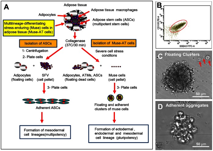
Advances in stem cell therapy face major clinical limitations, particularly challenged by low rates of post-transplant cell survival. Hostile host factors of the engraftment microenvironment such as hypoxia, nutrition deprivation, pro-inflammatory cytokines, and reactive oxygen species can each contribute to unwanted differentiation or apoptosis. In this report, we describe the isolation and characterization of a new population of adipose tissue (AT) derived pluripotent stem cells, termed Multilineage Differentiating Stress-Enduring (Muse) Cells, which are isolated using severe cellular stress conditions, including long-term exposure to the proteolytic enzyme collagenase, serum deprivation, low temperatures and hypoxia. Under these conditions, a highly purified population of Muse-AT cells is isolated without the utilization of cell sorting methods. Muse-AT cells grow in suspension as cell spheres reminiscent of embryonic stem cell clusters. Muse-AT cells are positive for the pluripotency markers SSEA3, TR-1-60, Oct3/4, Nanog and Sox2, and can spontaneously differentiate into mesenchymal, endodermal and ectodermal cell lineages with an efficiency of 23%, 20% and 22%, respectively. When using specific differentiation media, differentiation efficiency is greatly enhanced in Muse-AT cells (82% for mesenchymal, 75% for endodermal and 78% for ectodermal). When compared to adipose stem cells (ASCs), microarray data indicate a substantial up-regulation of Sox2, Oct3/4, and Rex1. Muse-ATs also exhibit gene expression patterns associated with the down-regulation of genes involved in cell death and survival, embryonic development, DNA replication and repair, cell cycle and potential factors related to oncogenecity. Gene expression analysis indicates that Muse-ATs and ASCs are mesenchymal in origin; however, Muse-ATs also express numerous lymphocytic and hematopoietic genes, such as CCR1 and CXCL2, encoding chemokine receptors and ligands involved in stem cell homing. Being highly resistant to severe cellular stress, Muse-AT cells have the potential to make a critical impact on the field of regenerative medicine and cell-based therapy.


Conflict of interest statement

Competing Interests: The authors have declared that no competing interests exist.

Figures

Figure 1
Figure 1. Isolation and morphologic characterization of Muse-ATs.
(A) Schematic of Muse-AT isolation and activation from their quiescent state by exposure to cellular stress. Muse-AT cells were obtained after 16 hours, with incubation with collagenase in DMEM medium without FCS at 4°C under very low O2 (See Methods). (B) FACS analysis demonstrates that 90% of isolated cells are both SSEA3 and CD105 positive. (C) Muse-AT cells can grow in suspension, forming spheres or cell clusters as well as individual cells (see red arrows) or (D) Muse-AT cells can adhere to the dish and form cell aggregates. Under both conditions, individual Muse-AT cells reached a diameter of approximately 10µm and cell clusters reached a diameter of up to 50µm, correlating to stem cell proliferative size capacity.
Figure 2
Figure 2. Muse-ATs express pluripotent stem cell markers.
Immunofluorescence microscopy demonstrates that Muse-AT aggregates, along with individual Muse-AT cells, express characteristic pluripotent stem cell markers, including SSEA3, Oct3/4, Nanog, Sox2, and TRA1-60. Comparatively, ASCs (right panel) derived from the same lipoaspirate under standard conditions (see above, were negative for these pluripotent stem cell markers. Nuclei were stained with DAPI (blue). Original magnification, 600 X.
Figure 3
Figure 3. Muse-AT cells can differentiate to mesodermal cell lineages.
Isolated Muse-ATs were grown as adherent cells in the presence of DMEM/10%FCS for 6 days. (A) Immunostaining indicates that Muse-AT have the capacity to spontaneously differentiate to mesodermal cell lineages. DLK is a specific marker for preadipocytes; BODIPY-C16 recognized lipid drops present in adipocytes; Myosin D is specific marker for myocytes. Comparatively, ASCs (right panel) which are mesenchymal stem cells derived from the same lipoaspirate under standard conditions (see above, were slightly positive for DLK; nuclei were stained with DAPI (blue), original magnification, 600 X. (B) Isolated Muse-AT cells were grown as adherent cells in the presence of adipocyte differentiation medium (See Methods) for 3 or 6 days and formation of adipocytes was detected using BODIPY-C16; (C) Isolated Muse-AT cells were grown as adherent cells in the presence of myocyte differentiation medium and formation of myocytes was detected using anti human-MSA antibodies. Comparatively, ASCs (right panel) which are mesenchymal stem cells derived from the same lipoaspirate under standard conditions (See above, were slightly positive for BODIPY-C16 or MSA (B and C, right panel). Nuclei were stained with DAPI (blue). Original magnification was 200 X (first three rows) or 600 X (last two rows).
Figure 4
Figure 4. Muse-ATs can differentiate to endodermal cell lineages.
Isolated Muse-ATs were grown as adherent cells in the presence of DMEM/10%FCS for 6 days. (A) Immunostaining indicates that Muse-AT have the capacity to spontaneously differentiate to endodermal cell lineages. α-fetoprotein and pan-keratin are specific markers for hepatocytes; Nuclei were stained with DAPI (blue). Original magnification, 600 X. Comparatively, ASCs (right panel) derived from the same lipoaspirate under standard conditions (see above, [16]) were negative for α-fetoprotein and pan-keratin. Nuclei were stained with DAPI (blue); original magnification, 600 X; (B) Isolated Muse-AT cells were grown as adherent cells in the presence of hepatocyte differentiation medium (see methods) for 3 or 6 days and formation of hepatocytes was detected using anti-human cytokeratin 7 or α-fetoprotein antibodies. Comparatively, ASCs (right panel) were completely negative for cytokeratin 7 and α-fetoprotein (B, right panel). Nuclei were stained with DAPI (blue). Original magnification was 200 X (first three rows) or 600 X (last two rows).
Figure 5
Figure 5. Muse- ATs can differentiate to ectodermal cell lineages.
(A) Isolated Muse-ATs were grown as adherent cells in the presence of DMEM/10%FCS for 6 days. Immunostaining indicates that Muse-AT cells have the capacity to spontaneously differentiate to ectodermal cell lineages. NSE, Glu-R and Neuro D are specific markers for neural progenitor or neural-like cells. Comparatively, ASCs (right panel) were negative for NSE, Glu-R and NeuroD. Nuclei were stained with DAPI (blue); original magnification was 600X. (B) Isolated Muse-AT cells were grown as non-adherent cells in the presence of Neurobasal medium/B-27 supplement serum free/kanamycin/glutamine/bFGF and EGF for 7 days. Formation of neural cell spheres was detected at different times during this incubation. Cells were then grown as adherent cells for an additional 7 days in a DMEM 2% FCS/bFGF/BDNF. Formation of neural-like cells is indicated by red arrows. (C) Isolated Muse-AT cells were grown for 7 days as non-adherent cells and then cultured for another 7 days as adherent cells (See Methods). Neural-like cells were detected by immunofluorescence using anti-human nestin and MAP2 antibodies. Comparatively, ASCs (right panel) were negative for nestin and MAP2 (B, right panel). Nuclei were stained with DAPI (blue). Original magnification was 200 X (first three rows) or 600X (last two rows).
Figure 6
Figure 6. Identification of functional groups and canonical pathways in Muse-ATs vs ASCs.
Identification of the top ten (A) functional groups (B) canonical pathways of all differentially expressed genes (2 fold or higher) in Muse-AT cells versus ASCs. Fischer’s exact test was used to calculate a p-value determining the probability of the association between the genes in the data set with functional groups and canonical pathways. Both (A) functional groups (B) canonical pathways are displayed along the x-axis, while the y-axis displays logarithm of p values calculated by Fisher exact between the ratio of the number of genes differentially expressed genes (2 fold or higher) in Muse-AT versus ASCs in a given functional group or pathway divided by total number of genes that make up that functional group or pathway with a threshold for statistical significance set at 0.05. The analysis was performed by Ingenuity Pathways analysis software.

References

    1. Dayen AA, Choi H, Kim J, Cho S (2010) Role of Oxidate Stress in Stem, Cancer, and Cancer Stem Cells. Cancers 2: 859–884. - PMC - PubMed
    1. Fulda S, Gorman AM, Hori O, Samali A (2009) Cellular Stress Responses: Cell Survival and Cell Death. International Journal of Cell Biology 2010. - PMC - PubMed
    1. Hodgetts SI, Beilharz MW, Scalzo AA, Grounds MD (2000) Why do cultured transplanted myoblasts die in vivo? DNA quantification shows enhanced survival of donor male myoblasts in host mice depleted of CD4+ and CD8+ cells or Nk1.1+ cells. Cell Transplant 9: 489–502. - PubMed
    1. Oh JS, Kim KN, An SS, Pennant WA, Kim HJ, et al. (2010) Cotransplantation of mouse neural stem cells (mNSCs) with adipose tissue-derived mesenchymal stem cells improves mNSC survival in a rat spinal cord injury model. Cell Transplant 20: 837–849. - PubMed
    1. Mingliang R, Bo Z, Zhengguo W (2011) Stem cells for cardiac repair: status, mechanisms, and new strategies. Stem Cells Int 2011: 310928. - PMC - PubMed

Publication types

MeSH terms

Substances