

Specific and evolving resting-state network alterations in post-concussion syndrome following mild traumatic brain injury
- PMID: 23755237
- PMCID: PMC3675039
- DOI: 10.1371/journal.pone.0065470
Specific and evolving resting-state network alterations in post-concussion syndrome following mild traumatic brain injury
Erratum in
-
Correction: Specific and Evolving Resting-State Network Alterations in Post-Concussion Syndrome Following Mild Traumatic Brain Injury.PLoS One. 2013 Oct 25;8(10):10.1371/annotation/fd9f9796-b42d-480d-b9f4-0adfbb919148. doi: 10.1371/annotation/fd9f9796-b42d-480d-b9f4-0adfbb919148. eCollection 2013. PLoS One. 2013. PMID: 29364946 Free PMC article.
Abstract
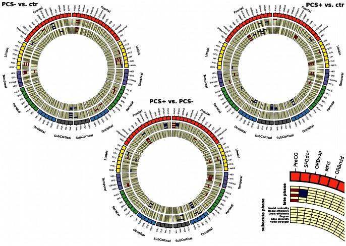
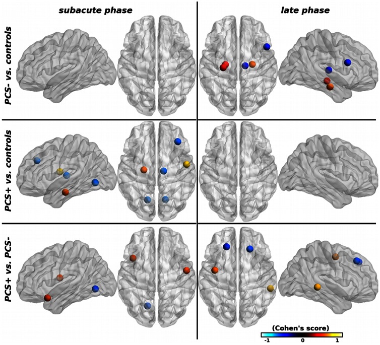
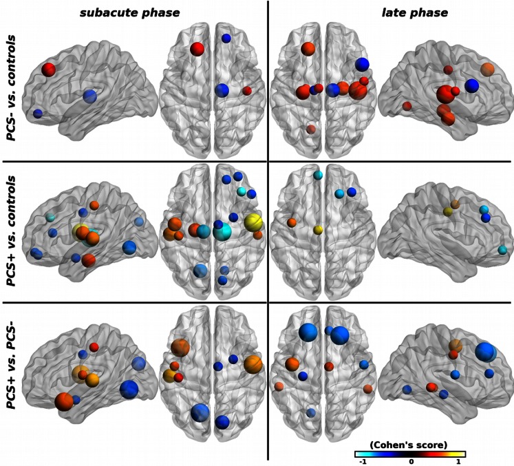
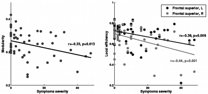
Post-concussion syndrome has been related to axonal damage in patients with mild traumatic brain injury, but little is known about the consequences of injury on brain networks. In the present study, our aim was to characterize changes in functional brain networks following mild traumatic brain injury in patients with post-concussion syndrome using resting-state functional magnetic resonance imaging data. We investigated 17 injured patients with persistent post-concussion syndrome (under the DSM-IV criteria) at 6 months post-injury compared with 38 mild traumatic brain injury patients with no post-concussion syndrome and 34 healthy controls. All patients underwent magnetic resonance imaging examinations at the subacute (1-3 weeks) and late (6 months) phases after injury. Group-wise differences in functional brain networks were analyzed using graph theory measures. Patterns of long-range functional networks alterations were found in all mild traumatic brain injury patients. Mild traumatic brain injury patients with post-concussion syndrome had greater alterations than patients without post-concussion syndrome. In patients with post-concussion syndrome, changes specifically affected temporal and thalamic regions predominantly at the subacute stage and frontal regions at the late phase. Our results suggest that the post-concussion syndrome is associated with specific abnormalities in functional brain network that may contribute to explain deficits typically observed in PCS patients.
Conflict of interest statement
Figures
Similar articles
-
A prospective biopsychosocial study of the persistent post-concussion symptoms following mild traumatic brain injury.J Neurotrauma. 2015 Apr 15;32(8):534-47. doi: 10.1089/neu.2014.3339. Epub 2015 Feb 25. J Neurotrauma. 2015. PMID: 25363626
-
Postconcussional disorder and PTSD symptoms of military-related traumatic brain injury associated with compromised neurocircuitry.Hum Brain Mapp. 2014 Jun;35(6):2652-73. doi: 10.1002/hbm.22358. Epub 2013 Sep 13. Hum Brain Mapp. 2014. PMID: 24038816 Free PMC article.
-
Small world properties changes in mild traumatic brain injury.J Magn Reson Imaging. 2017 Aug;46(2):518-527. doi: 10.1002/jmri.25548. Epub 2016 Nov 30. J Magn Reson Imaging. 2017. PMID: 27902865 Free PMC article.
-
Brain dysfunction underlying prolonged post-concussive syndrome: A systematic review.J Affect Disord. 2020 Feb 1;262:71-76. doi: 10.1016/j.jad.2019.10.058. Epub 2019 Nov 4. J Affect Disord. 2020. PMID: 31710931 Free PMC article.
-
[Mild traumatic brain injury and postconcussive syndrome: a re-emergent questioning].Encephale. 2012 Sep;38(4):329-35. doi: 10.1016/j.encep.2011.07.003. Epub 2011 Aug 31. Encephale. 2012. PMID: 22980474 Review. French.
Cited by
-
Network dysfunction after traumatic brain injury.Nat Rev Neurol. 2014 Mar;10(3):156-66. doi: 10.1038/nrneurol.2014.15. Epub 2014 Feb 11. Nat Rev Neurol. 2014. PMID: 24514870 Review.
-
Detecting functional connectivity disruptions in a translational pediatric traumatic brain injury porcine model using resting-state and task-based fMRI.Sci Rep. 2021 Jun 11;11(1):12406. doi: 10.1038/s41598-021-91853-5. Sci Rep. 2021. PMID: 34117318 Free PMC article.
-
Mapping brain recovery after concussion: From acute injury to 1 year after medical clearance.Neurology. 2019 Nov 19;93(21):e1980-e1992. doi: 10.1212/WNL.0000000000008523. Epub 2019 Oct 16. Neurology. 2019. PMID: 31619480 Free PMC article.
-
Connectomic markers of symptom severity in sport-related concussion: Whole-brain analysis of resting-state fMRI.Neuroimage Clin. 2018 Feb 17;18:518-526. doi: 10.1016/j.nicl.2018.02.011. eCollection 2018. Neuroimage Clin. 2018. PMID: 29560308 Free PMC article.
-
Traumatic Brain Injury Severity in a Network Perspective: A Diffusion MRI Based Connectome Study.Sci Rep. 2020 Jun 4;10(1):9121. doi: 10.1038/s41598-020-65948-4. Sci Rep. 2020. PMID: 32499553 Free PMC article.
References
-
- Levin HS, Mattis S, Ruff RM, Eisenberg HM, Marshall LF, et al. (1987) Neurobehavioral outcome following minor head injury: a three-center study. Journal of Neurosurgery 66: 234–43. - PubMed
-
- Iverson GL (2005) Outcome from mild traumatic brain injury. Current Opinion in Psychiatry 18: 301–17. - PubMed
-
- Stålnacke BM, Björnstig U, Karlsson K, Sojka P (2005) One-year follow-up of mild traumatic brain injury: post-concussion symptoms, disabilities and life satisfaction in relation to serum levels of S-100B and neurone-specific enolase in acute phase. Journal of Rehabilitation Medicine 37: 300–5. - PubMed
-
- Willer B, Leddy JJ (2006) Management of concussion and post-concussion syndrome. Current Treatment Options in Neurology 8: 415–26. - PubMed
Publication types
MeSH terms
LinkOut - more resources
Full Text Sources
Other Literature Sources
