. 2013 Jun 6;8(6):e65548.
doi: 10.1371/journal.pone.0065548. Print 2013.

Signal peptide cleavage from GP5 of PRRSV: a minor fraction of molecules retains the decoy epitope, a presumed molecular cause for viral persistence

Affiliations

Signal peptide cleavage from GP5 of PRRSV: a minor fraction of molecules retains the decoy epitope, a presumed molecular cause for viral persistence

Bastian Thaa et al. PLoS One. .

Abstract

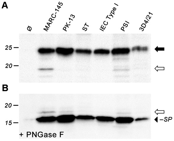
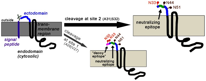
Porcine reproductive and respiratory syndrome virus (PRRSV) is the major pathogen in the pig industry. Variability of the antigens and persistence are the biggest challenges for successful control and elimination of the disease. GP5, the major glycoprotein of PRRSV, is considered an important target of neutralizing antibodies, which however appear only late in infection. This was attributed to the presence of a "decoy epitope" located near a hypervariable region of GP5. This region also harbors the predicted signal peptide cleavage sites and (dependent on the virus strain) a variable number of potential N-glycosylation sites. Molecular processing of GP5 has not been addressed experimentally so far: whether and where the signal peptide is cleaved and (as a consequence) whether the "decoy epitope" is present in virus particles. We show that the signal peptide of GP5 from the American type 2 reference strain VR-2332 is cleaved, both during in vitro translation in the presence of microsomes and in transfected cells. This was found to be independent of neighboring glycosylation sites and occurred in a variety of porcine cells for GP5 sequences derived from various type 2 strains. The exact signal peptide cleavage site was elucidated by mass spectrometry of virus-derived and recombinant GP5. The results revealed that the signal peptide of GP5 is cleaved at two sites. As a result, a mixture of GP5 proteins exists in virus particles, some of which still contain the "decoy epitope" sequence. Heterogeneity was also observed for the use of glycosylation sites in the hypervariable region. Lastly, GP5 mutants were engineered where one of the signal peptide cleavage sites was blocked. Wildtype GP5 exhibited exactly the same SDS-PAGE mobility as the mutant that is cleavable at site 2 only. This indicates that the overwhelming majority of all GP5 molecules does not contain the "decoy epitope".


Conflict of interest statement

Competing Interests: The Boehringer Ingelheim Vetmedica PRRSV Research Award does not alter their adherence to all the PLOS ONE policies on sharing data and materials.

Figures

Figure 1
Figure 1. Schematic representation of PRRSV GP5 and its possible processing.
(A), Topology of unprocessed PRRSV GP5 with signal peptide (purple, ∼30 amino acids), ectodomain (blue, ∼30 residues), a hydrophobic transmembrane stretch (∼70 amino acids, black) and a cytosolic/virus-internal endodomain (∼70 residues, black). The position of the C-terminal HA tag used in this study is indicated (dark grey). Membrane in light grey. (B), Signal peptide cleavage is predicted to occur at two sites: either at A26|V27 (site 1) or A31|S32 (site 2) as indicated. The neutralizing epitope (green) is present in all predicted variants, but the “decoy epitope” (magenta) is not preserved in the mature protein by cleavage at site 2. However, bioinformatic prediction tools rely on the amino acid sequence alone and do not take into account possible carbohydrate attachment to non-conserved glycosylation sites (red) located in the hypervariable region (yellow) and to conserved glycosylation sites (brown). The situation for PRRSV type 2 reference strain VR-2332 is depicted.
Figure 2
Figure 2. in vitro-transcription/translation of GP5–HA to assess processing (glycosylation/signal peptide cleavage).
(A), Plasmids encoding GP5–HA was subjected to in vitro-transcription/translation with rabbit reticulocyte lysate in the absence (–) or presence (+) of porcine pancreatic microsomes. The products were analysed by SDS-PAGE and Western blot (anti-HA tag). Wildtype (wt) and mutants with deleted or added glycosylation sites near the signal peptide cleavage site were employed; Ø, empty plasmid control. Molecular weight marker is indicated on the left-hand side, arrows on the right-hand side show the positions of unprocessed GP5–HA (white), fully glycosylated GP5–HA (black), and GP5–HA lacking one glycan (grey). (B), Glycans from the products of (A) were removed with PNGase F prior to SDS-PAGE and Western blot. Deglycosylated protein lacking the signal peptide (black arrowhead) is smaller than unprocessed GP5–HA and deglycosylated protein containing the signal peptide (white arrow), indicating signal peptide cleavage.
Figure 3
Figure 3. Processing analysis of cell expressed GP5–HA to reveal glycan-independent signal peptide cleavage.
CHO-K1 (A, B) and MARC-145 (C, D; permissive for PRRSV) were transfected with plasmids encoding GP5–HA as wildtype (wt) and mutants with deleted or added glycosylation sites near the signal peptide cleavage site; Ø, empty plasmid control. Cell lysates were subjected to SDS-PAGE and Western blot (anti-HA tag) before (A, C) and after (B, D) deglycosylation with PNGase F. Molecular weight marker given on the left-hand side; arrows indicate sizes of unprocessed GP5–HA (white), glycosylated protein (black: wildtype glycosylation; grey: lacking one glycan; black/+: with one additional glycan), and deglycosylated protein without signal peptide (black arrowhead). In B and D, in vitro-generated GP5–HA (in the absence of microsomes, thus intrinsically unprocessed, i.e. not glycosylated and still containing the signal peptide, cf. Fig. 2) is shown in the rightmost lane for size comparison. Deglycosylated GP5–HA and all variants mostly ran faster than unprocessed protein.
Figure 4
Figure 4. Limited PNGase F digestion of GP5–HA to show modification with three glycans.
MARC-145 cells were transfected with the GP5–HA wt construct. Aliquots of the cell lysates were treated with decreasing concentrations of PNGase F as indicated or left undigested (Ø), then analyzed by SDS-PAGE and Western blot (anti-HA tag). The number of carbohydrates cleaved by PNGase F decreases with decreasing concentration. This causes a ladder-like appearance of bands that allows counting of the total number of carbohydrates linked to GP5–HA (arrowheads). The band denoted with the asterisk must not be counted since it is also present in untreated GP5–HA and probably represents unprocessed/non-translocated protein (see also Fig. 3C, white arrow).
Figure 5
Figure 5. The signal peptide of GP5–HA is cleaved in different porcine cells.
GP5–HA wt was expressed in MARC-145 cells as well as different porcine cells, which were lyzed and analyzed by SDS-PAGE and Western blot (anti-HA tag) before (A) and after (B) deglycosylation with PNGase F. Labels as in Fig. 3. GP5–HA is processed in the same manner in all cell types tested: PK-13 (porcine kidney), ST (testis), IEC Type I (intestine), PSI (small intestine), and 3D4/21 (alveolar monocytic cell line that can be rendered permissive to PRRSV by expression of the receptor CD163 [56]).
Figure 6
Figure 6. The signal peptide of GP5 from different PRRSV type 2 strains is cleaved.
(A), N-terminal sequences (residues 1–60) of representative PRRSV type 2 strains (VR-2332: prototype strain, MLV: modified live vaccine “RespPRRS” (containing just one exchange – R13Q – in the signal peptide/ectodomain region relative to VR-2332), JXA-1: Chinese virulent strain, Neb-1: US-American virulent strain) with potential glycosylation sites (grey), predicted signal peptide (black), and propensity of signal peptide cleavage (D score according to SignalP 4.0, www.cbs.dtu.dk/services/SignalP/). Note that the difference in signal peptide cleavage at site 1 and 2 was small as shown in detail in Table S1. (B/C), MARC-145 cells were transfected with GP5–HA with the signal peptide/ectodomain sequence as depicted in (A), or with empty plasmid (Ø), subsequently lysed and assessed by SDS-PAGE and Western blot (anti-HA tag) before (B) and after (C) PNGase F digestion to remove glycans. Labels as in Fig. 2; the thin black arrow denotes additional glycosylation partially achieved in JXA-1; the number of glycans in the mature proteins is indicated on the bottom. The black arrowhead in (C) indicates the position of deglycosylated GP5–HA without signal peptide.
Figure 7
Figure 7. Identification of the signal peptide cleavage site of GP5 (virus-derived and recombinant) by mass spectrometry.
(A), PRRSV (strain VR-2332) was grown in MARC-145 cells, precipitated with PEG-8000, pelleted and subjected to sucrose density gradient centrifugation. The virus-containing fraction was left untreated or deglycosylated with PNGase F and separated by reducing SDS-PAGE followed by Coomassie staining (left-hand side) or Western blot (anti-GP5 antiserum, right-hand side). The deglycosylated band corresponding to GP5 (black box) was cut out of the gel, digested with trypsin or chymotrypsin and analyzed by LC-MS/MS. (B), PRRSV GP5 (with His tag) and M (with HA tag) were co-expressed in Sf9 insect cells by infection with recombinant baculovirus. Following cell harvesting and lysis, GP5–M was enriched using Ni-NTA agarose (binding to GP5–His). The eluated protein was left untreated or digested with PNGase F and subjected to reducing SDS-PAGE and Coomassie staining (left-hand side) or Western blot (anti-His-tag antibody, right-hand side). The deglycosylated GP5 band (black box; coinciding with M) was cut out and treated as in (A). (C), representative result from mass spectrometry of virus-derived GP5 (as in A). The first 61 residues of GP5 are shown with the positions of the predicted chymotrypsin cleavage sites (black lines) and the putative signal peptide cleavage sites (broken lines). Chymotryptic peptides that were identified are represented as black bars. The pattern of peptides is evidence for signal peptide cleavage at sites 1 and 2. No peptides corresponding to the signal peptide region (1–26) were identified. (D), Conclusion from mass spectrometry, showing the N-terminal sequence of GP5 with signal peptide (black), glycosylations (grey) and the positions of the neutralizing and the “decoy epitope”. Two GP5 species exist with signal peptide cleavage at sites 2 (top) and 1 (bottom), respectively.
Figure 8
Figure 8. The majority of GP5–HA molecules in transfected cells is cleaved at site 2.
MARC-145 cells were transfected with variants of GP5–HA or with empty plasmid (Ø), subsequently lysed and assessed by SDS-PAGE and Western blot (anti-HA tag) before (A) and after (B) PNGase F digestion to remove glycans. wt: GP5–HA wildtype; uncl.: Signal peptide (SP) cleavage completely blocked by mutation A26F, A29Y, A31F; cl.1: SP cleavage possible at site 1 (A26|V27) only (mutation A29S, A31Y); cl.2: SP cleavage at site 2 (A31|S32) only (mutation A26F, A29S). Black arrow: GP5–HA with cleaved SP, carrying three glycans, and/or GP5–HA comprising SP and two glycans; black arrow with plus sign: GP5–HA, SP cleaved, four glycans; white arrow: GP5–HA unprocessed/deglycosylated and containing the SP; grey and black arrowhead: GP5–HA with SP cleavage at site 1 or 2, respectively. (C), limited PNGase F digestion of GP5–HA uncl., performed and labeled as in Fig. 4. The protein carries two glycans.
Figure 9
Figure 9. Conclusion – Major fraction of GP5 from PRRSV type 2 does not contain the “decoy epitope”.
Schematic representation of GP5 fractions as evidenced in this study. The signal peptide of GP5 is predominantly cleaved at site 2 (A26|V27; black arrow), but is also cleaved in minor quantities at site 1 (A26|V27, thin grey arrow). The “decoy epitope” (magenta) is preserved only in GP5 cleaved at site 1. The neutralizing epitope (green) is present in either case. Heterogeneity occurs also at non-conserved glycosylation sites (red). The fraction of GP5 with the “decoy epitope” contains carbohydrates at either both N30 and N33, or only at N33 or none of these sites. A subfraction of site 2-cleaved GP5 does not contain a carbohydrate at N33. Conserved glycosylation sites are in brown.

References

    1. Snijder EJ, Meulenberg JJ (1998) The molecular biology of arteriviruses. J Gen Virol 79: 961–979. - PubMed
    1. Wensvoort G, Terpstra C, Pol JM, ter Laak EA, Bloemraad M, et al. (1991) Mystery swine disease in The Netherlands: the isolation of Lelystad virus. Vet Q 13: 121–130. - PubMed
    1. Collins JE, Benfield DA, Christianson WT, Harris L, Hennings JC, et al. (1992) Isolation of swine infertility and respiratory syndrome virus (isolate ATCC VR-2332) in North America and experimental reproduction of the disease in gnotobiotic pigs. J Vet Diagn Invest 4: 117–126. - PubMed
    1. Zhou L, Yang H (2010) Porcine reproductive and respiratory syndrome in China. Virus Res 154: 31–37. - PubMed
    1. Tian D, Wei Z, Zevenhoven-Dobbe JC, Liu R, Tong G, et al. (2012) Arterivirus minor envelope proteins are a major determinant of viral tropism in cell culture. J Virol 86: 3701–3712. - PMC - PubMed

Publication types