. 2013 Nov;22(4):92-102.
doi: 10.1016/j.jtv.2013.04.003. Epub 2013 Jun 10.

Connective tissue ulcers

Affiliations

Connective tissue ulcers

Ganary Dabiri et al. J Tissue Viability. 2013 Nov.

Abstract

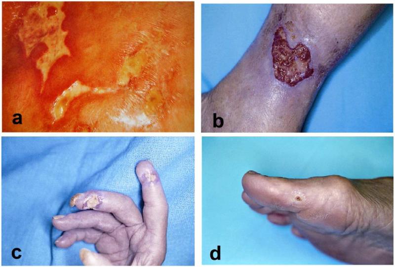
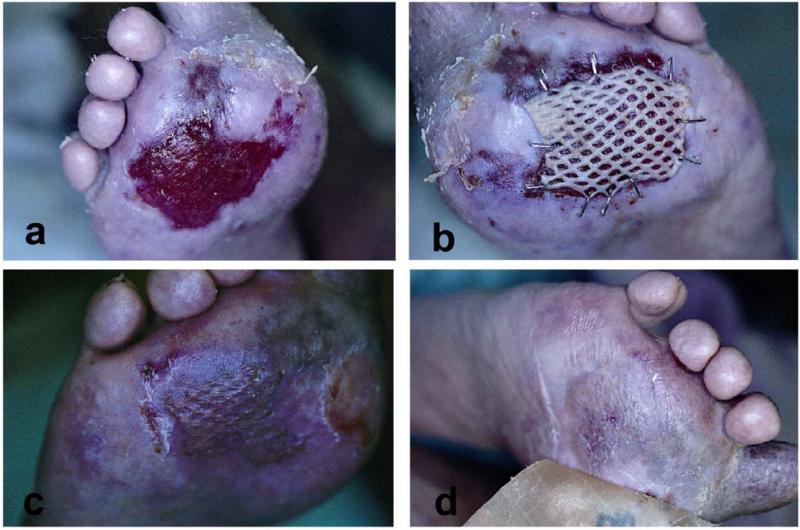
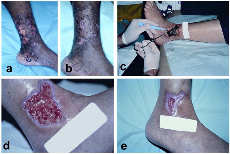
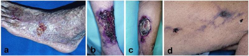
Connective tissue disorders (CTD), which are often also termed collagen vascular diseases, include a number of related inflammatory conditions. Some of these diseases include rheumatoid arthritis, systemic lupus erythematosus, systemic sclerosis (scleroderma), localized scleroderma (morphea variants localized to the skin), Sjogren's syndrome, dermatomyositis, polymyositis, and mixed connective tissue disease. In addition to the systemic manifestations of these diseases, there are a number of cutaneous features that make these conditions recognizable on physical exam. Lower extremity ulcers and digital ulcers are an infrequent but disabling complication of long-standing connective tissue disease. The exact frequency with which these ulcers occur is not known, and the cause of the ulcerations is often multifactorial. Moreover, a challenging component of CTD ulcerations is that there are still no established guidelines for their diagnosis and treatment. The morbidity associated with these ulcerations and their underlying conditions is very substantial. Indeed, these less common but intractable ulcers represent a major medical and economic problem for patients, physicians and nurses, and even well organized multidisciplinary wound healing centers.

Keywords: Connective tissue disease; Connective tissue ulcers; Ulcers; Vasculitis.


Figures

Figure 1
Figure 1. Examples of rheumatoid and systemic sclerosis ulcers
A: “angular” ulcers-the unusual irregular and “angular” appearance may at first suggest factitial disease, however imaged shows a 56-year-old woman with CREST syndrome. B: rheumatoid ulcer with an angulating configuration or undulating border. C: Digital ulceration in a patient with systemic sclerosis. Ulcers located distally like the one shown here, are more likely to be the result of significant ischemia. Autoamputation is often the outcome. D: Calcinosis and a painful nonhealing ulcer on the big toe in a patient with systemic sclerosis. Patients with systemic sclerosis commonly develop calcinosis, especially in CREST syndrome.
Figure 2
Figure 2. Erosive lichen planus/systemic lupus erythematosus overlap
A: Lichen planus rarely presents on the palms and soles. However, in an overlap of lichen planus and SLE, palms and soles can be involved. B: After failure of occlusive dressings and corticosteroid injections, split-thickness autologous skin grafting was used to stimulate healing. C: 4 weeks after grafting, there is complete healing. Satellite lesions of lichen planus are still visible at the edges however. D: Several weeks after grafting, with the use of hydrocolloid dressings, the affected area is looking more like normal skin.
Figure 3
Figure 3. Examples of vasculitic ulcers
A-B: Biopsy proven periarteritis nodosa with destruction of medium-sized vessels. Notice the slight microlivedo pattern surrounding the ulcer as well as the necrotic appearance of some of the ulcers. Treatment included methotrexate, sulfamethoxazole for pnuemocystis pneumonia (PCP) prophylaxis, and with a gel that promoted a moist wound environment and helped with autolytic debridement. C-E: This patient has an established diagnosis of Wegners granulomatosis c-ANCA positive small-medium sized vessel vasculitis. C: This large necrotic ulcer needed surgical debridement. D Three weeks after initial debridement, the wound bed is optimal with evidence of re-epithelialization. E: Six weeks after initial presentation. These cases illustrate the importance of attention to proper wound care in vasculitic ulcers in addition to systemic therapy.
Figure 4
Figure 4. Examples of cutaneous microthrombotic ulcers
Necrosis and livedo reticularis should always raise the suspicion of an obstruction of small blood vessels as illustrated in the following examples. A: This Caucasian man with antiphospholipid syndrome has extensive livedo reticularis and very painful ulcers. B: In this case, the underlying condition is cryoglobulinemia. Concomitant hepatitis C infection should always be excluded. Patients often need therapy with systemic immunosuppressive agents. C: Cyrofibrinogenemia was diagnosed in this 87-year-old man with multiple medical problems by plasma measurements and histology. His ulcer healed with stanozolol (which is no longer commercially available, danazol is the alternative). D: This women’s thigh shows livedo reticularis and necrotic ulcers. Histology would show deposition of calcium crystals in the wall of blood vessels.

References

    1. Goslen JB. Autoimmune Ulceration of the leg. Leg Ulcers. 1990;3:92–117. - PubMed
    1. Falanga V, Lindholm C, Carson PA, et al. Text Atlas of Wound Managament (ed 2) Informa Healthcare; London, UK: 2012.
    1. Panuncialman J, Falanga V. Basic approach to inflammatory ulcers. Dermatologic Therapy. 2006;19:365–376. - PubMed
    1. Jorrizo JL, O AJ, Stanely RJ. Superficial Ulcerating Necrobiosis in Rheumatoid Arthritis. A Variant of the Necrobiosis Lipoidica-Rheumatoid Nodule Spectrum? Arch Dermatol. 1982;118 - PubMed
    1. Danning CL, Illei GG, Boumpas DT. Vasculitis associated with primary rheumatologic diseases. Curr opin Rheumatol. 1998;10:58–65. - PubMed

Publication types

MeSH terms