. 2013 Jul 3;32(13):1855-68.
doi: 10.1038/emboj.2013.135. Epub 2013 Jun 11.

Exonuclease hDIS3L2 specifies an exosome-independent 3'-5' degradation pathway of human cytoplasmic mRNA

Affiliations

Exonuclease hDIS3L2 specifies an exosome-independent 3'-5' degradation pathway of human cytoplasmic mRNA

Michal Lubas et al. EMBO J. .

Abstract

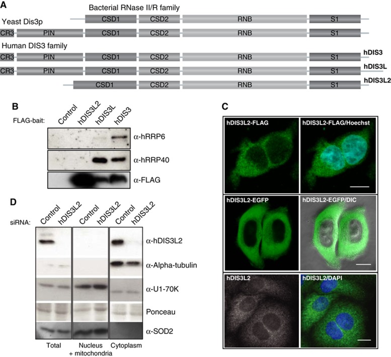
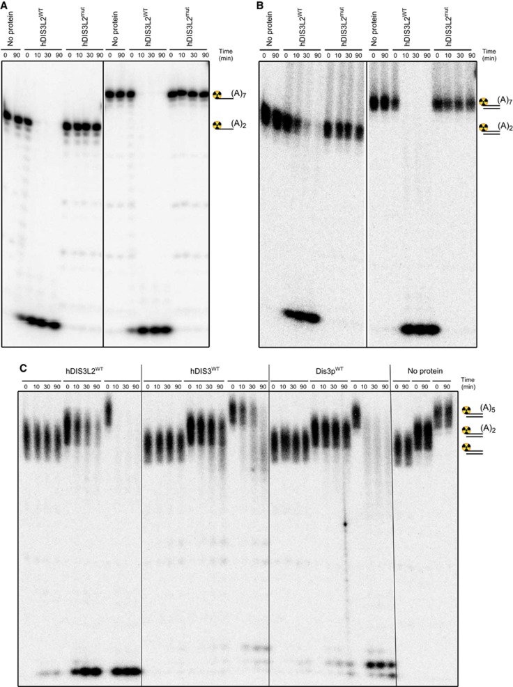
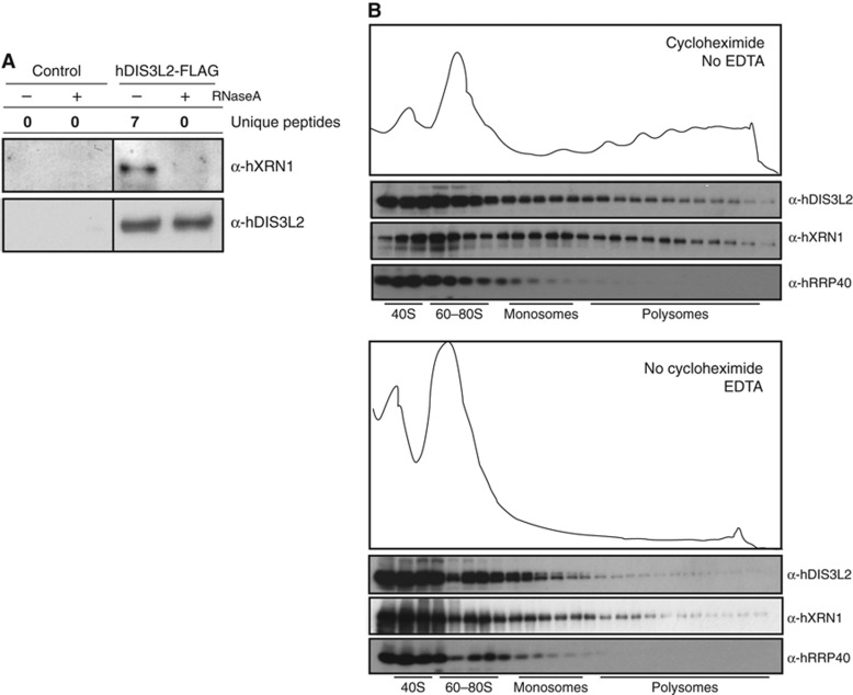
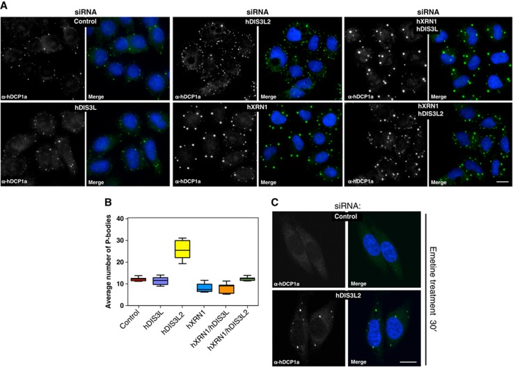
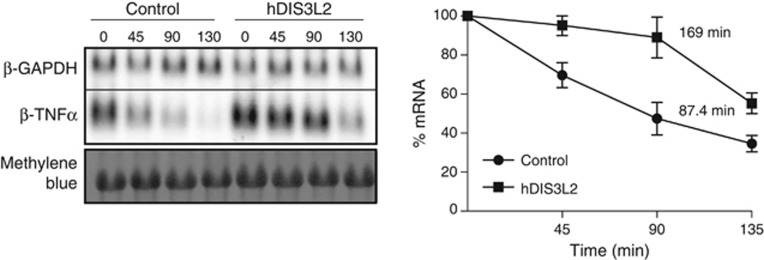
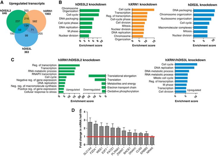
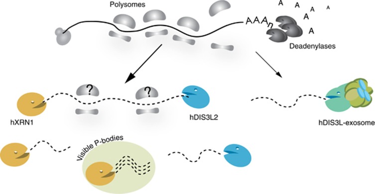
Turnover of mRNA in the cytoplasm of human cells is thought to be redundantly conducted by the monomeric 5'-3' exoribonuclease hXRN1 and the 3'-5' exoribonucleolytic RNA exosome complex. However, in addition to the exosome-associated 3'-5' exonucleases hDIS3 and hDIS3L, the human genome encodes another RNase II/R domain protein-hDIS3L2. Here, we show that hDIS3L2 is an exosome-independent cytoplasmic mRNA 3'-5' exonuclease, which exhibits processive activity on structured RNA substrates in vitro. hDIS3L2 associates with hXRN1 in an RNA-dependent manner and can, like hXRN1, be found on polysomes. The impact of hDIS3L2 on cytoplasmic RNA metabolism is revealed by an increase in levels of cytoplasmic RNA processing bodies (P-bodies) upon hDIS3L2 depletion, which also increases half-lives of investigated mRNAs. Consistently, RNA sequencing (RNA-seq) analyses demonstrate that depletion of hDIS3L2, like downregulation of hXRN1 and hDIS3L, causes changed levels of multiple mRNAs. We suggest that hDIS3L2 is a key exosome-independent effector of cytoplasmic mRNA metabolism.


Conflict of interest statement

The authors declare that they have no conflict of interest.

Figures

Figure 1
Figure 1
hDIS3L2 is an RNase II/R family-related and exosome-independent factor. (A) Domain composition of hDIS3L2 and its homologues. The RNB domain and the three RNA-binding domains (CSD1, CSD2, and S1) are present in all proteins. In contrast, the PIN domain and the CR3 motif are features characteristic for the Dis3 family, and absent from hDIS3L2, which instead harbours an extended CSD1 domain. (B) CoIP/western blotting analysis of hDIS3L2, hDIS3L, and hDIS3 associations with exosome subunits. hDIS3L2-, hDIS3L-, and hDIS3-FLAG IP eluates were probed with antibodies against hRRP6, hRRP40, and FLAG peptide as indicated. An empty host cell line was used as a negative control. (C) Subcellular localization analysis of hDIS3L2 in HEK293 (top) and HeLa (middle and bottom) cells. HEK293 Flp-In T-Rex cells were stably expressing C-terminal FLAG-tagged hDIS3L2, which was visualized by fluorescence microscopy. In the middle panel, HeLa cells were transiently transfected with a construct encoding hDIS3L2-EGFP, directly analysed by live-cell confocal microscopy and overlaid with phase contrast. The endogenous hDIS3L2 was detected in HeLa cells with rabbit α-hDIS3L2 antibodies (bottom). Blue colour indicates nuclei, stained with Hoechst or DAPI. Scale bars indicate 100 μm. (D) Analysis of hDIS3L2 distribution by subcellular fractionation of HeLa cells. Protein equivalents of total, nucleus/mitochondria, and cytoplasmic cell fractions were analysed by SDS–PAGE followed by western blotting analysis using α-hDIS3L2, α-alpha-tubulin (cytoplasmic marker), α-U1-70K (nuclear marker), and α-SOD2 (mitochondrial marker) antibodies as indicated. Ponceau staining was performed as a loading control.
Figure 2
Figure 2
hDIS3L2 is a processive 3′-5′ exonuclease active towards both ss- and ds-RNA. (A) hDIS3L2 displays 3′-5′ exoribonuclease activity towards ssRNA. Wild-type (hDIS3L2WT) or mutated (hDIS3L2mut) proteins were incubated in the presence of Mg2+ ions with 5′radiolabelled single-stranded 17nt RNA oligonucleotide substrates bearing oligoadenosine extensions of different length (A2 or A7) (see also Supplementary Figure S2E). Control reactions were performed in the absence of added protein (no protein). Reactions were terminated at the indicated time points and the products were analysed by 20% acrylamide/7 M urea PAGE followed by phosphorimaging. (B) hDIS3L2 is active towards dsRNA. RNase assays were performed as in A, but using 5′radiolabelled partially structured double-stranded 17nt RNA substrates in which the longer (labelled) oligoribonucleotide possessed 3′ss oligoadenosine overhangs of different length (A2 or A7) (see also Supplementary Figure S2H). (C) hDIS3L2 degrades partially structured RNA substrates bearing short single-stranded extensions more efficiently than hDIS3 and yeast Dis3p. Equal amounts of wild-type recombinant hDIS3L2, hDIS3 or S. cerevisiae Dis3p proteins were used for RNase assays performed as in B using radiolabelled blunt double-stranded 17nt RNA substrate or its counterparts with 3′ss oligoadenosine overhangs (A2 or A5).
Figure 3
Figure 3
hDIS3L2 interacts RNA dependently with hXRN1 and co-sediments with translating ribosomes. (A) In the presence of RNA, hXRN1 was detected as a specific interaction partner of hDIS3L2-FLAG as assayed by coIP/MS and coIP/western blotting analysis. MS results are shown as unique peptide counts from a representative biological replicate. For full data sets, see Supplementary Table S1. For IP/western blotting analysis, control (empty host cell line) and tetracycline-induced cell extracts were lysed in a buffer with or without RNAse A as indicated. Eluates from anti-FLAG columns were analysed by western blotting using α-hDIS3L2 and α-hXRN1 antibodies. (B) Both hDIS3L2 and hXRN1 co-purifies with polysomes. HeLa cells were treated with cycloheximide and subjected to 10–50% sucrose gradient fractionation followed by examination of the abundance of translating ribosomes (see Materials and methods). Collected fractions were analysed by western blotting using α-hXRN1, α-hDIS3L2, and α-hRRP40 antibodies as indicated. The bottom panel shows the control of disrupting polysomes. The sucrose gradient was performed in the absence of cycloheximide and in the presence of 12.5 mM EDTA. The five first fractions were diluted 2-fold prior to SDS–PAGE analysis.
Figure 4
Figure 4
hDIS3L2 depletion increases the numbers of PBs. (A) hDCP1a staining as a PB proxy of cells treated with siRNA against EGFP (control), hDIS3L, hDIS3L2, hXRN1, hXRN1/hDIS3L, or hXRN1/hDIS3L2 as indicated. Cells were inspected by fluorescence microscopy using a × 40 objective. Nuclei were stained with DAPI and merged with protein staining as indicated. Scale bar indicates 100 μm. (B) Boxplots displaying the average number of PBs per siRNA-treated cell from A. In all, 150 cells were inspected per experiment. Boxes indicate 5th and 95th percentiles, while thick lines indicate the mean values. (C) EGFP (control) or hDIS3L2 siRNA-treated HeLa cells subjected to 30 min emetine treatment to inhibit translation elongation. Cells were fixed and stained with α-hDCP1a antibodies. Control cells showed a decreased number of PBs while a 40% fraction of hDIS3L2-depleted cells retained PBs upon emetine treatment. Scale bar indicates 100 μm.
Figure 5
Figure 5
hDIS3L2 is required for ARE-mediated decay. Half-life measurements of β-globin reporter mRNA containing a 3′UTR element from TNF-α (ARE) performed in cells treated with siRNA against EGFP (control) or hDIS3L2. Transcription was induced and repressed by a tetracycline pulse-chase assay and RNA levels measured by northern blotting analysis (left panel). The graph shows the calculated decay rates and the average levels of β-TNF-α mRNA plotted over time. Mean values were quantified from three independent biological repeats normalized to the levels of β-GAPDH (internal transfection control) with error bars representing standard errors of the mean (right panel).
Figure 6
Figure 6
Transcriptome analysis of hDIS3L2-, hXRN1-, and hDIS3L-depleted cells. (A) Venn diagram showing distribution and overlap of upregulated transcripts from hDIS3L2-, hXRN1-, and hDIS3L-depleted cells as identified by DESeq in a given set of biological replicates (for full data sets, see Supplementary Figure S5 and Supplementary Tables S2–S4). Enrichment between sample sets was calculated using hypergeometric test, P-values scored below 10−30. (B) GO term analysis of upregulated mRNAs from each knockdown sample versus control clustered using DAVID (false discovery rate <0.05). Most enriched clusters for each condition are shown as barplots using enrichment score (−log(P-value)). (C) GO term analysis as in B for significantly upregulated or downregulated mRNAs from hDIS3L2/hXRN1 and hDIS3L/hXRN1 co-depletions (Supplementary Tables S5 and S6). (D) Changes in half-lives of selected mRNA caused by hDIS3L2 siRNA-mediated depletion. Half-lives of mRNAs were determined based on newly transcribed RNA/total RNA ratios (Dolken et al, 2008). The EGFP (control) and hDIS3L2 siRNA-treated HeLa cells were metabolically labelled with 4sU for 90 min and total cellular RNA was purified as described in Materials and methods. Changes in mRNA half-lives were calculated for DIS3L2-depleted cells relative to the control. For each condition, three biological replicates of each RNA subset were subjected to qRT-PCR analysis. Error bars calculated as standard error of the mean. * indicates AU-rich mRNAs (ARED3 database; Bakheet et al, 2006).
Figure 7
Figure 7
Schematic model for cytoplasmic mRNA degradation in human cells. The 5′-3′ exonuclease hXRN1, the 3′-5′ exonucleases hDIS3L2, and hDIS3L/exosome impact cytoplasmic mRNA turnover in human cells. A fraction of hXRN1 and hDIS3L2, but not the exosome, associates with ribosomes, however, it remains to be shown whether 3′-5′ degradation also occurs at these assemblies (indicated by question marks). Data indicate that more hXRN1, than hDIS3L2, activity may be localized to visible PBs (see text).

Comment in

References

    1. Allalou A, Wahlby C (2009) BlobFinder, a tool for fluorescence microscopy image cytometry. Comput Methods Prog Biomed 94: 58–65 - PubMed
    1. Allmang C, Kufel J, Chanfreau G, Mitchell P, Petfalski E, Tollervey D (1999) Functions of the exosome in rRNA, snoRNA and snRNA synthesis. EMBO J 18: 5399–5410 - PMC - PubMed
    1. Anders S, Huber W (2010) Differential expression analysis for sequence count data. Genome Biol 11: R106. - PMC - PubMed
    1. Anderson JS, Parker RP (1998) The 3′ to 5′ degradation of yeast mRNAs is a general mechanism for mRNA turnover that requires the SKI2 DEVH box protein and 3′ to 5′ exonucleases of the exosome complex. EMBO J 17: 1497–1506 - PMC - PubMed
    1. Astuti D, Morris MR, Cooper WN, Staals RH, Wake NC, Fews GA, Gill H, Gentle D, Shuib S, Ricketts CJ, Cole T, van Essen AJ, van Lingen RA, Neri G, Opitz JM, Rump P, Stolte-Dijkstra I, Muller F, Pruijn GJ, Latif F et al. (2012) Germline mutations in DIS3L2 cause the Perlman syndrome of overgrowth and Wilms tumor susceptibility. Nat Genet 44: 277–284 - PubMed

Publication types

MeSH terms