. 2013 Jun 15;27(12):1345-50.
doi: 10.1101/gad.219915.113. Epub 2013 Jun 11.

The structural basis of R-spondin recognition by LGR5 and RNF43

Affiliations

The structural basis of R-spondin recognition by LGR5 and RNF43

Po-Han Chen et al. Genes Dev. .

Abstract

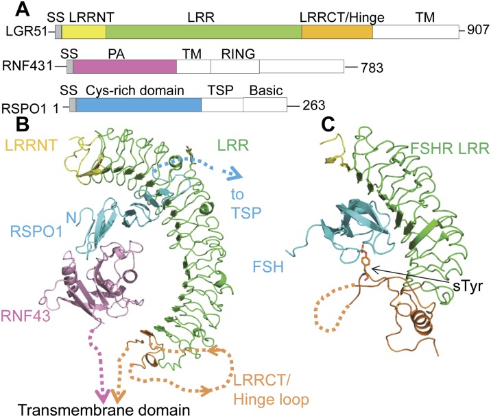
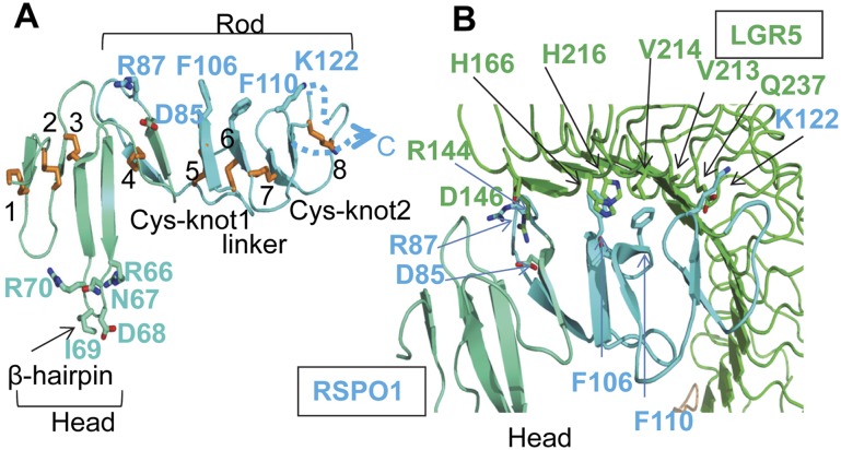
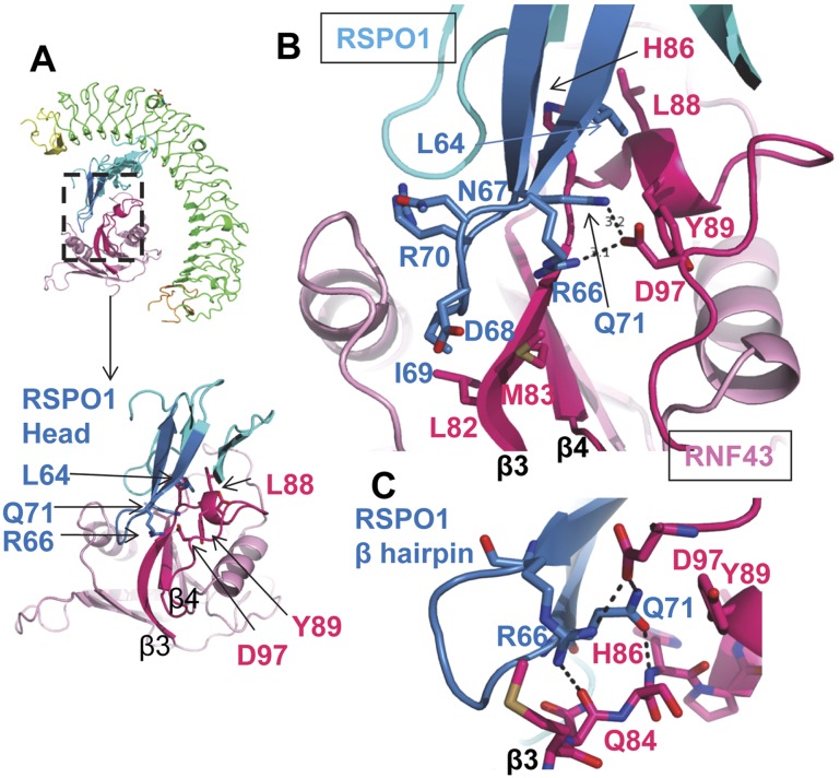
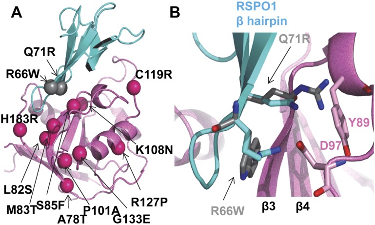
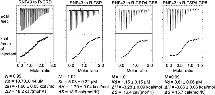
R-spondins (RSPOs) enhance Wnt signaling, affect stem cell behavior, bind to leucine-rich repeat-containing G-protein-coupled receptors 4-6, (LGR4-6) and the transmembrane E3 ubiquitin ligases RING finger 43/zinc and RING finger 3 (RNF43/ZNRF3). The structure of RSPO1 bound to both LGR5 and RNF43 ectodomains confirms their physical linkage. RSPO1 is sandwiched by LGR5 and RNF43, with its rod module of the cysteine-rich domain (CRD) contacting LGR5 and a hairpin inserted into RNF43. LGR5 does not contact RNF43 but increases the affinity of RSPO1 to RNF43, supporting LGR5 as an engagement receptor and RNF43 as an effector receptor. Disease mutations map to the RSPO1-RNF43 interface, which promises therapeutic targeting.

Keywords: E3 ubiquitin ligase; LGR5; R-spondin; RNF43; Wnt signaling; furin-like repeat.


Figures

Figure 1.
Figure 1.
Crystal structure of the LGR5–RSPO1–RNF43 ternary complex. (A) Domain organization of human LGR5, RNF43, and RSPO1. The domains included for crystallization are colored. (LRRNT) LRR N-terminal domain; (LRRCT) LRR C-terminal domain; (TM) transmembrane. (B) Ternary complex structure, color-coded as in A. The missing residues are depicted by dotted lines. N-linked glycosylation is shown as sticks. (Inset) Zoomed-in view of LRRCT/Hinge. (C) FSH–FSHR complex. The sulfated tyrosine is shown as sticks. Missing residues are depicted by dotted lines.
Figure 2.
Figure 2.
RSPO1 structure and interaction with LGR5. (A) The head module of RSPO1 is shown in blue-green, and the rod module is shown in cyan. The eight disulfide bridges are numbered and shown as sticks. The loop connected by disulfide 8 is disordered in the structure and is depicted as a dotted line. Selected hairpin loop residues and LGR5-interacting residues are shown as sticks. (B) The interface between LGR5 and the RSPO1 rod module. Key interacting residues are shown in sticks.
Figure 3.
Figure 3.
The head module of RSPO1 interaction with RNF43. For clarity, the RSPO1 β-hairpin protrusion is highlighted in marine, and key interacting loops and β strands from RNF43 are colored in hot pink. (A) Miniature view showing RSPO1 hairpin insertion into the RNF43 groove. (B) Magnified view, in the same orientation as A, of the boxed region in the ternary complex. RSPO1 R66/Q71 interaction with RNF43 D97 is shown as dashed lines. (C) Closer view of hydrogen bonding and salt bridge networks mediated between RSPO1 R66/Q71 and RNF43 Q84/H86/D97.
Figure 4.
Figure 4.
Selected known disease mutations mapped on RSPO1/RNF43 structure. (A) Same view as in Figure 3A. Mutations are shown in ball presentations. (B) RSPO1 Q71R and R66W superimposed onto wild-type structure.
Figure 5.
Figure 5.
Calorimetric measurements of RNF43 interaction with RSPO1 alone or the RSPO1–LGR5 binary complex. (R-CRD) RSPO1 CRD only; (R-RSP) RSPO1 CRD+TSP.

References

    1. Barker N, Clevers H 2010. Leucine-rich repeat-containing G-protein-coupled receptors as markers of adult stem cells. Gastroenterology 138: 1681–1696 - PubMed
    1. Barker N, Rookmaaker MB, Kujala P, Ng A, Leushacke M, Snippert H, van de Wetering M, Tan S, Van Es JH, Huch M et al. 2012. Lgr5+ve stem/progenitor cells contribute to nephron formation during kidney development. Cell Rep 2: 540–552 - PubMed
    1. Bella J, Hindle KL, McEwan PA, Lovell SC 2008. The leucine-rich repeat structure. Cell Mol Life Sci 65: 2307–2333 - PMC - PubMed
    1. Binnerts ME, Kim KA, Bright JM, Patel SM, Tran K, Zhou M, Leung JM, Liu Y, Lomas WE 3rd, Dixon M, et al. 2007. R-spondin1 regulates Wnt signaling by inhibiting internalization of LRP6. Proc Natl Acad Sci 104: 14700–14705 - PMC - PubMed
    1. Blaydon DC, Ishii Y, O'Toole EA, Unsworth HC, Teh MT, Ruschendorf F, Sinclair C, Hopsu-Havu VK, Tidman N, Moss C, et al. 2006. The gene encoding R-spondin 4 (RSPO4), a secreted protein implicated in Wnt signaling, is mutated in inherited anonychia. Nat Genet 38: 1245–1247 - PubMed

Publication types

MeSH terms

Associated data