. 2013 Aug 1;305(3):E439-50.
doi: 10.1152/ajpendo.00006.2013. Epub 2013 Jun 11.

Thrombospondin-1 regulates adiposity and metabolic dysfunction in diet-induced obesity enhancing adipose inflammation and stimulating adipocyte proliferation

Affiliations

Thrombospondin-1 regulates adiposity and metabolic dysfunction in diet-induced obesity enhancing adipose inflammation and stimulating adipocyte proliferation

Ping Kong et al. Am J Physiol Endocrinol Metab. .

Abstract

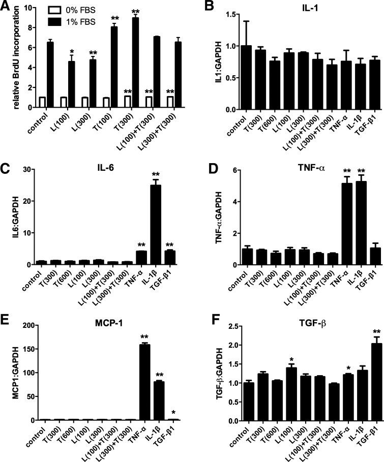
As a typical matricellular protein, thrombospondin (TSP)-1, binds to the structural matrix and regulates cellular behavior by modulating growth factor and cytokine signaling. Obesity and diabetes are associated with marked upregulation of TSP-1 in adipose tissue. We hypothesized that endogenous TSP-1 may play an important role in the pathogenesis of diet-induced obesity and metabolic dysfunction. Accordingly, we examined the effects of TSP-1 gene disruption on weight gain, adiposity, and adipose tissue inflammation in mice receiving a high-fat diet (HFD: 60% fat, 20% carbohydrate) or a high-carbohydrate low-fat diet (HCLFD: 10% fat, 70% carbohydrate). HFD mice had significantly higher TSP-1 expression in perigonadal adipose tissue; TSP-1 was predominantly localized in the adipose interstitium. TSP-1 loss attenuated weight gain and fat accumulation in HFD and HCLFD groups. Compared with corresponding wild-type animals, TSP-1-null mice had decreased insulin levels but exhibited elevated free fatty acid and triglyceride levels, suggesting impaired fatty acid uptake. TSP-1 loss did not affect adipocyte size and had no effect on adipose vascular density. However, TSP-1-null mice exhibited attenuated tumor necrosis factor-α mRNA expression and reduced macrophage infiltration, suggesting a role for TSP-1 in mediating obesity-associated inflammation. In vitro, TSP-1 enhanced proliferation of 3T3-L1 preadipocytes but did not modulate inflammatory cytokine and chemokine synthesis. In conclusion, TSP-1 upregulation contributes to weight gain, adipose growth, and the pathogenesis of metabolic dysfunction. The effects of TSP-1 may involve stimulation of adipocyte proliferation, activation of inflammatory signaling, and facilitated fatty acid uptake by adipocytes.

Keywords: adipocyte; inflammation; macrophage; matricellular proteins; obesity.


Figures

Fig. 1.
Fig. 1.
Thrombospondin-1 (TSP-1) expression in mouse adipose tissue. A: TSP-1 mRNA expression was assessed using qPCR in perigonadal (PG) and subcutaneous (SC) adipose tissue harvested from 2- and 6-mo-old wild-type (WT) C57BL/6J mice fed a regular chow diet (**P < 0.01 vs. 2 mo old). A significant increase in TSP-1 mRNA expression was noted at 6 mo of age. B: mice fed a high-fat diet (HFD) had higher TSP-1 mRNA expression in perigonadal adipose tissue than animals fed a high-carbohydrate low-fat diet (HCLFD) (*P < 0.05). In contrast, in subcutaneous fat, TSP-1 mRNA expression was comparable between groups (HFD, n = 8; HCLFD, n = 8; chow diet, n = 13). C: immunohistochemical staining localized TSP-1 immunoreactivity in adipose interstitium in HFD and HCLFD groups; however, staining was more intense in HFD animals (B). Adipose tissue harvested from TSP-1-null mice showed minimal staining for TSP-1. D: undifferentiated 3T3-L1 preadipocytes showed high levels of TSP-1 mRNA expression. Adipocyte differentiation markedly reduced TSP-1 mRNA expression in 3T3-L1 cells (**P < 0.01, n = 3). E: in differentiated 3T3-L1 adipocyte stimulation, TSP-1 expression was not induced by leptin (100–300 ng/ml) or proinflammatory cytokines IL-1β and TNF-α. In contrast, TGF-β significantly increased TSP-1 synthesis by 3T3-L1 cells (*P < 0.05 vs. control, n = 3). F–I: TSP-1-null mice exhibit reduced weight gain. Both female (F) and male TSP-1-null mice (H) fed HCLFD gained significantly less weight than WT controls. Male TSP-1-null mice fed HFD had reduced weight gain after 5–6 mo of feeding (I); in contrast, weight gain in female mice fed HFD was not affected by loss of TSP-1 (G) (female HCLFD: WT n = 21, −/− n = 9; male HCLFD: WT n = 19, −/− n = 8; female HFD: WT n = 24, −/− n = 8; male HFD: WT n = 15, −/− n = 5).
Fig. 2.
Fig. 2.
TSP-1 loss reduces adiposity without affecting food intake and energy expenditure. DEXA studies demonstrated that total body fat was significantly reduced in TSP-1-null mice fed HCLFD (A) or HFD (B). TSP-1 loss also attenuated abdominal adiposity in both HCLFD (C) and HFD (D) groups (*P < 0.05, **P < 0.01 vs. corresponding WT) (HFD: WT n = 15, −/− n = 19; HCLFD: WT n = 15, −/− n = 10). In a cohort of mice fed regular chow diet for 6 mo (n = 7), TSP-1 loss did not affect food intake (E) or energy expenditure (F).
Fig. 3.
Fig. 3.
TSP-1 loss attenuates insulin resistance and metabolic dysfunction in diet-induced obese mice. Serum glucose (A) and insulin (B) levels and HOMA-IR (C) were compared between WT and TSP-1-null animals (n = 10 per group). A: compared with WT animals, TSP-1−/− mice had reduced plasma glucose levels when fed HCLFD (but not when fed HFD). In HCLFD (B) and HFD (C) groups, TSP-1 loss was associated with reduced plasma insulin levels and HOMA IR, reflecting attenuated insulin resistance. D: serum cholesterol levels were also lower in TSP-1-null mice fed HCLFD compared with corresponding WT animals. E and F: serum FFA levels were significantly increased in TSP-1-null mice fed HCLFD or HFD (E); TSP-1 absence was associated with increased serum triglyceride levels only in animals fed HFD. G and H: serum leptin levels were markedly lower in TSP-1−/− mice fed HCLFD compared with WT animals (E); reduced serum leptin in TSP-1-null mice was associated with attenuated expression of adipose tissue leptin mRNA (F).
Fig. 4.
Fig. 4.
Effects of TSP-1 loss on adipocyte size. A: hematoxylin-eosin-stained sections were used to quantitate adipocyte size in perigonadal and subcutaneous fat in 6-mo-old animals (n = 10 per group). B: mice fed HFD had higher adipocyte size in perigonadal fat than those fed HCLFD in both WT and TSP-1-null groups (^P < 0.05, ^^P < 0.01 vs. corresponding HCLFD). However, TSP-1 loss did not affect adipocyte size in both HFD and HCLFD groups. C: in subcutaneous fat no significant differences in adipocyte size were noted.
Fig. 5.
Fig. 5.
TSP-1 loss does not affect adipose tissue vascular density. A: GS-I lectin histochemistry was used to identify vascular profiles in adipose tissue. B: in perigonadal fat, the microvessel:adipocyte ratio was not significantly affected by TSP-1 loss. C and D: to compare angiogenic responses between WT and TSP-1−/− animals, expression of endothelial-specific genes CD31 and VE-cadherin was assessed in perigonadal adipose tissue (HCLFD: WT n = 17, −/− n = 18; HFD: WT n = 20, −/− n = 14). Adipose tissue CD31 and VE-cadherin mRNA expression was significantly higher in mice fed HFD; however, TSP-1 loss did not affect endothelial gene transcription.
Fig. 6.
Fig. 6.
TSP-1 loss attenuates infiltration of adipose tissue with macrophages. A: immunofluorescent staining for macrophage marker Mac2 was used to quantitatively assess macrophage density in adipose tissue. B: macrophage-to-adipocyte ratio was significantly increased in animals fed HFD compared with HCLFD animals (*P < 0.05 vs. HCLFD). TSP-1 loss reduced macrophage infiltration in mice fed HCLFD but not in animals fed HFD (n = 7 for HFD mice; n = 10 for HCLFD groups). C: mRNA expression of macrophage-specific gene CD68 was assessed in perigonadal adipose tissue (HCLFD: WT n = 17, −/− n = 18; HFD: WT n = 20, −/− n = 14). Findings were consistent with immunohistochemical observations. TSP-1-null mice fed HCLFD exhibited significantly lower CD68 mRNA expression than corresponding WT mice, suggesting attenuated macrophage infiltration.
Fig. 7.
Fig. 7.
Effects of TSP-1 on adipose tissue inflammatory gene expression. A: TSP-1 loss significantly attenuated adipose TNF-α mRNA expression in HFD and HCLFD groups. In contrast, IL-6 (B), IL-1β (C), MCP-1 (D), and IP-10 (E) mRNA levels were comparable between WT and corresponding TSP-1-null animals. F and G: TSP-1 disruption significantly reduced expression of TGF-β1 (F) and collagen Iα1 (G) mRNA in HCLFD but not HFD mice (HCLFD: WT n = 17, −/− n = 18; HFD: WT n = 20, −/− n = 14).
Fig. 8.
Fig. 8.
TSP-1 enhances proliferative activity but does not modulate inflammatory gene expression in 3T3-L1 preadipocytes. A: stimulation with TSP-1 (T, 300 ng/ml) modestly but significantly increased proliferative activity of 3T3-L1 cells in the absence of FBS. In the presence of 1% FBS, TSP-1 increased 3T3-L1 proliferation by 20–30%; in contrast, leptin (L) stimulation exerted antiproliferative actions (n = 6). B: expression of IL-1β by 3T3-L1 cells was not modulated by TSP-1 or inflammatory cytokine stimulation. C: in contrast, IL-6 mRNA synthesis was markedly upregulated by IL-1β and significantly increased by TNF-α and TGF-β. TSP-1 and leptin stimulation had no significant effects on IL-6 transcription. D and E: TNF-α and IL-1β, but not TSP-1, increased TNF-α (D) and MCP-1 (E) mRNA expression by 3T3-L1 adipocytes. F: leptin (100 ng/ml) and TNF-α modestly but significantly increased TGF-β1 mRNA synthesis. TGF-β1 stimulation was the most potent stimulator, increasing its own expression by 100%. TSP-1 had no effects on TGF-β1 mRNA expression by 3T3-L1 cells (n = 3).

References

    1. Bornstein P. Diversity of function is inherent in matricellular proteins: an appraisal of thrombospondin 1. J Cell Biol 130: 503–506, 1995 - PMC - PubMed
    1. Bornstein P. Matricellular proteins: an overview. J Cell Commun Signal 3: 163–165, 2009 - PMC - PubMed
    1. Bradshaw AD, Graves DC, Motamed K, Sage EH. SPARC-null mice exhibit increased adiposity without significant differences in overall body weight. Proc Natl Acad Sci USA 100: 6045–6050, 2003 - PMC - PubMed
    1. Catalan V, Gomez-Ambrosi J, Rodriguez A, Ramirez B, Rotellar F, Valenti V, Silva C, Gil MJ, Salvador J, Fruhbeck G. Increased tenascin C and Toll-like receptor 4 levels in visceral adipose tissue as a link between inflammation and extracellular matrix remodeling in obesity. J Clin Endocrinol Metab 97: E1880–E1889, 2012 - PMC - PubMed
    1. Chen W, Saxena A, Li N, Sun J, Gupta A, Lee DW, Tian Q, Dobaczewski M, Frangogiannis NG. Endogenous IRAK-M attenuates postinfarction remodeling through effects on macrophages and fibroblasts. Arterioscler Thromb Vasc Biol 32: 2598–2608, 2012 - PMC - PubMed

Publication types

MeSH terms