Review
. 2013 Jul;140(13):2645-55.
doi: 10.1242/dev.087668.

Lineage-dependent circuit assembly in the neocortex

Affiliations
Review

Lineage-dependent circuit assembly in the neocortex

Peng Gao et al. Development. 2013 Jul.

Abstract

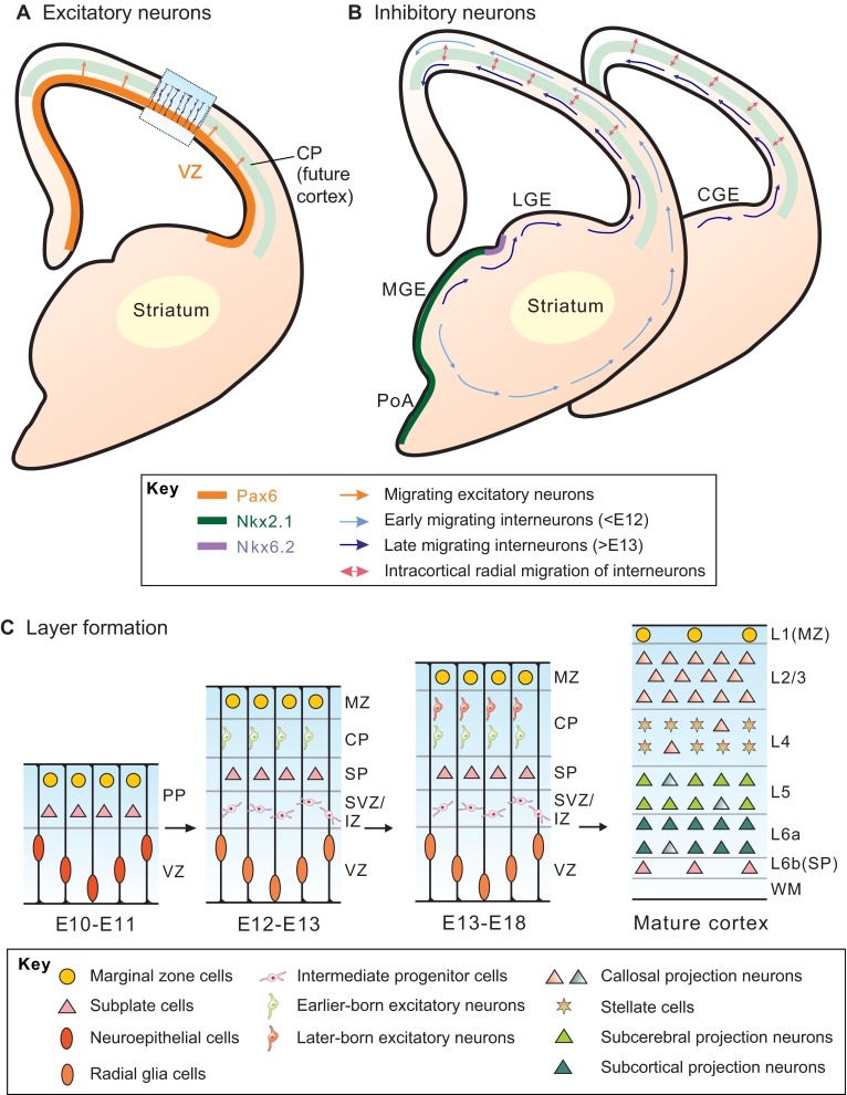
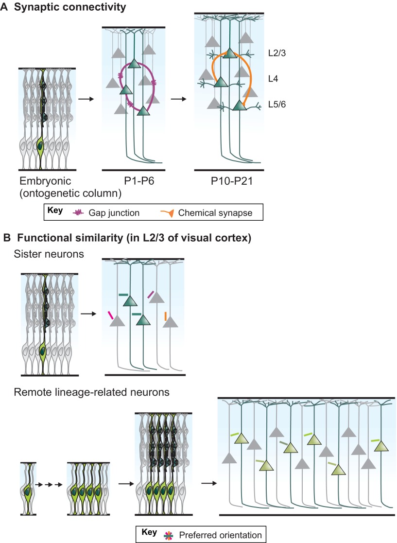
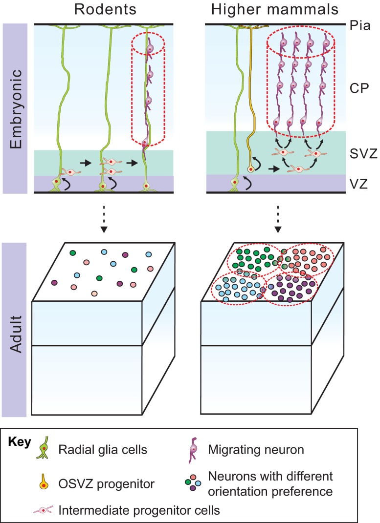
The neocortex plays a key role in higher-order brain functions, such as perception, language and decision-making. Since the groundbreaking work of Ramón y Cajal over a century ago, defining the neural circuits underlying brain functions has been a field of intense study. Here, we review recent findings on the formation of neocortical circuits, which have taken advantage of improvements to mouse genetics and circuit-mapping tools. These findings are beginning to reveal how individual components of circuits are generated and assembled during development, and how early developmental processes, such as neurogenesis and neuronal migration, guide precise circuit assembly.

Keywords: Lineage; Neocortex; Neuronal circuits.


Figures

Fig. 1.
Fig. 1.
Generation and migration of neocortical excitatory and inhibitory neurons. (A,B) Excitatory and inhibitory neurons originate from different germinal zones of the embryonic telencephalon. (A) Cortical excitatory neurons are generated from progenitor cells (Pax6+, orange) residing in the ventricular zone (VZ) of the dorsal telencephalon. Newborn excitatory neurons undergo radial glial fiber-guided radial migration (orange arrows) and settle into the developing cortical plate (CP, light green). (B) Cortical inhibitory interneurons are predominantly generated from progenitor cells located in the proliferative zone of the ventral telencephalon, mainly within the medial ganglionic eminence (MGE; contains Nkx2.1+ cells, dark green) and the caudal ganglionic eminence (CGE). A small population of cortical inhibitory interneurons is produced from the preoptic area (PoA). Newborn inhibitory interneurons follow two tangentially oriented migratory streams to enter the cortex: a superficially migrating early cohort (pale-blue arrows) migrates through the marginal zone, and a deeply migrating second and more prominent cohort (dark-blue arrows) migrates through the lower intermediate zone and subventricular zone. Upon reaching the cortex, they switch to radial migration (pink double-headed arrows) and settle into their final laminar position in the CP. (C) Inside-out fashion of cortical layer (L) formation. In early developmental stages (E10-E11), the neural tube is composed of a single layer of neuroepithelial cells. A small fraction of these undergo asymmetric division to generate the first wave of postmitotic neurons that migrate out radially and form the preplate (PP). As development proceeds (E12-E13), newborn excitatory neurons split the PP into a superficial marginal zone (MZ) and a deeper subplate (SP). Successive waves of newly generated excitatory neurons migrate past the existing neurons to occupy a more superficial region in the CP (E13-E18), creating the mature six-layered cortex. Excitatory neurons in the mature cortex are heterogeneous. IZ/SVZ, intermediate zone/subventricular zone; WM, white matter.
Fig. 2.
Fig. 2.
Lineage-dependent circuit assembly of neocortical excitatory neurons. (A) Sister neurons (dark green) derived from the same RGCs during embryonic development migrate along the radial glial fiber (light green) and form ontogenetic columns. Sister neurons are preferentially coupled through electrical synapses (gap junctions, purple) in the first postnatal week. In the second postnatal week, sister neurons develop preferential chemical synapses (orange) with each other, and the direction of connectivity resembles that in the mature circuits. (B) In layer (L)2/3 of the mouse visual cortex, lineage-related neurons have similar orientation tuning response properties (colored bars). Vertically aligned sister neurons (top) labeled by retrovirus at E15-E17 have similar orientation tuning response properties, unlike those of their non-sisters. Remote lineage-related clones of neurons (bottom), labeled by a sporadically expressing Cre driver line, still have slightly more similar orientation tuning response properties compared with those of non-related neurons.
Fig. 3.
Fig. 3.
Patterns of neocortical neurogenesis may influence the organization of functional maps. In the visual cortex of rodents (left), functional columns are thought to exist at single-cell resolution, and individual embryonic/ontogenetic columns (dotted red lines) composed of a few sister excitatory neurons may lead to this organization in adults. In higher mammals (right), by contrast, functional columns with clusters of neurons sharing similar properties are observed; the expansion of the subventricular zone (SVZ) through its greater proliferative potential may significantly increase the number of neurons in individual embryonic/ontogenetic columns and contribute to the development of these functional columns in adults. CP, cortical plate; VZ, ventricular zone.

Similar articles

Cited by

References

    1. Abeles M., Goldstein M. H., Jr (1970). Functional architecture in cat primary auditory cortex: columnar organization and organization according to depth. J. Neurophysiol. 33, 172–187 - PubMed
    1. Anderson S. A., Eisenstat D. D., Shi L., Rubenstein J. L. (1997). Interneuron migration from basal forebrain to neocortex: dependence on Dlx genes. Science 278, 474–476 - PubMed
    1. Anderson S. A., Kaznowski C. E., Horn C., Rubenstein J. L. R., McConnell S. K. (2002). Distinct origins of neocortical projection neurons and interneurons in vivo. Cereb. Cortex 12, 702–709 - PubMed
    1. Ang E. S. B. C., Jr, Haydar T. F., Gluncic V., Rakic P. (2003). Four-dimensional migratory coordinates of GABAergic interneurons in the developing mouse cortex. J. Neurosci. 23, 5805–5815 - PMC - PubMed
    1. Angevine J. B., Jr, Sidman R. L. (1961). Autoradiographic study of cell migration during histogenesis of cerebral cortex in the mouse. Nature 192, 766–768 - PubMed

Publication types

LinkOut - more resources