. 2013 Jul 9;52(27):4687-96.
doi: 10.1021/bi400540m. Epub 2013 Jun 26.

Conserved hydrogen bonding networks of MitoNEET tune Fe-S cluster binding and structural stability

Affiliations

Conserved hydrogen bonding networks of MitoNEET tune Fe-S cluster binding and structural stability

Daniel W Bak et al. Biochemistry. .

Abstract

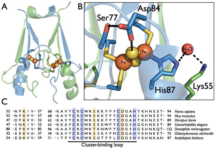
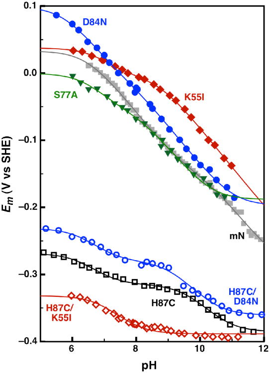
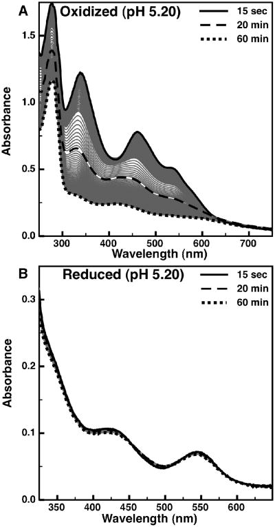
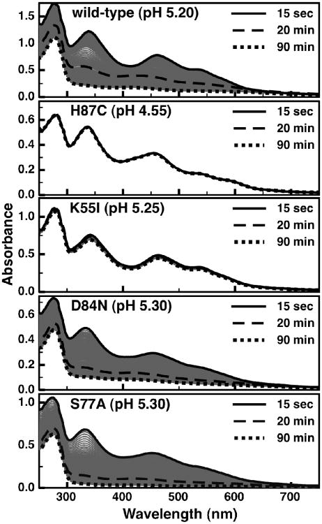
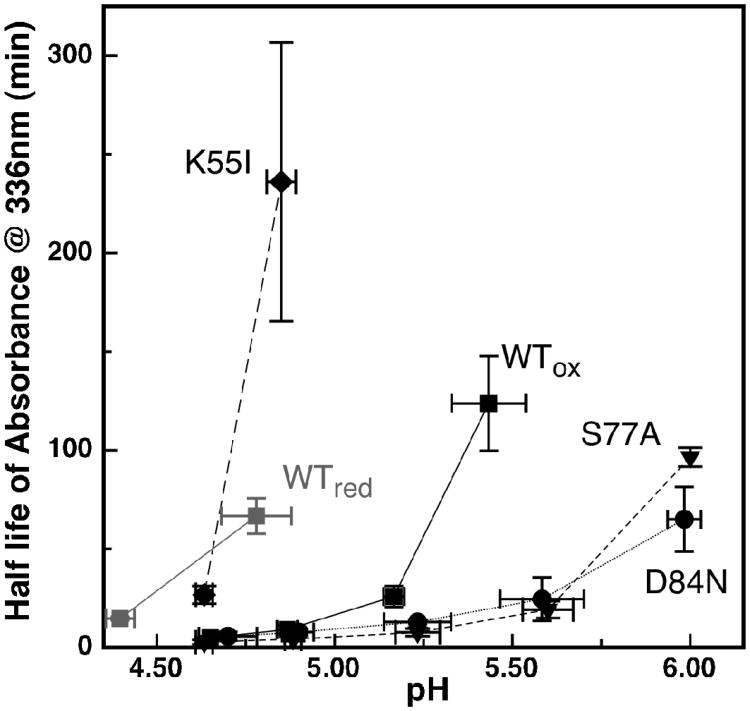
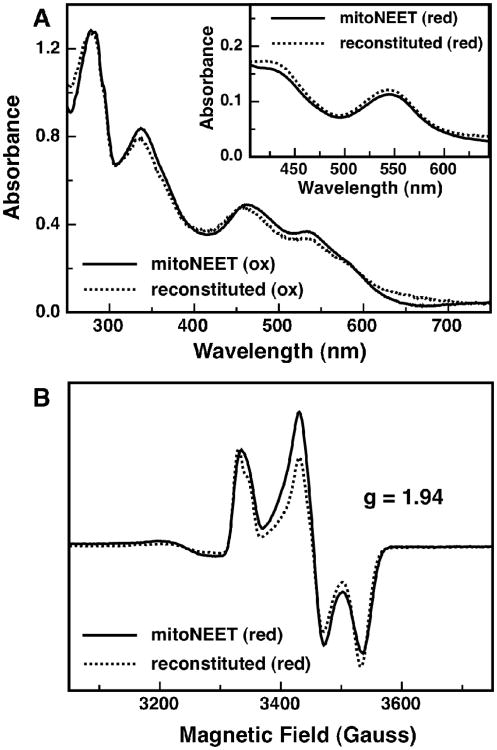
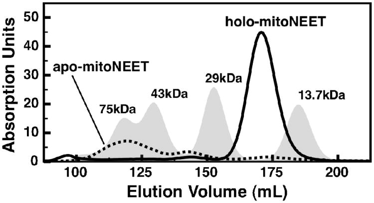
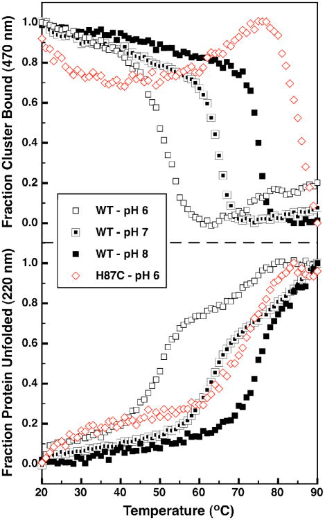
While its biological function remains unclear, the three-cysteine, one-histidine ligated human [2Fe-2S] cluster containing protein mitoNEET is of interest because of its interaction with the anti-diabetes drug pioglitazone. The mitoNEET [2Fe-2S] cluster demonstrates proton-coupled electron transfer (PCET) and marked cluster instability, which have both been linked to the single His ligand. Highly conserved hydrogen bonding networks, which include the His-87 ligand, exist around the [2Fe-2S] cluster. Through a series of site-directed mutations, PCET of the cluster has been examined, demonstrating that multiple sites of protonation exist in addition to the His ligand, which can influence redox potential. The mutations also demonstrate that while replacement of the His ligand with cysteine results in a stable cluster, the removal of Lys-55 also greatly stabilizes the cluster. We have also noted for the first time that the oxidation state of the cluster controls stability: the reduced cluster is stable, while the oxidized one is much more labile. Finally, it is shown that upon cluster loss the mitoNEET protein structure becomes less stable, while upon in vitro reconstitution, both the cluster and the secondary structure are recovered. Recently, two other proteins have been identified with a three-Cys(sulfur), one-His motif, IscR and Grx3/4-Fra2, both of which are sensors of iron and redox homeostatsis. These results lead to a model in which mitoNEET could sense the cellular oxidation state and proton concentration and respond through cluster loss and unfolding.


Figures

Figure 1
Figure 1
Structure and sequence comparison of human mitoNEET. (A) The dimeric mitoNEET protein is displayed with each monomer (colored blue or green) containing one [2Fe-2S] cluster, ligated by three Cys-ligands and a His-ligand. (B) The cluster binding site, showing two hydrogen bonding network, the first between the His-87-ligand, a conserved molecule of water, and Lys-55, and the second between a μ-sulfido ligand, Asp-84, and Ser-77. Proposed hydrogen bonds are represent by dashed black lines. (C) The partial sequence alignment of various eukaryotic mitoNEET homologs. Conserved cluster ligands are highlighted in purple, and conserved residues involved in hydrogen bonding networks around the cluster are highlighted in gold. The cluster-binding loop is underlined.
Figure 2
Figure 2
Comparison of the pH-dependent reduction potential for wild-type and site-directed variants of mitoNEET: wild-type (gray solid squares), H87C (black open squares), K55I (red solid diamonds), D84N (blue solid circles), S77A (green solid triangles), H87C/K55I (red open diamonds), and H87C/D84N (blue open circles). The wild-type, H87C, K55I, D84N, S77A, and H87C/D84N data were each fit to a Nernst equation for 4 pKa values (Eq 3), while the H87C/K55I data was fit to a Nernst equation for 2 pKa values (Eq 1).
Figure 3
Figure 3
The time-dependent loss of visible absorption signals for mitoNEET at pH 5.20: (A) oxidized mitoNEET, and (B) reduced mitoNEET. Time-points of 15 second (solid), 20 minutes (dashed), and 90 minutes (dotted) are shown in bold, while intermediate time-points are shown as thin gray traces.
Figure 4
Figure 4
The time-dependent loss of visible absorption signals for wild-type and site-directed variants of mitoNEET: wild-type at pH 5.20, H87C at pH 4.55, K55I at pH 5.25, D84N at pH 5.30, and S77A at pH 5.30. Timepoints of 15 second (solid), 20 minutes(dashed), and 90 minutes (dotted) are shown in bold, with intermediate time-points shown as thin gray traces.
Figure 5
Figure 5
A comparison of the stability of the [2Fe-2S] cluster optical signals as a function of pH for wild-type and site-directed variants of mitoNEET: wild-type mitoNEET oxidized (black square) and reduced (grey squares), K55I oxidized (diamonds), D84N oxidized (circles) S77A oxidized (triangles). Oxidized H87C is not shown due to its complete stability in this pH range. Plotted is the half-life of the optical signal at 336nm versus pH. Data points and error bars represent the average of three experiments.
Figure 6
Figure 6
Spectroscopic characterization of the reconstituted-mitoNEET. (A) Optical spectra of oxidized reconstituted mitoNEET (dotted line) and as-isolated mitoNEET (solid line). Inset: optical spectra of dithionite reduced reconstituted-mitoNEET (dotted line) and as-isolated mitoNEET (solid line). (B) Low temperature (10K) EPR spectra of dithionite-reduced reconstituted-mitoNEET (dotted line) and as-isolated mitoNEET (solid line) measured at 9.24 GHz.
Figure 7
Figure 7
Size-exclusion chromatograms for wild-type mitoNEET: holo-mitoNEET (solid trace) and apo-mitoNEET (dotted trace). A set of four protein standards is reported for comparison (gray peaks): Ribonuclease A (13.7 kDa), Carbonic Anhydrase (29 kDa), Ovalbumin (43 kDa), and Conalbumin (75 kDa). All chromatograms are calculated from absorbance at 280 nm.
Figure 8
Figure 8
CD melting curves of the wild-type and H87C variant of mitoNEET. The fraction of bound cluster as monitored by the 470 nm CD band (top panel) and the fraction of unfolded protein (bottom panel) for WT mitoNEET pH 6 (open black squares), pH 7 (half filled black squares), and pH 8 (solid black squares) and for the H87C mutant at pH 6 (open red diamonds).

References

    1. Paddock ML, Wiley SE, Axelrod HL, Cohen AE, Roy M, Abresch EC, Capraro D, Murphy AN, Nechushtai R, Dixon JE. MitoNEET is a uniquely folded 2Fe–2S outer mitochondrial membrane protein stabilized by pioglitazone. Proceedings of the National Academy of Sciences. 2007;104:14342–14347. - PMC - PubMed
    1. Colca JR, McDonald WG, Waldon DJ, Leone JW, Lull JM, Bannow CA, Lund ET, Mathews WR. Identification of a novel mitochondrial protein (“ mitoNEET”) cross-linked specifically by a thiazolidinedione photoprobe. American Journal of Physiology-Endocrinology And Metabolism. 2004;286:E252–E260. - PubMed
    1. Lin J, Zhou T, Ye K, Wang J. Crystal structure of human mitoNEET reveals distinct groups of iron–sulfur proteins. Proceedings of the National Academy of Sciences. 2007;104:14640–14645. - PMC - PubMed
    1. Hou X, Liu R, Ross S, Smart EJ, Zhu H, Gong W. Crystallographic studies of human MitoNEET. J Biol Chem. 2007;282:33242–33246. - PubMed
    1. Wiley SE, Murphy AN, Ross SA, Van Der Geer P, Dixon JE. MitoNEET is an iron-containing outer mitochondrial membrane protein that regulates oxidative capacity. Proceedings of the National Academy of Sciences. 2007;104:5318–5323. - PMC - PubMed

Publication types

LinkOut - more resources