. 2013 Jul 3;135(26):9885-97.
doi: 10.1021/ja4041412. Epub 2013 Jun 21.

Drug-induced conformational and dynamical changes of the S31N mutant of the influenza M2 proton channel investigated by solid-state NMR

Affiliations

Drug-induced conformational and dynamical changes of the S31N mutant of the influenza M2 proton channel investigated by solid-state NMR

Jonathan K Williams et al. J Am Chem Soc. .

Abstract

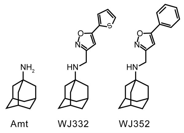
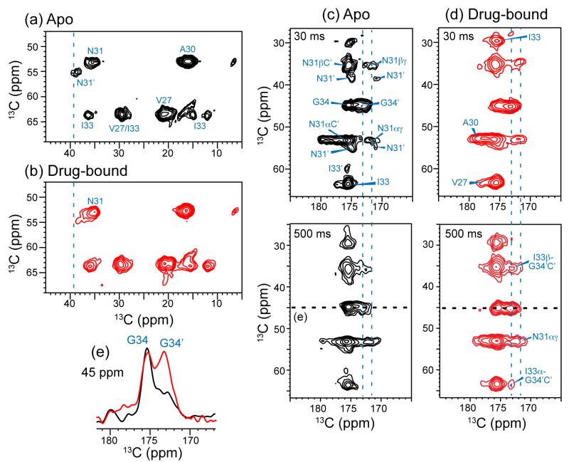
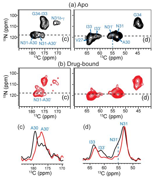
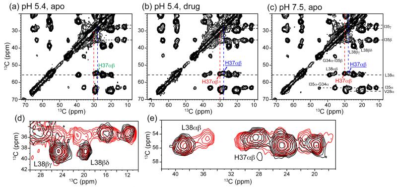
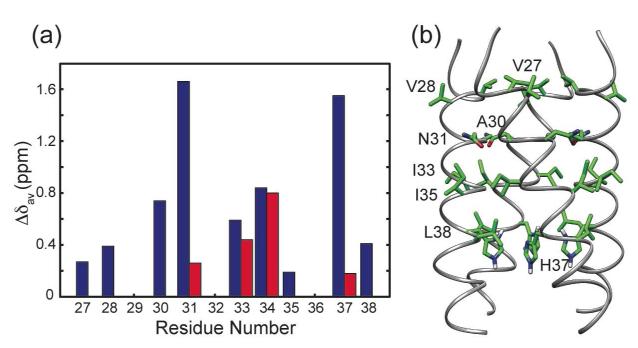
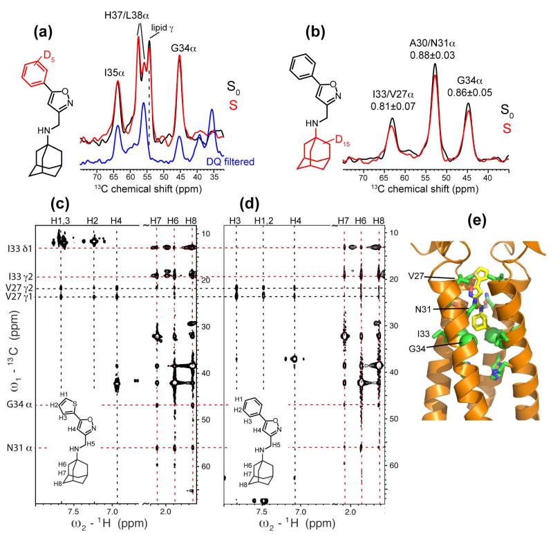
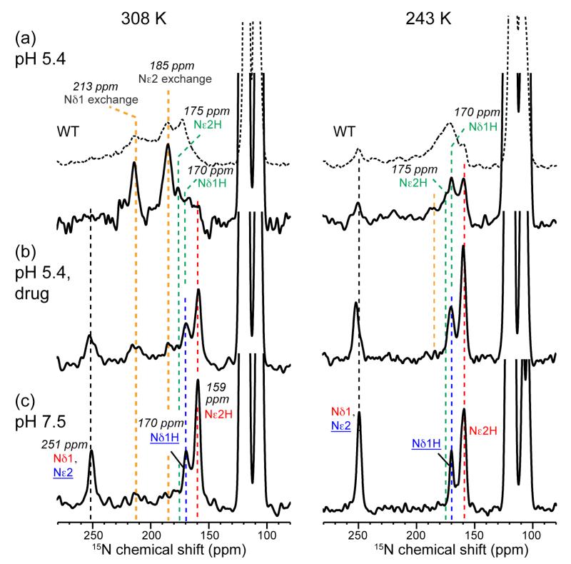
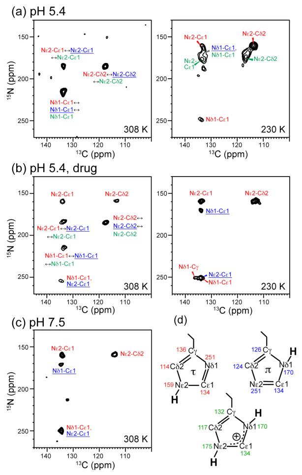
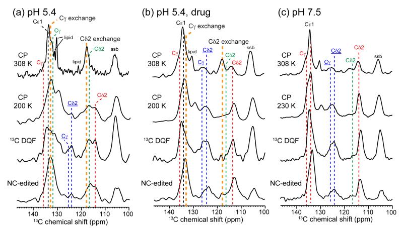
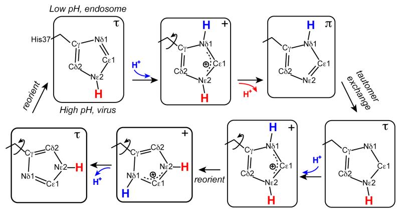
The M2 protein of influenza A viruses forms a tetrameric proton channel that is targeted by the amantadine class of antiviral drugs. A S31N mutation in the transmembrane (TM) domain of the protein has caused widespread amantadine resistance in most of the currently circulating flu viruses. Recently, a new family of compounds based on amantadine- and aryl-substituted isoxazole were discovered to inhibit the S31N channel activity and reduce replication of S31N-harboring viruses. We now use solid-state NMR spectroscopy to investigate the effects of one of these isoxazole compounds, WJ352, on the conformation of the S31N TM segment and the dynamics of the proton-selective residue, His37. Chemical shift perturbations show that WJ352 changes the conformational equilibrium of multiple TM residues, with the maximal perturbation occurring at the crucial Asn31. (13)C-(2)H distance measurements and (1)H-(1)H NOE cross peaks indicate that the adamantane moiety of the drug is bound in the spacious pore between Asn31 and Gly34 while the phenyl tail is located near Val27. Thus, the polar amine points to the channel exterior rather than to His37, in contrast to amantadine and rimantadine in the wild-type channel, suggesting that the drug is significantly stabilized by hydrophobic interactions between the adamantane and the TM peptide. (15)N and (13)C chemical shifts indicate that at low pH, His37 undergoes fast exchange among the τ tautomer, the π tautomer, and the cationic state due to proton transfer with water. The exchange rate is higher than the wild-type channel, consistent with the larger single-channel conductance of the mutant. Drug binding at acidic pH largely suppresses this exchange, reverting the histidines to a similar charge distribution as that of the high-pH closed state.


Figures

Figure 1
Figure 1
Chemical structures of adamantyl-based drugs against influenza M2 proton channels: amantadine (Amt) against the WT protein, and WJ332 and WJ352 against the S31N mutant.
Figure 2
Figure 2
2D 13C-13C correlation spectra of VANIG19-49, S31N in DMPC bilayers without and with WJ352 at pH 6.5. (a, b) Aliphatic region of the 30 ms spectra of the apo peptide (a) and drug-bound peptide (b). Drug binding changed the conformational equilibrium of the N31 Cα-Cβ peaks. (c, d) Carbonyl region of the 30 ms (top) and 500 ms (bottom) spectra of the (c) apo and (d) drug-bound peptide. N31, I33 and G34 exhibit two sets of chemical shifts. (e) 45-ppm cross section of the apo (black) and drug-bound (red) peptide, showing the G34 Cα-CO cross peak. All spectra were measured at 243 K under 7 kHz MAS.
Figure 3
Figure 3
2D 15N-13C correlation spectra of VANIG19-49, S31N in DMPC bilayers at pH 6.5. (a) Without drug (black). (b) With drug (red) at the 10:1 drug : tetramer ratio. (c) A30 CO cross section at the 15N chemical shift of 117.6 ppm. Drug binding changed the intensity distribution of two A30 CO peaks. (d) I33 and N31 Cα cross section at 119.1 ppm 15N chemical shift. Drug binding selected one of the two peaks of I33 and N31. The spectra were measured at 243 K under 7 kHz MAS.
Figure 4
Figure 4
Aliphatic region of the 2D 13C-13C correlation spectra of VM+ membrane bound VGIHL19-49, S31N. The spectra were measured at 243 K under 7 kHz MAS. (a) Apo peptide at pH 5.4. (b) Drug-bound peptide at pH 5.4. (c) Apo peptide at pH 7.5. The signals of τ, π, and cationic His37 are annotated in red, blue underline, and green, respectively. (d) Amplified L38 and I35 sidechain region of the apo (black) and drug-bound (red) spectra at low pH. (e) Amplified H37 and L38 Cα-Cβ region of the apo and drug-bound spectra at low pH. The low-pH drug-bound spectrum resembles the high-pH spectrum, indicating that WJ352 shifts the backbone conformation of S31N to that of the closed state.
Figure 5
Figure 5
(a) Residue-specific average chemical shift change in S31N M2TM induced by the drug WJ352. Residues with a single set of chemical shifts in both the apo and drug-bound states have a single blue bar for the average CSP, while residues with two sets of chemical shifts in the apo state but only one set in the drug-bound state have two CSP values. (b) Solution NMR structure of S31N M2TM (PDB: 2LY0), showing the positions of most residues whose CSPs were measured.
Figure 6
Figure 6
Binding site and orientation of WJ352 in S31N bound to DMPC bilayers (a, b) and DPC micelles (c, d). (a) 13C-2H REDOR S0 (black) and S (red) spectra of d5-WJ352 complexed GIHL22-46, S31N in DMPC bilayers. The mixing time was 14.2 ms and the drug : tetramer ratio was 8 : 1. The deuterated phenyl group did not dephase any peptide 13C signals, especially G34. The REDOR spectra were measured without DQ filtering, thus showed a lipid headgroup Cγ peak at 54 ppm. This is verified by the absence of the peak in a 13C DQ filtered spectrum (blue). (b) 13C-2H DQ filtered REDOR spectra of d15-WJ352 bound VANIG19-49, S31N in DMPC bilayers. The mixing time was 9.6 ms and the drug : tetramer ratio was 1 : 1. The deuterated adamantane dephased multiple 13C signals. (c, d) 2D 13C-(1H)-1H NOESY (150 ms) spectra of VANIG19-49, S31N in DPC micelles with bound WJ332 (c) or with bound WJ352 (d). Drug – peptide cross peaks are observed and consistent with the amine-up orientation. (e) Solution NMR structure of S31N-M2 with bound WJ332 (PDB: 2LY0).
Figure 7
Figure 7
1D 15N CP-MAS spectra of His37 in VM+ membrane bound VGIHL19-49, S31N at 308 K (left) and 243 K (right). The spectra were scaled to show roughly equal integrated intensities for the imidazole nitrogens. (a) Apo peptide at pH 5.4. (b) Drug-bound peptide at pH 5.4. (c) Apo peptide at pH 7.5. Assignments shown here and in subsequent figures are annotated in red for the τ tautomer, blue underline for the π tautomer, and green for cationic His. For comparison, the pH 5.2 spectra of WT His37 are shown (dotted line) above the S31N spectra in (a). At low pH and high temperature, S31N-M2 exhibited sharp 15N exchange peaks. WJ352 binding largely suppressed these peaks.
Figure 8
Figure 8
2D 15N-13C correlation spectra of VM+ bound VGIHL19-49, S31N at 308 K and 230 K under 10 kHz MAS. (a) Apo peptide at pH 5.4. (b) Drug-bound peptide at pH 5.4. (c) Apo peptide at pH 7.5. The Cγ signal was not detected due to rotational resonance with Cβ, thus allowing the distinction between Nδ1 and Nε2 signals. (d) Imidazole chemical shifts for the τ tautomer, π tautomer, and cationic His.
Figure 9
Figure 9
Aromatic region of the 1D 13C MAS spectra of His37 in VM+ bound VGIHL19-49, S31N. From top to bottom are the 308 K 13C CP-MAS spectra, the low-temperature CP-MAS (200 or 230 K) spectra, the low-temperature 13C-13C DQ filtered (DQF) spectra, and the low-temperature 15N-13C filtered spectra. (a) Apo peptide at pH 5.4. (b) Drug-bound peptide at pH 5.4. (c) Apo peptide at pH 7.5. Spinning sidebands are denoted as ssb. All spectra were measured under 7 kHz MAS.
Figure 10
Figure 10
Proposed mechanism of His37-water proton exchange for S31N and WT M2 channels. His37 residues undergo rapid interconversion among τ, π, and cationic states during proton transfer with water. Ring reorientations facilitate this proton shuttling by pointing the unprotonated nitrogen towards the low pH exterior.

Similar articles

Cited by

References

    1. Lamb RA, Holsinger KJ, Pinto LH. In: Cellular Receptors of Animal Viruses. Wemmer E, editor. Cold Spring Harbor Lab Press; Plainview, NY: 1994. pp. 303–321.
    1. Pinto LH, Lamb RA. J. Biol. Chem. 2006;281:8997–9000. - PubMed
    1. Sugrue RJ, Bahadur G, Zambon MC, Hall-Smith M, Douglas AR, Hay AJ. EMBO J. 1990;9:3469–3476. - PMC - PubMed
    1. Wang C, Lamb RA, Pinto LH. Biophys. J. 1995;69:1363–1371. - PMC - PubMed
    1. Hong M, Degrado WF. Protein Sci. 2012;21:1620–1633. - PMC - PubMed

Publication types

MeSH terms