. 2013 Jun 11:11:146.
doi: 10.1186/1741-7015-11-146.

Phenotype, donor age and gender affect function of human bone marrow-derived mesenchymal stromal cells

Affiliations

Phenotype, donor age and gender affect function of human bone marrow-derived mesenchymal stromal cells

Georg Siegel et al. BMC Med. .

Abstract

Background: Mesenchymal stromal cells (MSCs) are attractive for cell-based therapies ranging from regenerative medicine and tissue engineering to immunomodulation. However, clinical efficacy is variable and it is unclear how the phenotypes defining bone marrow (BM)-derived MSCs as well as donor characteristics affect their functional properties.

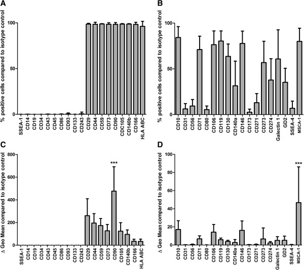
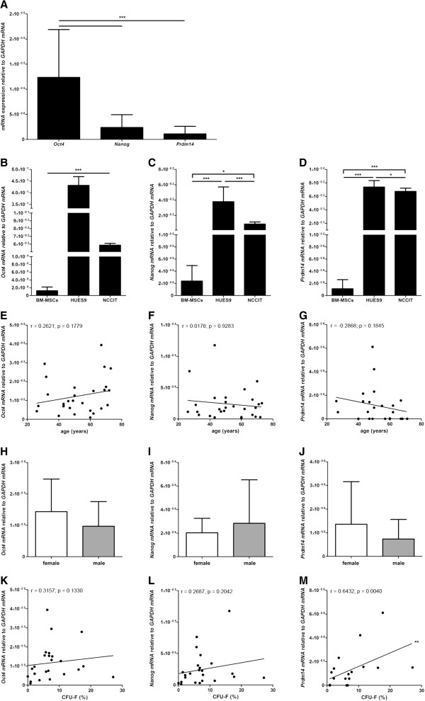
Methods: BM-MSCs were isolated from 53 (25 female, 28 male; age: 13 to 80 years) donors and analyzed by: (1) phenotype using flow cytometry and cell size measurement; (2) in vitro growth kinetics using population doubling time; (3) colony formation capacity and telomerase activity; and (4) function by in vitro differentiation capacity, suppression of T cell proliferation, cytokines and trophic factors secretion, and hormone and growth factor receptor expression. Additionally, expression of Oct4, Nanog, Prdm14 and SOX2 mRNA was compared to pluripotent stem cells.

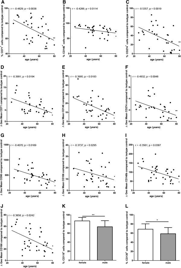
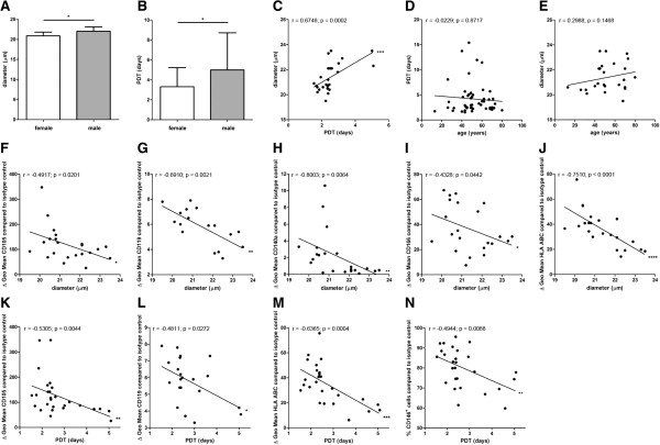
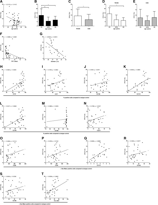
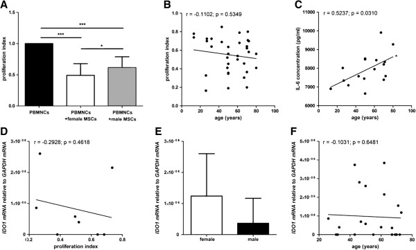
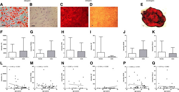
Results: BM-MSCs from younger donors showed increased expression of MCAM, VCAM-1, ALCAM, PDGFRβ, PDL-1, Thy1 and CD71, and led to lower IL-6 production when co-cultured with activated T cells. Female BM-MSCs showed increased expression of IFN-γR1 and IL-6β, and were more potent in T cell proliferation suppression. High-clonogenic BM-MSCs were smaller, divided more rapidly and were more frequent in BM-MSC preparations from younger female donors. CD10, β1integrin, HCAM, CD71, VCAM-1, IFN-γR1, MCAM, ALCAM, LNGFR and HLA ABC were correlated to BM-MSC preparations with high clonogenic potential and expression of IFN-γR1, MCAM and HLA ABC was associated with rapid growth of BM-MSCs. The mesodermal differentiation capacity of BM-MSCs was unaffected by donor age or gender but was affected by phenotype (CD10, IFN-γR1, GD2). BM-MSCs from female and male donors expressed androgen receptor and FGFR3, and secreted VEGF-A, HGF, LIF, Angiopoietin-1, basic fibroblast growth factor (bFGF) and NGFB. HGF secretion correlated negatively to the expression of CD71, CD140b and Galectin 1. The expression of Oct4, Nanog and Prdm14 mRNA in BM-MSCs was much lower compared to pluripotent stem cells and was not related to donor age or gender. Prdm14 mRNA expression correlated positively to the clonogenic potential of BM-MSCs.

Conclusions: By identifying donor-related effects and assigning phenotypes of BM-MSC preparations to functional properties, we provide useful tools for assay development and production for clinical applications of BM-MSC preparations.


Figures

Figure 1
Figure 1
Flow cytometric analyses. The expression of CD10, CD14, CD19, CD29, CD31, CD34, CD43, CD44, CD45, CD56, CD59, CD71, CD73, CD80, CD86, CD90, CD93, CD105, CD106, CD119, CD130, CD133, CD140a, CD140b, CD146, CD173, CD166, CD243, CD271, CD273, CD274, Galectin 1, GD2, MSCA-1, SSEA-1, SSEA-4 and HLA class I was analyzed by flow cytometry in MSC preparations from multiple donors identifying three groups of marker expression. Markers that were expressed on all/most of the cells or on none/very few of the cells within the respective MSC preparation (A) and markers that identified MSC subpopulations by presence or absence of the respective marker (B) (n = 33 except for CD80 (n = 30), CD86 (n = 30), CD119 (n = 27), CD130 (n = 34), CD273 (n = 30), CD274 (n = 30), Galectin 1 (n = 24), and SSEA-4 (n = 36)). Analyses of specific antibody mediated fluorescence per cell (ΔGeo Mean) identified markers that were expressed at high and low density per cell (C, D) (n = 36 except for CD80 (n = 32), CD86 (n = 32), CD119 (n = 29), CD130 (n = 37), CD273 (n = 32), CD274 (n = 32), Galectin 1 (n = 26), and SSEA-4 (n = 39)). ANOVA analysis of variance followed by Tukey’s Multiple Comparison Test (***P <0.001). Error bars: SD.
Figure 2
Figure 2
Correlation analyses of marker expression to donor age and gender distribution. The percentage of CD71+, CD146+, and CD274+ cells and the specific antibody mediated fluorescence per cell (ΔGeo Mean) of CD71, CD90, CD106, CD140b, CD146, CD166 and CD274 correlated negatively with the donor age (n = 34 except for CD274 (n = 31)) (A-J). Spearman two-tailed correlation test (*P <0.05; **P <0.01). MSC preparations from female donors harbored significantly more CD119+ cells (n = 11) and CD130+ cells (n = 16) compared to MSC preparations from male donors (CD119+ cells: n = 17; CD130+ cells: n = 19) (K, L). Two-tailed Student’s t-test (*P <0.05; **P <0.01). Error bars: SD.
Figure 3
Figure 3
Analyses of cell size and growth kinetics. The diameter of MSCs from female donors (n = 14) was slightly but significantly smaller compared to male donors (n = 11) (A). The PDT of MSCs from female donors (n = 25) was significantly reduced compared to MSCs from male donors (n = 27) (B). Two-tailed Student’s t-test (*P <0.05). Error bars: SD. The cell diameter correlated positively to the PDT (n = 25) (C). No correlation was found between the donor age and the proliferation capacity of the MSCs (n = 52) (D) and between the donor age to the cell size (n = 25) (E). The specific antibody mediated fluorescence per cell (ΔGeo Mean) of CD105, CD119, CD140a, CD166 and HLA ABC correlated negatively with the cell size (n = 22 except for CD119 (n = 17)) (F-J). The specific antibody mediated fluorescence per cell (ΔGeo Mean) of CD105, CD119, and HLA ABC correlated negatively with the PDT (n = 27 except for CD119 (n = 21)) (K-M). The PDT correlated negatively with the percentage of CD146+ cells within the MSC preparations (n = 27) (N). Spearman two-tailed correlation test (*P <0.05; **P <0.01; ***P <0.001; ****P <0.0001).
Figure 4
Figure 4
Analyses of clonogenic potential. Comparing all ages and both genders, MSC preparations from the BM of younger donors trended toward more colony forming cells than MSC preparations from older donors (n = 50) (A). Spearman two-tailed correlation test. Comparing age groups including both genders, significantly more colonies could be detected in young donors (<45 years) (n = 11) compared to middle-aged (n = 26) and older donors (n = 13) (B). ANOVA analysis of variance followed by Tukey’s Multiple Comparison Test (*P <0.05; **P <0.01). Error bars: SD. In BM-MSC preparations from female donors more colony forming cells could be detected (n = 23) compared to BM-MSC preparations from male donors (n = 27) (C). Two-tailed Student’s t-test (*P <0.05). Error bars: SD. Age-group specific analyses for each gender confirmed that more colonies could be detected in young female donors (<45 years) compared to older donors (D, E). ANOVA analysis of variance followed by Tukey’s Multiple Comparison Test (*P <0.05). Error bars: SD. The clonogenic potential of the cells within the BM-MSC preparations was negatively correlated to the population doubling time (n = 42) and the cell diameter (n = 22) (F, G). Spearman two-tailed correlation test (**P <0.01; ****P <0.0001). The percentage of CD10+, CD71+, CD106+, CD119+ CD146+, CD166+ and CD271+ cells correlated positively with the clonogenic potential of the BM-MSCs (n = 31 except for CD119 (n = 25)) (H-N). The specific antibody mediated fluorescence per cell (ΔGeo Mean) of CD29, CD44, CD119, CD146, CD166 and HLA ABC correlated positively with the clonogenic potential of the MSCs (n = 31 except for CD119 (n = 25)) (O-T). Spearman two-tailed correlation test (*P <0.05; **P <0.01; ***P <0.001; ****P <0.0001).
Figure 5
Figure 5
Analyses of the immunosuppressive potential of MSCs. MSCs from both female and male donors significantly suppressed the proliferation of activated allogeneic T cells. MSCs from female donors (n = 18) showed a significantly higher potency to suppress T cell proliferation than MSCs from male donors (n = 16). Data are normalized to the proliferation index of PBMNCs alone (A). ANOVA analysis of variance followed by Tukey’s Multiple Comparison Test (*P <0.05; ***P <0.001). Error bars: SD. No correlation of the donor age to the potential to suppress T cell proliferation could be detected (B). Upon co-culturing of MSCs from older donors with activated allogeneic T cells more IL-6 could be detected in the supernatant compared to MSCs from younger donors that were co-cultured with activated T cells (n = 17) (C). Spearman two-tailed correlation test (*P <0.05). Although a trend toward a negative correlation was observed, no significant correlation of IDO 1 mRNA expression to T cell proliferation could be detected (n = 8) (D). Spearman two-tailed correlation test. No difference of IDO 1 mRNA expression from male MSC donors compared to female MSC donors could be detected (n = 22) (E). Two-tailed Student’s t-test. Error bars: SD. No correlation of the IDO 1 mRNA expression to the donor age could be observed (n = 22) (F). Spearman two-tailed correlation test.
Figure 6
Figure 6
Mesodermal differentiation potential of MSCs. No donor age or gender related differences could be detected for the adipogenic, osteogenic and chondrogenic in vitro differentiation capacity of the MSC preparations as analysed by lineage specific staining (Oil Red O for adipogensis; Alizarin Red for osteogenesis, and Safranain O for chondrogenesis (n = 40); differentiation: A, C, E; negative controls: B, D) and no statistically significant differences could be detected for the lineage specific mRNA expression (LPL (n = 44) and PPARγ (n = 48) for adipogenesis; OPN (n = 17) and AP (n = 41) for osteogenesis; SOX9 (n = 47) and COLL2 (n = 32) for chondrogenesis; F-Q). Two-tailed Student’s t-test and Spearman two-tailed correlation test. Error bars: SD. Scale bars: 100 μm.
Figure 7
Figure 7
Expression of Oct4, Nanog and Prdm14 mRNA and functional relevance. The MSCs expressed significantly more Oct4 mRNA than Nanog or Prdm14 mRNA; however, no significant difference in Nanog mRNA expression compared to Prdm14 mRNA expression could be detected (n = 28 except for Prdm14 (n = 23)) (A). Compared to pluripotent HUES9 cells and pluripotent NCCIT cells the expression of Oct4 mRNA, Nanog mRNA and Prdm14 mRNA was much lower in MSCs (B-D). Compared to NCCIT cells the HUES9 cells expressed more Oct4 mRNA, Nanog mRNA and Prdm14 mRNA (B-D). ANOVA analysis of variance followed by Tukey’s Multiple Comparison Test (*P <0.05; ***P <0.001). Error bars: SD. No correlation of the expression of Oct4, Nanog and Prdm14 mRNA to donor age (n = 28 except for Prdm14 (n = 23)) (E-G) or any gender-related differences could be detected (n = 28 except for Prdm14 (n = 23)) (H-J). The clonogenic potential of MSCs correlated positively to the expression of Prdm14 mRNA (n = 18) but not to Oct4 (n = 24) or Nanog mRNA (n = 24) (K-M). Two-tailed Student’s t-test and Spearman two-tailed correlation test (**P <0.01). Error bars: SD.

References

    1. Friedenstein AJ, Petrakova KV, Kurolesova AI, Frolova GP. Heterotopic of bone marrow. Analysis of precursor cells for osteogenic and hematopoietic tissues. Transplantation. 1968;6:230–247. doi: 10.1097/00007890-196803000-00009. - DOI - PubMed
    1. Friedenstein AJ, Chailakhyan RK, Latsinik NV, Panasyuk AF, Keiliss-Borok IV. Stromal cells responsible for transferring the microenvironment of the hemopoietic tissues. Cloning in vitro and retransplantation in vivo. Transplantation. 1974;17:331–340. doi: 10.1097/00007890-197404000-00001. - DOI - PubMed
    1. Muguruma Y, Yahata T, Miyatake H, Sato T, Uno T, Itoh J, Kato S, Ito M, Hotta T, Ando K. Reconstitution of the functional human hematopoietic microenvironment derived from human mesenchymal stem cells in the murine bone marrow compartment. Blood. 2006;107:1878–1887. doi: 10.1182/blood-2005-06-2211. - DOI - PubMed
    1. Miura Y, Miura M, Gronthos S, Allen MR, Cao C, Uveges TE, Bi Y, Ehirchiou D, Kortesidis A, Shi S, Zhang L. Defective osteogenesis of the stromal stem cells predisposes CD18-null mice to osteoporosis. Proc Natl Acad Sci U S A. 2005;102:14022–14027. doi: 10.1073/pnas.0409397102. - DOI - PMC - PubMed
    1. Horwitz EM, Gordon PL, Koo WK, Marx JC, Neel MD, McNall RY, Muul L, Hofmann T. Isolated allogeneic bone marrow-derived mesenchymal cells engraft and stimulate growth in children with osteogenesis imperfecta: Implications for cell therapy of bone. Proc Natl Acad Sci U S A. 2002;99:8932–8937. doi: 10.1073/pnas.132252399. - DOI - PMC - PubMed

Publication types