. 2013 Jun 10;13(1):59.
doi: 10.1186/1475-2867-13-59.

Role of relaxin-2 in human primary osteosarcoma

Affiliations

Role of relaxin-2 in human primary osteosarcoma

Jinfeng Ma et al. Cancer Cell Int. .

Abstract

Background: The aim of this study was to clarify the clinicopathological outcome of serum relaxin-2 and tissues relaxin-2 expression levels in human primary osteosarcoma (OS), and to explore the roles of relaxin-2 inhibition and determine its possibility as a therapeutic target in human osteosarcoma.

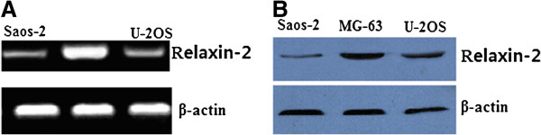
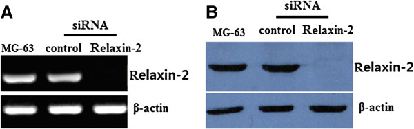
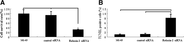
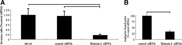
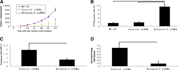
Methods: Real-time quantitative RT-PCR assay was performed to detect the expression of relaxin-2 mRNA in 36 cases of human osteosarcoma tissue samples. Serum relaxin-2 levels was measured in ELISA-based method in the 36 cases of osteosarcoma and 50 cases of controls. MTT and TUNEL assay was used to detect cell proliferation and apoptosis after relaxin-2 knockdown with siRNA transfection for 48 hs in vitro. Matrigel invasion and angiogenesis formation assay was used to detect cell metastasis and angiogenesis with HMEC-1 endothelial cells after relaxin-2 knockdown with siRNA transfection for 48 hs in vitro. The effects of relaxin-2 knockdown with anti- relaxin-2 mAb treatment on growth, apoptosis angiogenesis formation and lung metastasis in vivo was analyzed.

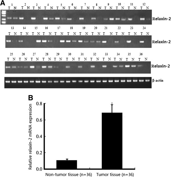
Results: The results showed the levels of relaxin-2 mRNA expression in osteosarcoma tissue samples were significantly higher than those in the corresponding non-tumor tissue samples (P < 0.01), and the serum relaxin-2 levels were significantly higher in OS patients than in healthy controls (P < 0.01). The incidence of advanced stage cancer and hematogenous metastasis cancer in the high relaxin-2 mRNA expression group and high serum relaxin-2 levels groups was significantly higher than that in the low relaxin-2 expression group and low serum relaxin-2 levels groups, respectively. Knockdown of relaxin-2 by siRNA transfection in vitro inhibited proliferation, invasion and angiogenesis in vitro in MG-63 OS cells. In vivo, knockdown of relaxin-2 with anti- relaxin-2 mAb treatment inhibited tumor growth by 62% (P < 0.01) and the formation of lung metastases was inhibited by 72.4% (P < 0.01). Microvascular density was reduced more than 60% due to anti- relaxin-2 mAb treatment (P < 0.01).

Conclusions: Our study suggests that overexpression of relaxin-2 is critical for the metastasis of human osteosarcoma. Detection of relaxin-2 mRNA expression or serum relaxin-2 levels may provide the first biological prognostic marker for OS. Furthermore, relaxin-2 is the potential molecular target for osteosarcoma therapy.


Figures

Figure 1
Figure 1
Detection of relaxin-2 mRNA expression in tissue samples. A. Gel images of electrophoresis. B. Real-time quantitative RT-PCR assay. Osteosarcoma tissues showed obviously higher relaxin-2 mRNA expression than corresponding non-tumor tissues. β-actin was used to normalize for any differences in mRNA between lanes. Student’s t-test showed a significant difference (*P < 0.01).
Figure 2
Figure 2
Detection of relaxin-2 mRNA and proterin expression in human osteosarcoma cell lines. A. Gel images of electrophoresis. B. Western blot assay. MG-63 cell line had the highest expression level of relaxin-2 mRNA and protein. Osteosarcoma tissues showed obviously higher relaxin-2 mRNA expression than corresponding non-tumor tissues. β-actin was used to normalize for any differences in mRNA between lanes.
Figure 3
Figure 3
Effects of relaxin-2 mRNA on relaxin-2 mRNA and protein assessed by quantitative RT-PCR and western blot assays. Relaxin-2 mRNA (A) and protein (B) was completely inhibited by relaxin-2 siRNA transfection for 48 hs than those of MG-63-non-transfection or MG-63-siRNA control cells respectively.
Figure 4
Figure 4
Effect of relaxin-2 knockdown on osteosarcoma MG-63 cell proliferation and apoptosis. A, Cell viability was determined by MTT. The growth rate of MG-63 cells after relaxin-2 knockdown with siRNA for 48 hs was significantly decreased in MTT assay. B, Apoptotic cells was determined by TUNEL analysis in MG-63 cells following relaxin-2 knockdown with siRNA for 48 hs. The apoptotic cells in relaxin-2 siRNA transfectants were significantly higher compared to MG-63-non-transfection or MG-63-siRNA control cells respectively. Columns, mean values from triplicate experiments; Each bar represents mean ± SE (n = 3); *, P < 0.05 versus MG-63-non-transfection or MG-63-siRNA control.
Figure 5
Figure 5
Effect of relaxin-2 knockdown with siRNA on invasion and angiogenesis. A, invasiveness of MG-63 cells transfected with relaxin-2 siRNA in Matrigel invasion assay. Cells were transfected with mock siRNA or relaxin-2 siRNA. After 48 h, 1 × 105 cells were allowed to invade through transwell inserts (8 Am) coated with Matrigel. The cells on lower surface of chambers were stained, counted, and photographed under a light microscope. The in vitro inhibition of invasiveness was calculated in five random 200-fold fields using the following formula: percent inhibition = (mock siRNA-relaxin-2 siRNA) /mock. Columns, mean from three separate experiments; bars, (*P < 0.01). B, HMEC endothelial cells treated with conditional medium from MG-63 cells after siRNA transfection by in vitro angiogenesis assay. The conditioned medium of MG-63 cells was collected after 48-h transfection following filtering of medium. HMEC-1endothelial cells seeded in eight-chamber slides were cultured with the above medium for 72 h until the formation of capillary network was observed. In the end of the experiment, angiogenesis was assessed by H&E staining and photographed under a microscope. columns, mean from three separate experiments; bars, SD (*P < 0.01).
Figure 6
Figure 6
Tumor growth inhibition and prevention of metastasis and angiogenesis formation by relaxin-2 inhibition. A, treatment with anti- relaxin-2 mAb at 100 ug weekly i.p for 42 d. inhibited primary tumor growth by 73 ± 6% compared with controls (n = 6; *P <0.01). B, treatment with anti- relaxin-2 mAb at 100 ug weekly i.p for 42 d. promotes apoptosis in primary tumor cells by 8.2 ± 1.2% compared with controls (n = 6; *P <0.01). C, inhibition of tumor angiogenesis by anti- relaxin-2 mAb treatment as measured by MVD. The vascular density of the anti- relaxin-2 mAb–treated tumor was 0.57 ± 0.22%, which was significantly lower than the controls 1.84 ± 0.26%.( n = 6). *, P < 0.01. D, MG-63 cells transfected with Relaxin-2 siRNA (1 × 105 cells/100 uL in PBS) were injected into the tail vein of SCID mice, followed by analysis of settlement of tumor cells to lung after 2 weeks. The number of macroscopic tumor nodules in Relaxin-2 siRNA–treated mice was 9.46 ± 1.8 lung nodules per mouse versus 62.4 ± 7.4 nodules in the controls (n = 5 mice/group). One way ANOVA analysis was performed to compare the number of micro-metastasis in the lung of mice. *, P < 0.05.

References

    1. Broadhead ML, Clark JC, Myers DE, Dass CR, Choong PF. The molecular pathogenesis of osteosarcoma: a review. Sarcoma. 2011;2011:959248–959252. - PMC - PubMed
    1. Jaffe N. Osteosarcoma: review of the past, impact on the future. The American experience. Cancer Treat Res. 2009;152:239–262. doi: 10.1007/978-1-4419-0284-9_12. - DOI - PubMed
    1. Chou AJ, Geller DS, Gorlick R. Therapy for osteosarcoma: where do we go from here? Paediatr Drugs. 2008;10:315–327. doi: 10.2165/00148581-200810050-00005. - DOI - PubMed
    1. Trent JC. Rapid evolution of the biology and treatment of sarcoma. Curr Opin Oncol. 2008;20:393–394. doi: 10.1097/CCO.0b013e328303ba31. - DOI - PubMed
    1. Siegel HJ, Pressey JG. Current concepts on the surgical and medical management of osteosarcoma. Expert Rev Anticancer Ther. 2008;8:1257–1269. doi: 10.1586/14737140.8.8.1257. - DOI - PubMed

LinkOut - more resources