. 2013 Aug;13(8):1989-2005.
doi: 10.1111/ajt.12310. Epub 2013 Jun 11.

Regulatory dendritic cell infusion prolongs kidney allograft survival in nonhuman primates

Affiliations

Regulatory dendritic cell infusion prolongs kidney allograft survival in nonhuman primates

M B Ezzelarab et al. Am J Transplant. 2013 Aug.

Abstract

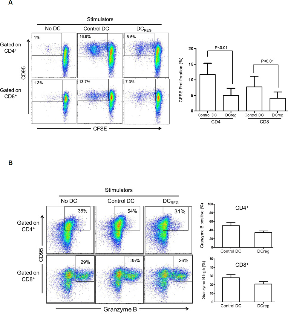
We examined the influence of regulatory dendritic cells (DCreg), generated from cytokine-mobilized donor blood monocytes in vitamin D3 and IL-10, on renal allograft survival in a clinically relevant rhesus macaque model. DCreg expressed low MHC class II and costimulatory molecules, but comparatively high levels of programmed death ligand-1 (B7-H1), and were resistant to pro-inflammatory cytokine-induced maturation. They were infused intravenously (3.5-10 × 10(6) /kg), together with the B7-CD28 costimulation blocking agent CTLA4Ig, 7 days before renal transplantation. CTLA4Ig was given for up to 8 weeks and rapamycin, started on Day -2, was maintained with tapering of blood levels until full withdrawal at 6 months. Median graft survival time was 39.5 days in control monkeys (no DC infusion; n = 6) and 113.5 days (p < 0.05) in DCreg-treated animals (n = 6). No adverse events were associated with DCreg infusion, and there was no evidence of induction of host sensitization based on circulating donor-specific alloantibody levels. Immunologic monitoring also revealed regulation of donor-reactive memory CD95(+) T cells and reduced memory/regulatory T cell ratios in DCreg-treated monkeys compared with controls. Termination allograft histology showed moderate combined T cell- and Ab-mediated rejection in both groups. These findings justify further preclinical evaluation of DCreg therapy and their therapeutic potential in organ transplantation.

Keywords: Costimulation blockade; dendritic cells; memory T cells; rapamycin; renal transplant; rhesus macaques.


Conflict of interest statement

Disclosure

The authors of this manuscript have conflicts of interest to disclose as described by The American Journal of Transplantation. AWT and AJD are inventors of US patents for generation of dendritic cells to promote organ transplant survival.

Figures

Figure 1
Figure 1. Phenotypic and functional characteristics of control DC and DCreg propagated from CD14+ blood monocytes mobilized in rhesus monkey leukapheresis products
(A), DC were propagated in GM-CSF + IL-4 from CD14+ monocytes, in the absence (control DC) or presence of VitD3/IL-10 (DCreg), as described in the Materials and Methods. Phenotypic data obtained using flow cytometry are shown for unstimulated control DC and DCreg, and also for both populations stimulated for 24 h with a potent, pro-inflammatory cytokine cocktail. (B), DCreg induce minimal allogeneic CD4+ and CD8+ T cell proliferation compared with control DC. Left: Proliferative responses of normal allogeneic CD4+ and CD8+ T cells in response to control DC or DCreg were evaluated. CFSE-labeled rhesus PBMC (0.2×10⁶) were cocultured with either control DC or DCreg (0.01×10⁶) at 20:1 ratio for 5 days. After gating on CD4+ and CD8+ T cells, proliferation was evaluated by CFSE dilution analysis. Right: Mean CFSE proliferation of allogeneic CD4+ and CD8+ T cells was significantly less in response to DCreg compared to control DC. Data are representative of 5 independent experiments using different monkeys.
Figure 2
Figure 2. Rhesus DCreg enhance apoptosis of alloreactive T memory cells
As in Figure 1, CFSE-labeled rhesus PBMC were cocultured with control DC or DCreg at 20:1 ratio for 5 days, followed by evaluation of T cell proliferation by CFSE dilution analysis. (A) Left: DCreg elicit less CD4+/CD8+ CD95+ T memory cell proliferation. Right: Mean of 5 experiments showing significantly less proliferation of CD95+ T cells in response to DCreg. (B) Left: DCreg elicit less granzyme B+ CD95+ effector memory T cells in comparison to control DC in 5-day MLR. Right: Mean of 3 independent experiments using different donor:responder pairs, showing less induction of granzyme B+ T cells after coculture with DCreg. Gating for CD4+ T cells was based on isotype controls. As the vast majority of CD8+ T cells were Granzyme B+ under all culture conditions, gating was based on granzyme Bhi populations. (C) Rhesus DCreg induce less allogeneic CD4+ and CD8+ T cell proliferation in 5-day CFSE-MLR, but enhance apoptosis of non-proliferating T cells compared with control DC. (D), DCreg induce higher incidences of CD4+/CD8+ CD95+ Annexin+ memory T cells than control DC after 5-day MLR culture. Data in C and D are representative of 2 independent experiments using different stimulator-responder pairs.
Figure 3
Figure 3. DCreg infusion prolongs MHC-mismatched renal allograft survival in rhesus monkeys
(A), Costimulation blockade/rapamycin-based immunosuppression protocol. Monkeys received DCreg (3–10×106/kg i.v.) (n=6) or no cells (n=6) on day −7 relative to kidney transplantation on day 0. Each group of monkeys was given CTLA4Ig (25 mg/kg i.v.), either from day −7 to day 10 post-transplant (n=4 per group) or from day −7 to day 56 (n=2 per group). Intramuscular rapamycin was commenced on day −2 and whole blood trough levels maintained at 10–15 ng/ml (day 0-day 28); 5–10 ng/ml (day 29-day 150) and 1–5 ng/ml (day 151-day 180). Immunosuppressive treatment was stopped at day 180. (B), serum creatinine levels in control (n=6) and DCreg-treated animals (n=6) at various times post transplant; (C), body weight loss in each group at various times post-transplant (D), urinary protein/creatinine ratios in the same groups of monkeys, 6–8 weeks post-transplant and (E), actuarial graft survival data.
Figure 4
Figure 4. DCreg infusion exerts no consistent effect on anti-donor T cell proliferative responses
(A), Ex vivo anti-donor and anti-third party proliferative responses of circulating CD4+ and CD8+ T cells from control or DCreg-treated rhesus renal allograft recipients before, and 4–6 and 8–12 weeks post-transplant. Each graph represents the response of one recipient from the DCreg group and its concurrent recipient from the control group against the same donor cells or 3rd party cells. CFSE-MLRs were performed as described in the Materials and Methods. PBMC from each recipient were cocultured with T cell-depleted PBMC from either donor or third party at 1:1 ratio for 5 days. (B), proliferative responses of T cells from a DCreg-treated monkey (M113) whose transplant survived 300 days, showing initial depression (day 180) of anti-donor and anti-third party responses, but later recovery of anti-donor reactivity (day 240) after withdrawal of all immunosuppression, followed by enhanced anti-donor reactivity at the time of rejection (day 300).
Figure 5
Figure 5. DCreg infusion suppresses CD4+ and CD8+ Tmem in renal-allografted monkeys and enhances PD-1 and CTLA4 expression on donor-reactive Tmem
(A), Absolute numbers of circulating CD4+ and CD8+ CD95+ Tmem in control and DCreg-treated renal allograft recipients at various times pre- and post-transplant. (B, C) Incidences of PD1+CTLA4+ Tmem in ex-vivo-stimulated CD4+ and CD8+ T cell populations from (B) M112 and M143 (control) monkeys, and (C) M147 and M148 (DCreg-treated) monkeys. Recipient PBMC obtained 4 weeks (and 8 weeks in the DCreg group monkeys) post transplant, were cocultured with either donor or third party stimulators for 5 days as in Figure 4. CD4+ and CD8+ CD95+ T cells were then analyzed for cell surface PD-1 and CTLA4 expression. POD = post-operative day.

Comment in

Similar articles

Cited by

References

    1. Wood KJ, Bushell A, Hester J. Regulatory immune cells in transplantation. Nat Rev Immunol. 2012;12(6):417–430. - PubMed
    1. Morelli AE, Thomson AW. Tolerogenic dendritic cells and the quest for transplant tolerance. Nat Rev Immunol. 2007;7(8):610–621. - PubMed
    1. Lombardi G, Sagoo P, Scotta C, Fazekasova H, Smyth L, Tsang J, et al. Cell therapy to promote transplantation tolerance: a winning strategy? Immunotherapy. 2011;3(4 Suppl):28–31. - PubMed
    1. Stenger EO, Turnquist HR, Mapara MY, Thomson AW. Dendritic cells and regulation of graft-versus-host disease and graft-versus-leukemia activity. Blood. 2012;119(22):5088–5103. - PMC - PubMed
    1. Steptoe RJ, Thomson AW. Dendritic cells and tolerance induction. Clin Exp Immunol. 1996;105:397–402. - PMC - PubMed

Publication types

MeSH terms