. 2013 Jun 10;11(1):41.
doi: 10.1186/1478-811X-11-41.

Down-regulation of the cancer/testis antigen 45 (CT45) is associated with altered tumor cell morphology, adhesion and migration

Affiliations

Down-regulation of the cancer/testis antigen 45 (CT45) is associated with altered tumor cell morphology, adhesion and migration

Anja Koop et al. Cell Commun Signal. .

Abstract

Background: Due to their restricted expression in male germ cells and certain tumors, cancer/testis (CT) antigens are regarded as promising targets for tumor therapy. CT45 is a recently identified nuclear CT antigen that was associated with a severe disease score in Hodgkin's lymphoma and poor prognosis in multiple myeloma. As for many CT antigens, the biological function of CT45 in developing germ cells and in tumor cells is largely unknown.

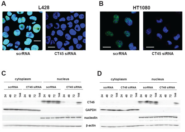
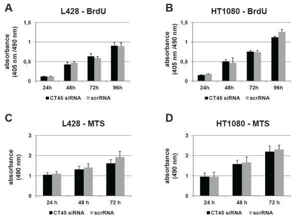
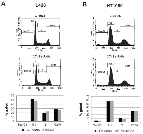
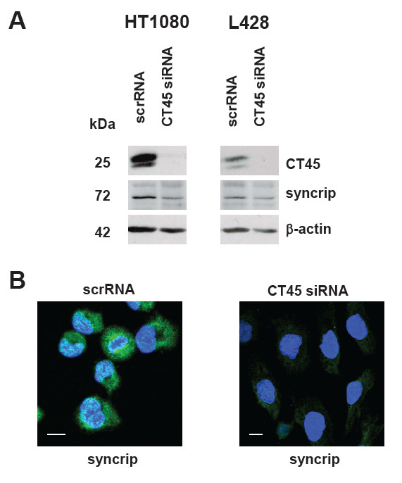
Methods: CT45 expression was down-regulated in CT45-positive Hodgkin's lymphoma (L428), fibrosarcoma (HT1080) and myeloma (U266B1) cells using RNA interference. An efficient CT45 knock-down was confirmed by immunofluorescence staining and/or Western blotting. These cellular systems allowed us to analyze the impact of CT45 down-regulation on proliferation, cell cycle progression, morphology, adhesion, migration and invasive capacity of tumor cells.

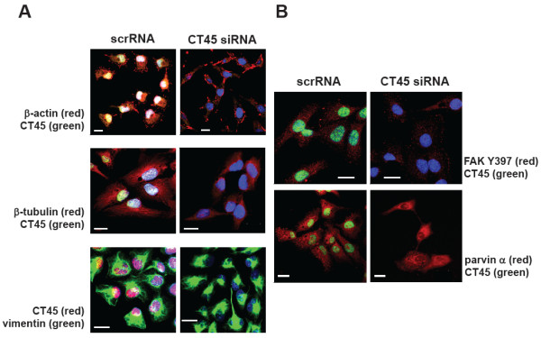
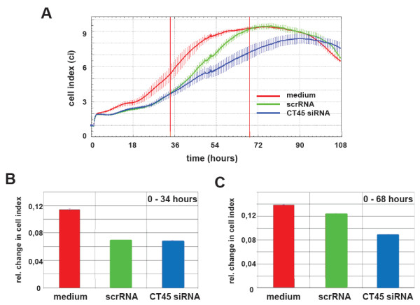
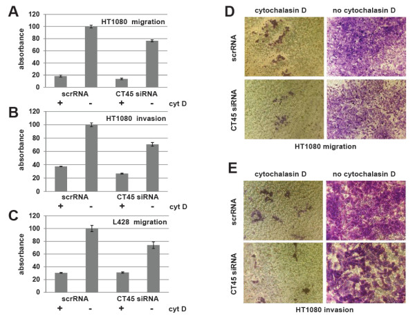
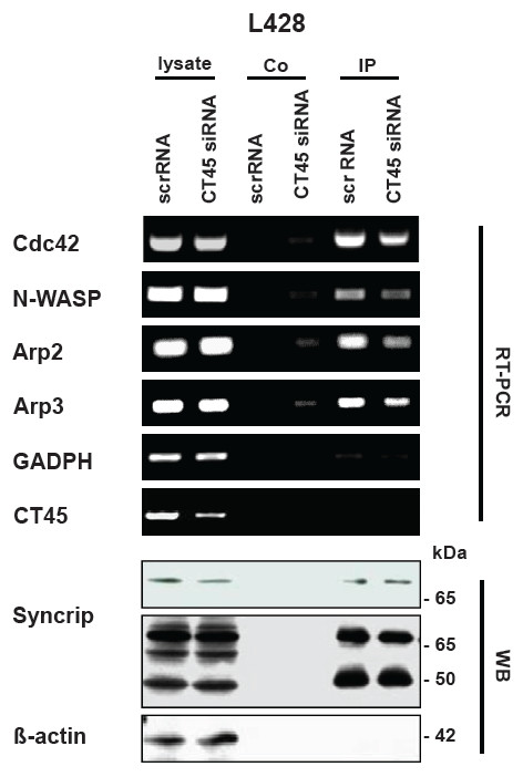
Results: Reduced levels of CT45 did not coincide with changes in cell cycle progression or proliferation. However, we observed alterations in cell adherence, morphology and migration/invasion after CT45 down-regulation. Significant changes in the distribution of cytoskeleton-associated proteins were detected by confocal imaging. Changes in cell adherence were recorded in real-time using the xCelligence system with control and siRNA-treated cells. Altered migratory and invasive capacity of CT45 siRNA-treated cells were visualized in 3D migration and invasion assays. Moreover, we found that CT45 down-regulation altered the level of the heterogeneous nuclear ribonucleoprotein syncrip (hnRNP-Q1) which is known to be involved in the control of focal adhesion formation and cell motility.

Conclusions: Providing first evidence of a cell biological function of CT45, we suggest that this cancer/testis antigen is involved in the modulation of cell morphology, cell adherence and cell motility. Enhanced motility and/or invasiveness of CT45-positive cells could contribute to the more severe disease progression that is correlated to CT45-positivity in several malignancies.


Figures

Figure 1
Figure 1
Subcellular localization of CT45 and down-regulation of CT45 by RNA interference. Immunofluorescence staining of cytospin preparations of L428 cells (A) and of adherently growing HT1080 cells (B) treated with scrambled (scrRNA) or with CT45-specific (CT45 siRNA) with mab Ki-A10. DAPI was used for DNA staining. Down-regulation of CT45 using CT45 siRNA was analyzed after 72 h (A, B) or over a period of up to 144 h in L428 (C) and HT1080 cells (D). Cytoplasmic and nuclear fractions of the cells were prepared and CT45 expression was analyzed with mab Ki-A10 in L428 and mab Ki-CT45-2 in HT1080 cells. β-actin staining was performed to demonstrate equal loading, GAPDH was used as a cytoplasmic and nucleolin as a nuclear marker. All experiments were performed at least three times. White bars indicate 20 μm.
Figure 2
Figure 2
Impact of CT45 down-regulation on cell proliferation. BrdU (A, B) and MTS (C, D) assays were performed in quadruplicates to determine the effect of CT45 down-regulation on cell metabolism and proliferation of L428 (A, C) and HT1080 (B, D) cells. Data are shown for representative experiments of two to six independent assays. In all cases, inhibition of CT45 expression had no significant effect on cell proliferation and cell growth in the tested cell lines.
Figure 3
Figure 3
Impact of down-regulation of CT45 on cell cycle progression. 72 h after transfection with scrRNA or CT45 siRNA, cells were harvested and labeled with propidium iodide. The analysis of the different cell cycle phases of L428 (A) and HT1080 (B) cells was performed by flow cytometry. Representative histograms for scrRNA- or CT45 siRNA-treated cells are shown in the upper and middle panels. A quantification using the indicated regions is depicted in the lower panels. Down-regulation of CT45 had no apparent impact on cell cycle progression. All experiments were performed at least three times.
Figure 4
Figure 4
Down-regulation of CT45 alters cell morphology. (A) 72 h after transfection, double immunofluorescence staining of adherently growing HT1080 cells with mab Ki-A10 and antibodies specific for actin, ß-tubulin and vimentin was performed. ScrRNA-treated cells demonstrate normal cell morphology, whereas CT45 siRNA-treated cells show a significantly altered cell morphology (right panels in A). (B) Double immunofluorescence staining of focal adhesion proteins FAK Y397 and parvin alpha was performed in HT1080 cells 72 h after siRNA transfection. Focal adhesion sites in scrRNA-treated cells are indicated by arrows. Down-regulation of CT45 resulted in a markedly reduced number of FAK Y397- and parvin α-positive focal adhesion sites. White bars indicate 20 μm.
Figure 5
Figure 5
Influence of CT45 down-regulation on the time-dependent adherence profiles of HT1080 cells. (A) To analyze the effect of down-regulation of CT45 on cell adhesion, we performed real-time measurements of the cell impedance over a period of 108 h with HT1080 cells as a control (red line), scrRNA-transfected cells (green line) and CT45 siRNA-transfected cells (blue line). Starting after 34 h, the cellular impedance of CT45 siRNA-treated cells decreased significantly reaching a maximal difference (18%) at 68–72 h after transfection. The changes in the cell indices were calculated for the time intervals 0–34 h (B) and 0–68 h (C), respectively using the RTCA-integrated software provided by the manufacturer. Data of one representative experiment (with four individual biological replicates) of four are shown.
Figure 6
Figure 6
CT45 affects cell invasion and migration. (A-C) Cell migration and cell invasion capacities of scrRNA- or CT45 siRNA-treated HT1080 or L428 cells were analyzed in colorimetric or fluorometric transwell systems as described in the experimental procedures (A, D: HT1080 migration, B, E: HT1080 invasion, C: L428 migration). As a control, cell motility was inhibited with 2 μM Cytochalasin D. The data analysis revealed that cell migration of siRNA-treated HT1080 cells was down-regulated by 23%. Staining of cells collected from the lower chamber revealed that less siRNA-transfected and only very few cytochalasin D-treated cells had passed the membrane (D). The cell invasion assay of HT1080 (B, E) and the migration assay for L428 cells (C) revealed similar results. All experiments were performed at least three times.
Figure 7
Figure 7
CT45 influences the expression of syncrip. (A) L428 and HT1080 cells were transfected with scrRNA or CT45 siRNA, respectively. 72 h after transfection, Western blot analyses of cell lysates were performed using antibodies specific for CT45 (Ki-A10 for L428 cells, and Ki-CT45-2 for HT1080 cells), syncrip and ß-actin (as loading control). 72 h after down-regulation of CT45, syncrip is also down-regulated in both cell lines. (B) In parallel, immunofluorescence staining for syncrip of scrRNA-transfected and CT45 siRNA-transfected HT1080 cells was performed. DAPI was used for visualization of the nuclei. White bars indicate 20 μm.
Figure 8
Figure 8
Syncrip selectively co-precipitates mRNAs of Cdc42, N-WASP, Arp2, Arp3 but not GAPDH and CT45. L428 cells were transfected with scrRNA or CT45 siRNA, respectively. 72 h after transfection, cell lysates were prepared and syncrip immunoprecipitations performed as detailed in Material and Methods. Protein A beads used for preclearing were used as internal control (co) for the RT-PCR or Western blot. In addition, respective RT-PCRs were also performed using unseparated cell lysates as starting material. The whole cell lysates were also used to document the mild reduction of syncrip protein by Western blotting (WB, upper panel for syncrip, short exposure; lower panel, long exposure). The RT-PCR revealed that mRNAs for Cdc42, N-WASP, Arp2 and Arp3 (but not CT45 or GAPDH) could be amplified from syncrip-immunoprecpitations. In addition, the amount of co-precipitated mRNA was reduced in CT45 siRNA-treated cells.

Similar articles

Cited by

References

    1. Scanlan MJ, Simpson AJ, Old LJ. The cancer/testis genes: review, standardization, and commentary. Cancer Immun. 2004;4:1. - PubMed
    1. Hofmann O, Caballero OL, Stevenson BJ, Chen YT, Cohen T, Chua R, Maher CA, Panji S, Schaefer U, Kruger A, Lehvaslaiho M, Carninci P, Hayashizaki Y, JongeneeL CV, Simpson AJG, Old LJ, Hide W. Genome-wide analysis of cancer/testis gene expression. PNAS. 2008;105:20422–27. doi: 10.1073/pnas.0810777105. - DOI - PMC - PubMed
    1. Almeida LG, Sakabe NJ, DeOliveira AR, Silva MCC, Mundstein AS, Cohen T, Chen YT, Chua R, Gurung S, Gnjatic S, Jungbluth AA, Caballero OL, Bairoch A, Kiesler E, White SL, Simpson AJG, Old LJ, Camargo AA, Vasconcelos AT. CTdatabase: a knowledge-base of high-throughput and curated data on cancer-testis antigens. Nucleic Acids Res. 2009;37:D816–D819. doi: 10.1093/nar/gkn673. - DOI - PMC - PubMed
    1. Boon T, Old LJ. Cancer Tumor antigens. Curr Opin Immunol. 1997;9:681–683. doi: 10.1016/S0952-7915(97)80049-0. - DOI - PubMed
    1. Fratta E, Coral S, Covre A, Parisi G, Collizzi F, Danielli R, Nicolay HJM, Sigalotti L, Maio M. The biology of cancer testis antigens: Putative function, regulation and therapeutic potential. Mol Oncol. 2011;5:164–182. doi: 10.1016/j.molonc.2011.02.001. - DOI - PMC - PubMed