. 2013 Jun 10;3(1):46.
doi: 10.1186/2191-219X-3-46.

High correlation between in vivo [123I]β-CIT SPECT/CT imaging and post-mortem immunohistochemical findings in the evaluation of lesions induced by 6-OHDA in rats

Affiliations

High correlation between in vivo [123I]β-CIT SPECT/CT imaging and post-mortem immunohistochemical findings in the evaluation of lesions induced by 6-OHDA in rats

Susanne Bäck et al. EJNMMI Res. .

Abstract

Background: 6-Hydroxydopamine (6-OHDA) is widely used in pre-clinical animal studies to induce degeneration of midbrain dopamine neurons to create animal models of Parkinson's disease. The aim of our study was to evaluate the potential of combined single-photon emission computed tomography/computed tomography (SPECT/CT) for the detection of differences in 6-OHDA-induced partial lesions in a dose- and time-dependent manner using the dopamine transporter (DAT) ligand 2β-carbomethoxy-3β-(4-[123I]iodophenyl)tropane ([123I]β-CIT).

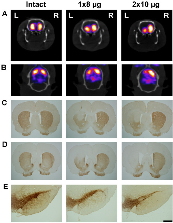
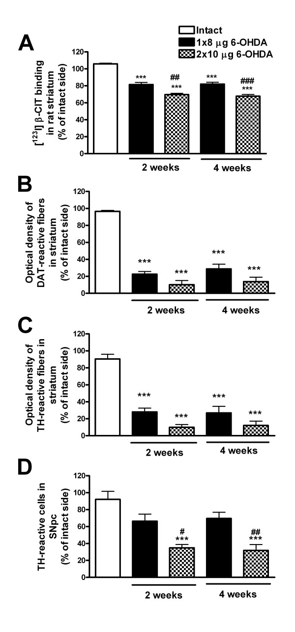
Methods: Rats were unilaterally lesioned with intrastriatal injections of 8 or 2 × 10 μg 6-OHDA. At 2 or 4 weeks post-lesion, 40 to 50 MBq [123I]β-CIT was administered intravenously and rats were imaged with small-animal SPECT/CT under isoflurane anesthesia. The striatum was delineated and mean striatal activity in the lesioned side was compared to the intact side. After the [123I]β-CIT SPECT/CT scan, the rats were tested for amphetamine-induced rotation asymmetry, and their brains were immunohistochemically stained for DAT and tyrosine hydroxylase (TH). The fiber density of DAT- and TH-stained striata was estimated, and TH-immunoreactive cells in the rat substantia nigra pars compacta (SNpc) were stereologically counted.

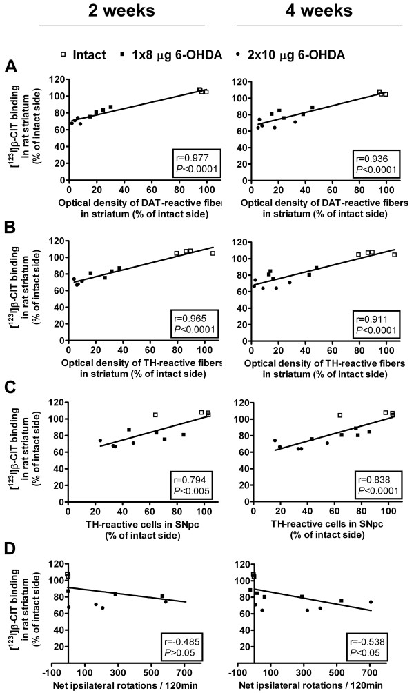
Results: The striatal uptake of [123I]β-CIT differed significantly between the lesion groups and the results were highly correlated to both striatal DAT- and TH-immunoreactive fiber densities and to TH-immunoreactive cell numbers in the rat SNpc. No clear progression of the lesion could be seen.

Conclusions: [123I]β-CIT SPECT/CT is a valuable tool in predicting the condition of the rat midbrain dopaminergic pathway in the unilateral partial 6-OHDA lesion model of Parkinson's disease and it offers many advantages, allowing repeated non-invasive analysis of living animals.


Figures

Figure 1
Figure 1
Representative pictures of striatal binding of [123I]β-CIT and immunohistochemical staining with dopaminergic markers. Striatal activity of [123I]β-CIT detected with SPECT/CT (coronal view (A) and transversal view (B)), DAT immunoreactivity in the striatum (C), TH immunoreactivity in the striatum (D), and nigral TH-immunoreactive cells (E) in intact and 6-OHDA-lesioned rats (8 and 2 × 10 μg 6-OHDA) at 4 weeks post-lesion. The orientation of the pictures is denoted in (A) with left (L) and right (R). Scale bar in (E) is 500 μm.
Figure 2
Figure 2
Unilateral 6-OHDA-induced changes in [123I]β-CIT SPECT/CT and immunohistochemical findings. All results are presented as percentage of the lesioned/left side versus the intact/right side. (A) The 6-OHDA injections caused a significant decrease in striatal [123I]β-CIT uptake as compared to intact rats at both 2 and 4 weeks post-lesion. Furthermore, the two different lesions (8 and 2 × 10 μg) could be distinguished with [123I]β-CIT SPECT/CT. Intrastriatal injection of 8 or 2 × 10 μg 6-OHDA resulted in extensive loss of (B) DAT- and (C) TH-immunoreactive fibers in the striatum, but there was no significant difference between the two lesion groups. However, when counting (D) TH-reactive cells in the substantia nigra pars compacta, the cell loss significantly differed between the lesion types. All results are shown as mean ± SEM. ***P < 0.001 as compared to intact rats; #P < 0.05, ##P < 0.01, and ###P < 0.001 as compared to 8-μg 6-OHDA lesion group (one-way ANOVA followed by Tukey post hoc test, n = 4 to 5 per group).
Figure 3
Figure 3
Correlations between [123I]β-CIT SPECT/CT and immunohistochemical and behavioral data at 2 and 4 weeks post-lesion. Striatal binding of [123I]β-CIT showed high correlation to striatal (A) DAT- and (B) TH-reactive fiber densities and (C) nigral TH-reactive cell numbers at both time points. At 4 weeks, [123I]β-CIT SPECT/CT correlated moderately to the amount of (D) amphetamine-induced rotations, whereas no significant correlation was found at 2 weeks post-lesion. Correlation coefficients (r) and statistical significance (P) were determined with Pearson correlation test (n = 12 (2 weeks) and n = 14 (4 weeks)).

References

    1. Dauer W, Przedborski S. Parkinson's disease: mechanisms and models. Neuron. 2003;3:889–909. doi: 10.1016/S0896-6273(03)00568-3. - DOI - PubMed
    1. Ungerstedt U. 6-Hydroxy-dopamine induced degeneration of central monoamine neurons. Eur J Pharmacol. 1968;3:107–110. doi: 10.1016/0014-2999(68)90164-7. - DOI - PubMed
    1. Ungerstedt U, Arbuthnott GW. Quantitative recording of rotational behavior in rats after 6-hydroxy-dopamine lesions of the nigrostriatal dopamine system. Brain Res. 1970;3:485–493. doi: 10.1016/0006-8993(70)90187-3. - DOI - PubMed
    1. Schwarting RK, Huston JP. The unilateral 6-hydroxydopamine lesion model in behavioral brain research. Analysis of functional deficits, recovery and treatments. Prog Neurobiol. 1996;3:275–331. doi: 10.1016/S0301-0082(96)00040-8. - DOI - PubMed
    1. Schwarting RK, Huston JP. Unilateral 6-hydroxydopamine lesions of meso-striatal dopamine neurons and their physiological sequelae. Prog Neurobiol. 1996;3:215–266. doi: 10.1016/S0301-0082(96)00015-9. - DOI - PubMed

LinkOut - more resources