. 2013 Jun 10:14:391.
doi: 10.1186/1471-2164-14-391.

Archaeal nucleosome positioning in vivo and in vitro is directed by primary sequence motifs

Affiliations

Archaeal nucleosome positioning in vivo and in vitro is directed by primary sequence motifs

Narasimharao Nalabothula et al. BMC Genomics. .

Abstract

Background: Histone wrapping of DNA into nucleosomes almost certainly evolved in the Archaea, and predates Eukaryotes. In Eukaryotes, nucleosome positioning plays a central role in regulating gene expression and is directed by primary sequence motifs that together form a nucleosome positioning code. The experiments reported were undertaken to determine if archaeal histone assembly conforms to the nucleosome positioning code.

Results: Eukaryotic nucleosome positioning is favored and directed by phased helical repeats of AA/TT/AT/TA and CC/GG/CG/GC dinucleotides, and disfavored by longer AT-rich oligonucleotides. Deep sequencing of genomic DNA protected from micrococcal nuclease digestion by assembly into archaeal nucleosomes has established that archaeal nucleosome assembly is also directed and positioned by these sequence motifs, both in vivo in Methanothermobacter thermautotrophicus and Thermococcus kodakarensis and in vitro in reaction mixtures containing only one purified archaeal histone and genomic DNA. Archaeal nucleosomes assembled at the same locations in vivo and in vitro, with much reduced assembly immediately upstream of open reading frames and throughout the ribosomal rDNA operons. Providing further support for a common positioning code, archaeal histones assembled into nucleosomes on eukaryotic DNA and eukaryotic histones into nucleosomes on archaeal DNA at the same locations. T. kodakarensis has two histones, designated HTkA and HTkB, and strains with either but not both histones deleted grow normally but do exhibit transcriptome differences. Comparisons of the archaeal nucleosome profiles in the intergenic regions immediately upstream of genes that exhibited increased or decreased transcription in the absence of HTkA or HTkB revealed substantial differences but no consistent pattern of changes that would correlate directly with archaeal nucleosome positioning inhibiting or stimulating transcription.

Conclusions: The results obtained establish that an archaeal histone and a genome sequence together are sufficient to determine where archaeal nucleosomes preferentially assemble and where they avoid assembly. We confirm that the same nucleosome positioning code operates in Archaea as in Eukaryotes and presumably therefore evolved with the histone-fold mechanism of DNA binding and compaction early in the archaeal lineage, before the divergence of Eukaryotes.


Figures

Figure 1
Figure 1
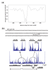
Archaeal nucleosomes protect ~60 bp chromosomal DNA fragments from micrococcal nuclease (MN) digestion. (a). Ethidium bromide stained electrophoretic separation of DNA molecules protected from MN digestion of T. kodakarensis TS517 chromatin. The control lane (C) contained double-stranded DNA size standards (bp). As indicated, MN digestion generated DNA molecules that migrated to form a band consistent with ~60 bp molecules. These were isolated and sequenced. (b). The number and size profile of the sequences of the ~60 bp DNA fragment generated by ABI-SoLiD deep sequencing.
Figure 2
Figure 2
Archaeal nucleosomes assembled in vivo contain offset helical repeats of AA/AT/TA/TT and CC/GG/GC/CG dinucleotides and lack oligo A/T-rich sequences. The frequencies of occurrence of AA/AT/TA/TT (red line) and CC/GG/GC/CG (blue line) dinucleotides, at each position relative to the center of archaeal nucleosomes assembled (a) in M. thermautotrophicus and (b) in T. kodakarensis (c and d). The ratios of the presence and absence of all pentamers in the DNA molecules protected from MN digestion by nucleosome assembly in M. thermoautotrophicus and in T. kodakarensis, respectively. The graphs show the ratio of occurrence of each of the 1024 possible pentamers in nucleosomal DNA (Pnucleosome) versus in non-nucleosomal DNA (P). As examples, the specific locations on the curves of representative G and/or C only, and A and/or T only pentamers are indicated. As noted, all 32 G and/or C-only pentamers were located preferentially within nucleosomal DNA (blue shaded region), whereas all 32 A and/or T-only pentamers were preferentially excluded from nucleosome incorporation (red shaded region).
Figure 3
Figure 3
Depletion of archaeal nucleosomes in intergenic regions. (a). The occurrence of nucleosomes relative to the start codons of all open reading frames (ORFs), as documented in the Archaeal Genome Browser [39]. The frequency of occurrence of each nucleosome read is plotted relative to the average value of occurrence of all nucleosomal reads sequenced from the T. kodakarensis TS517 genome. (b) The sequence of the intergenic region separating TK1760 and TK1761 is positioned above the profiles of nucleosomes assembled in vivo at this locus and downstream, within the well-established TK1761-TK1762-TK1763 operon [40].
Figure 4
Figure 4
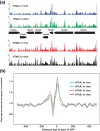
Archaeal nucleosome profiles in vivo are reproduced in vitro. (a) Profiles of the archaeal nucleosomes assembled in vivo and in vitro by HTkA and HTkB between nucleotide positions 5,000 and 10,000 on the T. kodakarensis genome. The organization of T. kodakarensis genes in this region is shown between upper and lower panels [34,39,42]. (b). The occurrence of nucleosome positions relative to the start codon of all open reading frames (ORFs), assembled by HTkA in vivo (blue line) and in vitro (green line), and by HTkB in vivo (red line) and in vitro (black line) normalized to the total number of nucleosomal reads from each sequencing experiment.
Figure 5
Figure 5
Archaeal nucleosomes do not assemble on the ribosomal DNA operon. Profiles of the archaeal nucleosomes assembled by HTkA and HTkB in vivo in T. kodakarensis LC125 and LC124, respectively, and in vitro on a 10 Kbp region of the T. kodakarensis genome. As illustrated, this region encodes the 16S and 23S rRNA (rDNA operon; red arrows) and several protein-encoding flanking genes [34,39].
Figure 6
Figure 6
Archaeal and eukaryotic nucleosomes are positioned by conserved sequences. The frequencies of occurrence of AA/AT/TA/TT and CC/GG/GC/CG dinucleotides, at each position relative to the center of (a) eukaryotic nucleosomes assembled by chicken histones on T. kodakarensis genomic DNA, and (b) of archaeal nucleosomes assembled by T. kodakarensis histones on yeast genomic DNA. (c) The ratio of the presence and absence of all pentamers in the T. kodakarensis DNA protected from MN digestion by assembly into eukaryotic nucleosomes, and (d) in yeast DNA assembled into archaeal nucleosomes. As indicated, all pentamers that contained only G and/or C were preferentially incorporated into both eukaryotic and archaeal nucleosomes (blue regions) whereas all pentamers that contained only A and/or T (red region) were preferentially excluded nucleosomes.
Figure 7
Figure 7
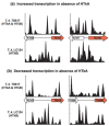
Nucleosome profiles, related to transcription changes that result from archaeal histone deletion. The profiles of nucleosomes assembled in vivo in T. kodakarensis TS517 and LC124 are respectively shown above and below the 1-kbp genomic region of interest [34,39]. Intergenic regions are depicted as a single line and the genes that (a) had increased transcription or (b) had decreased transcription in the absence of HTkA [13] are shaded red.

References

    1. Field Y, Kaplan N, Fondufe-Mittendorf Y, Moore IK, Sharon E, Lubling Y, Widom J, Segal E. Distinct modes of regulation by chromatin encoded through nucleosome positioning signals. PLoS Comput Biol. 2008;4:e1000216. doi: 10.1371/journal.pcbi.1000216. - DOI - PMC - PubMed
    1. Shivaswamy S, Bhinge A, Zhao Y, Jones S, Hirst M, Iyer VR. Dynamic remodeling of individual nucleosomes across a eukaryotic genome in response to transcriptional perturbation. PLoS Biol. 2008;6:e65. doi: 10.1371/journal.pbio.0060065. - DOI - PMC - PubMed
    1. Jiang C, Pugh BF. Nucleosome positioning and gene regulation: advances through genomics. Nature Rev Genet. 2009;10:161–172. - PMC - PubMed
    1. Guan Y, Yao V, Tsui K, Gebbia M, Dunham MJ, Nislow C, Troyanskaya OG. Nucleosome-coupled expression differences in closely-related species. BMC Genomics. 2011;12:466. doi: 10.1186/1471-2164-12-466. - DOI - PMC - PubMed
    1. Segal E, Fondufe-Mittendorf Y, Chen L, Thastrom A, Field Y, Moore IK, Wang JP, Widom J. A genomic code for nucleosome positioning. Nature. 2006;442:772–778. doi: 10.1038/nature04979. - DOI - PMC - PubMed

Publication types