. 2013 Jun 7:8:136.
doi: 10.1186/1748-717X-8-136.

Changes in the micromorphology of the corneal subbasal nerve plexus in patients after plaque brachytherapy

Changes in the micromorphology of the corneal subbasal nerve plexus in patients after plaque brachytherapy

Andrey Zhivov et al. Radiat Oncol. .

Abstract

Background: To quantify the development of radiation neuropathy in corneal subbasal nerve plexus (SNP) after plaque brachytherapy, and the subsequent regeneration of SNP micromorphology and corneal sensation.

Methods: Nine eyes of 9 melanoma patients (ciliary body: 3, iris: 2, conjunctiva: 4) underwent brachytherapy (ruthenium-106 plaque, dose to tumour base: 523 ± 231 Gy). SNP micromorphology was assessed by in-vivo confocal microscopy. Using software developed in-house, pre-irradiation findings were compared with those obtained after 3 days, 1, 4 and 7 months, and related to radiation dose and corneal sensation.

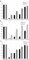
Results: After 3 days nerve fibres were absent from the applicator zone and central cornea, and corneal sensation was abolished. The earliest regenerating fibres were seen at the one-month follow-up. By 4 months SNP structures had increased to one-third of pre-treatment status (based on nerve fibre density and nerve fibre count), and corneal sensation had returned to approximately two-thirds of pre-irradiation values. Regeneration of SNP and corneal sensation was nearly complete 7 months after plaque brachytherapy.

Conclusions: The evaluation of SNP micromorphology and corneal sensation is a reliable and clinically useful method for assessing neuropathy after plaque brachytherapy. Radiation-induced neuropathy of corneal nerves develops quickly and is partly reversible within 7 months. The clinical impact of radiation-induced SNP damage is moderate.


Figures

Figure 1
Figure 1
Clinical situation and isodose distribution by Ru-106 brachytherapy. (a) Schematic diagram of the corneal zones: applicator zone (A), central zone (B) and distal zone (C), each representing approximately one-third of the corneal vertical diameter. Note that vertical diameter of the cornea is approx. 10.5 mm, and the horizontal diameter 11.5 mm. (b) Photograph of a patient’s eye during brachytherapy: the Ru-106 plaque is located in the upper third of the cornea. (c) Diagram of isodose distribution: Simulation of CCB plaque with a dose of 522 Gy to the tumour base. The directly adjacent cornea receives 2.29 Gy (0.44% isodose), the central zone 0.63 Gy (0.12%) and the distal zone 0.08 Gy (0.015%) over the entire time course of plaque brachytherapy.
Figure 2
Figure 2
Imaging and automated quantification of SNP. (a) Representative in-vivo confocal image of the corneal SNP (distal zone, 4 months after plaque brachytherapy). (b) Results of automated image segmentation and analysis: total fibre length 1578 μm, nerve fibre density 9.863 mm/mm2, and single nerve fibre count (n) 19. Image size: 400 x 400 μm.
Figure 3
Figure 3
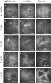
Representative in-vivo confocal images of the corneal SNP of patients over time. Image size: 400 × 400 μm. Each image also shows data on corneal sensation (in mm).
Figure 4
Figure 4
Automated quantification of SNP structures and corneal sensation over time (percentage changes compared with pre-irradiation level): (a) Automated quantification of nerve fibre density. (b) Automated quantification of single nerve fibres. (c) Corneal sensation.

Similar articles

Cited by

References

    1. Char DH, Quivey JM, Castro JR, Kroll S, Phillips T. Helium ions versus iodine 125 brachytherapy in the management of uveal melanoma. A prospective, randomized, dynamically balanced trial. Ophthalmology. 1993;100:1547–1554. - PubMed
    1. Bergman L, Nilsson B, Lundell G, Lundell M, Seregard S. Ruthenium brachytherapy for uveal melanoma, 1979–2003: survival and functional outcomes in the Swedish population. Ophthalmology. 2005;112:834–840. doi: 10.1016/j.ophtha.2004.11.038. - DOI - PubMed
    1. Guthoff R, Frischmuth J, Jensen OA, Bjerrum K, Prause JU. Choroid melanoma. A retrospective randomized comparative study of ruthenium irradiation vs enucleation. Klin Monbl Augenheilkd. 1992;200:257–261. doi: 10.1055/s-2008-1045748. - DOI - PubMed
    1. Lommatzsch PK, Werschnik C, Schuster E. Long-term follow-up of Ru-106/Rh-106 brachytherapy for posterior uveal melanoma. Graefes Arch Clin Exp Ophthalmol. 2000;238:129–137. doi: 10.1007/PL00007880. - DOI - PubMed
    1. Lommatzsch PK, Alberti W, Lommatzsch R, Rohrwacher F. Radiation effects on the optic nerve observed after brachytherapy of choroidal melanomas with 106Ru/106Rh plaques. Graefes Arch Clin Exp Ophthalmol. 1994;232:482–487. doi: 10.1007/BF00195358. - DOI - PubMed

MeSH terms

Substances