. 2013 Sep;11(2):743-57.
doi: 10.1016/j.scr.2013.05.002. Epub 2013 May 16.

Astroglial cells regulate the developmental timeline of human neurons differentiated from induced pluripotent stem cells

Affiliations

Astroglial cells regulate the developmental timeline of human neurons differentiated from induced pluripotent stem cells

Xin Tang et al. Stem Cell Res. 2013 Sep.

Abstract

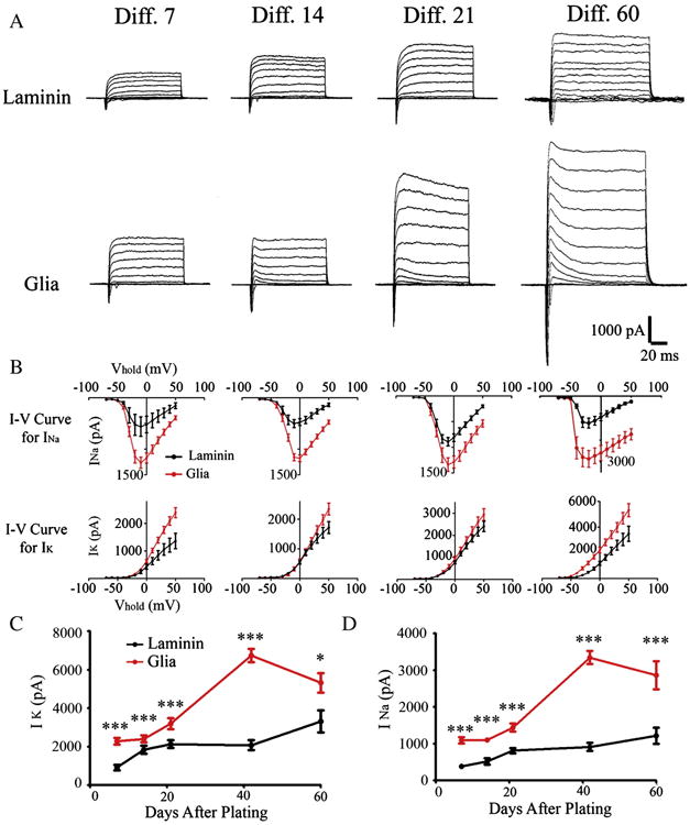
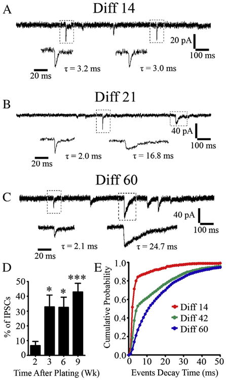
Neurons derived from human induced-pluripotent stem cells (hiPSCs) have been used to model a variety of neurological disorders. Different protocols have been used to differentiate hiPSCs into neurons, but their functional maturation process has varied greatly among different studies. Here, we demonstrate that laminin, a commonly used substrate for iPSC cultures, was inefficient to promote fully functional maturation of hiPSC-derived neurons. In contrast, astroglial substrate greatly accelerated neurodevelopmental processes of hiPSC-derived neurons. We have monitored the neural differentiation and maturation process for up to two months after plating hiPSC-derived neuroprogenitor cells (hNPCs) on laminin or astrocytes. We found that one week after plating hNPCs, there were 21-fold more newly differentiated neurons on astrocytes than on laminin. Two weeks after plating hNPCs, there were 12-fold more dendritic branches in neurons cultured on astrocytes than on laminin. Six weeks after plating hNPCs, the Na(+) and K(+) currents, as well as glutamate and GABA receptor currents, were 3-fold larger in neurons cultured on astrocytes than on laminin. And two months after plating hNPCs, the spontaneous synaptic events were 8-fold more in neurons cultured on astrocytes than on laminin. These results highlight a critical role of astrocytes in promoting neural differentiation and functional maturation of human neurons derived from hiPSCs. Moreover, our data presents a thorough developmental timeline of hiPSC-derived neurons in culture, providing important benchmarks for future studies on disease modeling and drug screening.


Figures

Figure 1
Figure 1
Astrocytes promote neuronal differentiation of hiPSCs. (A) Representative images showing neuronal differentiation of human hNPCs within first week after plating under different conditions including laminin, laminin + GCM, and astroglial cells. Human nuclei (red) label hiPSC-derived cells, and doublecortin (green) labels newly differentiated neurons. Scale bar = 10 μm. (B) Quantification of doublecortin positive (DCX+) cells (20× imaging field, 427 × 341 μm) in different experiment groups. Data are presented as mean ± SEM. n > 7 for each bar graph. *p < 0.05 (one-way ANOVA followed by Bonferroni correction). (C) Quantification of total cell number (human nuclei labeled cells) under different conditions. N = 7–10 independent replicates. *p < 0.05, **p < 0.01 (one-way ANOVA followed by Bonferroni correction).
Figure 2
Figure 2
Astrocytes promote morphological development of hiPSC-derived neurons. (A) Representative dendritic tree of hiPSC-derived neurons growing on laminin or glial cells at different time points. (B) Schematic diagram showing the dendritic branch points (red dots) and intersections (green dots) used for Sholl analysis. (C) Quantification of the average number of intersections that neurites cross on a series of concentric Sholl circles. For C–E: *p < 0.05, ***p < 0.001 (Student's t test). (D) Quantification of the average number of dendritic branch points. (E) Quantification of the cell body size of hiPSC-derived neurons cultured on glial cells or laminin. N = 20 for each data point.
Figure 3
Figure 3
Rapid functional development of hiPSC-derived neurons cocultured with astrocytes. (A) Phase image of cells (WT 126) cultured on astrocytes for four days. (B) Representative action potentials detected 4 days after plating hNPCs on astrocytes. (C & D) Phase image of cells (WT 126) after six days on astrocytes (C), and spontaneous synaptic events (D) detected from hiPSC-derived neurons. (E–H) Cells from a different cell line (WT 33) also showed rapid action potential firing (E–F) and spontaneous synaptic events (G–H) after coculture with astrocytes. Scale bar = 10 μm.
Figure 4
Figure 4
Astrocytes promote the development of action potential firing ability and passive membrane properties. (A) Representative action potentials recorded from 2 week hiPSC-derived neurons growing on laminin versus glial cells. (B) Representative action potentials recorded from 2 month hiPSC-derived neurons growing with or without glial support. (C–E) The developmental curves for the threshold (C), amplitude (D), and half-width (E) of action potentials recorded from hiPSC-derived neurons growing with or without glial support. *p < 0.05, **p < 0.01, ***p < 0.001 (Student's t test). (F–H) The developmental curves for passive membrane properties including membrane capacitance (F), membrane resistance (G), and resting membrane potential (H) recorded from hiPSC-derived neurons growing on laminin versus astrocytes. *p < 0.05, **p < 0.01, ***p < 0.001 (Student's t test).
Figure 5
Figure 5
Astrocytes increase the expression of Na and K channels. (A) Representative whole-cell Na and K currents recorded from hiPSC-derived neurons cultured up to two months on laminin versus astrocytes. (B) The I–Vcurves for peak Na (INa) and K (IK) currents recorded from hiPSC-derived neurons cultured with or without glial cells. (C) The developmental curves for peak K currents in human neurons cultured for up to two months on laminin or astrocytes. *p < 0.05, **p < 0.01, ***p < 0.001 (Student's t test). (D) The developmental curves for peak Na currents in human neurons cultured for up to two months on laminin or astrocytes.
Figure 6
Figure 6
Astrocytes increase the expression of neurotransmitter receptors. (A) Representative whole-cell GABA response traces recorded from hiPSC-derived neurons cultured up to two months on laminin versus astrocytes. (B) Representative whole-cell glutamate (Glu) response traces recorded from hiPSC-derived neurons growing with or without glial support. (C) The developmental curves for peak GABA currents in human neurons cultured for two months on laminin or astrocytes. *p < 0.05, **p < 0.01, ***p < 0.001 (Student's t test). (D) The developmental curves for peak glutamate currents in human neurons cultured for two months on laminin or astrocytes. (E–G) Representative traces showing the development of whole-cell NMDA current in hiPSC-derived neurons cultured with or without glial cells. (E) NMDA current recorded from human neuron cultured on astrocyte for six weeks. (F) NMDA current recorded from human neuron cultured on astrocyte for two months. Note that the NMDA current can be largely blocked by AP5 (red trace). (G) NMDA current recorded from 2 month human neuron cultured on laminin.
Figure 7
Figure 7
Astrocytes are essential for synaptic maturation of human neurons. (A) Representative images showing SV2-labeled synaptic puncta along the DCX+ neurites of hiPSC-derived neurons developing for two months on laminin or astrocytes. Scale bar = 10 μm. (B) Representative traces showing spontaneous synaptic events recorded from hiPSC-derived neurons after plating hNPCs on laminin or astrocytes for three weeks and two months. (C & D) Quantified results illustrating that hiPSC-derived neurons supported by glial cells consistently show higher frequency (C) and larger amplitude (D) of spontaneous synaptic events than those on laminin. *p < 0.05, **p < 0.01, ***p < 0.001 (student's t-test). (E) Representative traces showing spontaneous synaptic events recorded from human neurons derived from a different iPSC line (WT 33) after plating on astrocytes for six weeks.
Figure 8
Figure 8
Excitatory synaptic transmission precedes inhibitory synaptic transmission in hiPSC-derived neurons. (A–C) Representative traces showing spontaneous synaptic events recorded from human neurons at 14 (A), 21 (B), and 60 (C) days after plating on astrocytes. Note that the synaptic responses which initially appeared were usually fast-decaying glutamatergic events (A), but slow-decaying GABAergic events appeared later. (D) Quantified results showing that the percentage of spontaneous IPSCs among all events increased as the human neurons mature. *p < 0.05, ***p < 0.001 (one-way ANOVA followed by Bonferroni correction). (E) Cumulative probability plot of decay time of spontaneous synaptic events recorded from hiPSC-derived neurons 14 (red), 42 (green) or 60 (blue) days in culture (1 ms bins, p < 0.001 by Kolmogorov–Smirnov test).
Figure 9
Figure 9
Human neurons can be incorporated into neural networks. (A–B) CFDA-labeled hiPSC-derived neurons (green) received synaptic input as early as one week after co-cultured with primary mouse neurons. (C–D) Dual whole-cell recordings on a pair of human neurons derived from hiPSCs (WT 126) revealed action potential-evoked synaptic responses after one month in coculture with astrocytes. (E–F) Dual whole-cell recordings revealed functional synaptic connection between human neurons derived from a different hiPSC line (WT 33). Glutamate receptor antagonist CNQX blocked the evoked synaptic responses (red trace).
Figure 10
Figure 10
Generation of human glial cells from hiPSC-derived NPCs. (A) After the two month culture on laminin, a significant portion of HuNu+ cells (blue) were immunopositive for astroglial marker GFAP (green, indicated by arrows). (B) These astroglial cells survived up to 3–4 months in culture, and were both GFAP+ and S100B+. (C) hNPCs derived from low passage of hiPSCs differentiated into MAP2+ neurons after being cultured on glial cells (indicated by *). (D) A significant portion of high-passage (>20 passages) NPCs differentiated into GFAP+ glial cells (indicated by arrows).

Similar articles

Cited by

References

    1. Aarum J, Sandberg K, Haeberlein SL, Persson MA. Migration and differentiation of neural precursor cells can be directed by microglia. Proc Natl Acad Sci U S A. 2003;100:15983–15988. - PMC - PubMed
    1. Ballas N, Lioy DT, Grunseich C, Mandel G. Non-cell autonomous influence of MeCP2-deficient glia on neuronal dendritic morphology. Nat Neurosci. 2009;12:311–317. - PMC - PubMed
    1. Banker GA. Trophic interactions between astroglial cells and hippocampal neurons in culture. Science. 1980;209:809–810. - PubMed
    1. Barres BA. The mystery and magic of glia: a perspective on their roles in health and disease. Neuron. 2008;60:430–440. - PubMed
    1. Bilican B, Serio A, Barmada SJ, Nishimura AL, Sullivan GJ, Carrasco M, Phatnani HP, Puddifoot CA, Story D, Fletcher J, Park IH, Friedman BA, Daley GQ, Wyllie DJ, Hardingham GE, Wilmut I, Finkbeiner S, Maniatis T, Shaw CE, Chandran S. Mutant induced pluripotent stem cell lines recapitulate aspects of TDP-43 proteinopathies and reveal cell-specific vulnerability. Proc Natl Acad Sci U S A. 2012;109(15):5803–5808. - PMC - PubMed

Publication types