Review
. 2013 May 28;14(6):11319-46.
doi: 10.3390/ijms140611319.

Danger control programs cause tissue injury and remodeling

Affiliations
Review

Danger control programs cause tissue injury and remodeling

Jan H Hagemann et al. Int J Mol Sci. .

Abstract

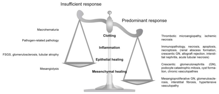
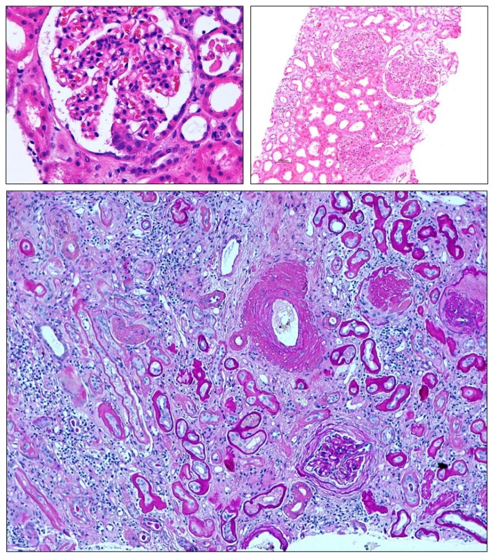
Are there common pathways underlying the broad spectrum of tissue pathologies that develop upon injuries and from subsequent tissue remodeling? Here, we explain the pathophysiological impact of a set of evolutionary conserved danger control programs for tissue pathology. These programs date back to the survival benefits of the first multicellular organisms upon traumatic injuries by launching a series of danger control responses, i.e., 1. Haemostasis, or clotting to control bleeding; 2. Host defense, to control pathogen entry and spreading; 3. Re-epithelialisation, to recover barrier functions; and 4. Mesenchymal, to repair to regain tissue stability. Taking kidney pathology as an example, we discuss how clotting, inflammation, epithelial healing, and fibrosis/sclerosis determine the spectrum of kidney pathology, especially when they are insufficiently activated or present in an overshooting and deregulated manner. Understanding the evolutionary benefits of these response programs may refine the search for novel therapeutic targets to limit organ dysfunction in acute injuries and in progressive chronic tissue remodeling.


Figures

Figure 1
Figure 1
Insufficient and predominant danger response programs determine kidney pathology. Distinct entities of clinical syndromes or kidney injury patterns are consequences of insufficient or overshooting danger control programs.
Figure 2
Figure 2
Overshooting clotting in thrombotic microangiopathy. Local activation microvascular endothelial cells and lack of distinct inhibitory factors can lead to thrombotic microangiopathy which is characterized by microthrombi obstructing arteriolar and glomerular vessels, a lesion associated with significant inflammatory cell infiltrates (left). Fibrin immunostaining displayes clot formation within glomeruli (right). Original magnification 200×.
Figure 3
Figure 3
Interstitial nephritis in polyoma (BK) virus nephropathy. Local activation of inflammation can destroy renal parenchyma and cause acute kidney injury. In this example of BK virus reactivation in a kidney allograft was proven by immunostaining for BK viral protein with positivity in affected tubular epithelial cells (right). Viral replication activates antiviral immunity and immunopathology as evidenced by the dense leukocyte infiltrates in the renal interstitium (left). Original magnification 200×.
Figure 4
Figure 4
Overshooting epithelial regeneration in crescentic glomerulonephritis. Massive and uncoordinated proliferation of parietal epithelial cells leads to crescent formation in Bowman’s space, e.g., in necrotizing renal vasculitis. Original magnification 100×.
Figure 5
Figure 5
Insufficient epithelial repair results in mesenchymal repair. Loss of glomerular epithelial cells (podocytes) cannot be easily repaired in adults leading to focal-segmental glomerulosclerosis (upper left). Global glomerulosclerosis (upper right) results from the progression of focal-segmental lesions or as a consequence of diffuse and persistent disease mechanisms such as diabetes, hypertension, or immune complex glomerulonephritis. Here also, mesangial cells contribute to the sclerosis by excess production of mesangial matrix. In chronic kidney disease insufficient tubular regeneration results in tubular atrophy, which is usually associated concomitant tubulointerstitial fibrosis. These complex lesions make it difficult to appreciate the individual contributing danger response programs. Magnification 100×–400×.

Similar articles

Cited by

References

    1. Schilmiller A.L., Howe G.A. Systemic signaling in the wound response. Curr. Opin. Plant Biol. 2005;8:369–377. - PubMed
    1. Gurtner G.C., Werner S., Barrandon Y., Longaker M.T. Wound repair and regeneration. Nature. 2008;453:314–321. - PubMed
    1. Singer A.J., Clark R.A. Cutaneous wound healing. N. Engl. J. Med. 1999;341:738–746. - PubMed
    1. Clark R.A. Cutaneous tissue repair: Basic biologic considerations. I. J. Am. Acad. Dermatol. 1985;13:701–725. - PubMed
    1. Martin P. Wound healing—Aiming for perfect skin regeneration. Science. 1997;276:75–81. - PubMed

Publication types

LinkOut - more resources