. 2013 Jul 26;288(30):21909-23.
doi: 10.1074/jbc.M112.444364. Epub 2013 Jun 11.

Low density lipoprotein receptor-related protein 1 (LRP1) modulates N-methyl-D-aspartate (NMDA) receptor-dependent intracellular signaling and NMDA-induced regulation of postsynaptic protein complexes

Affiliations

Low density lipoprotein receptor-related protein 1 (LRP1) modulates N-methyl-D-aspartate (NMDA) receptor-dependent intracellular signaling and NMDA-induced regulation of postsynaptic protein complexes

Chikako Nakajima et al. J Biol Chem. .

Abstract

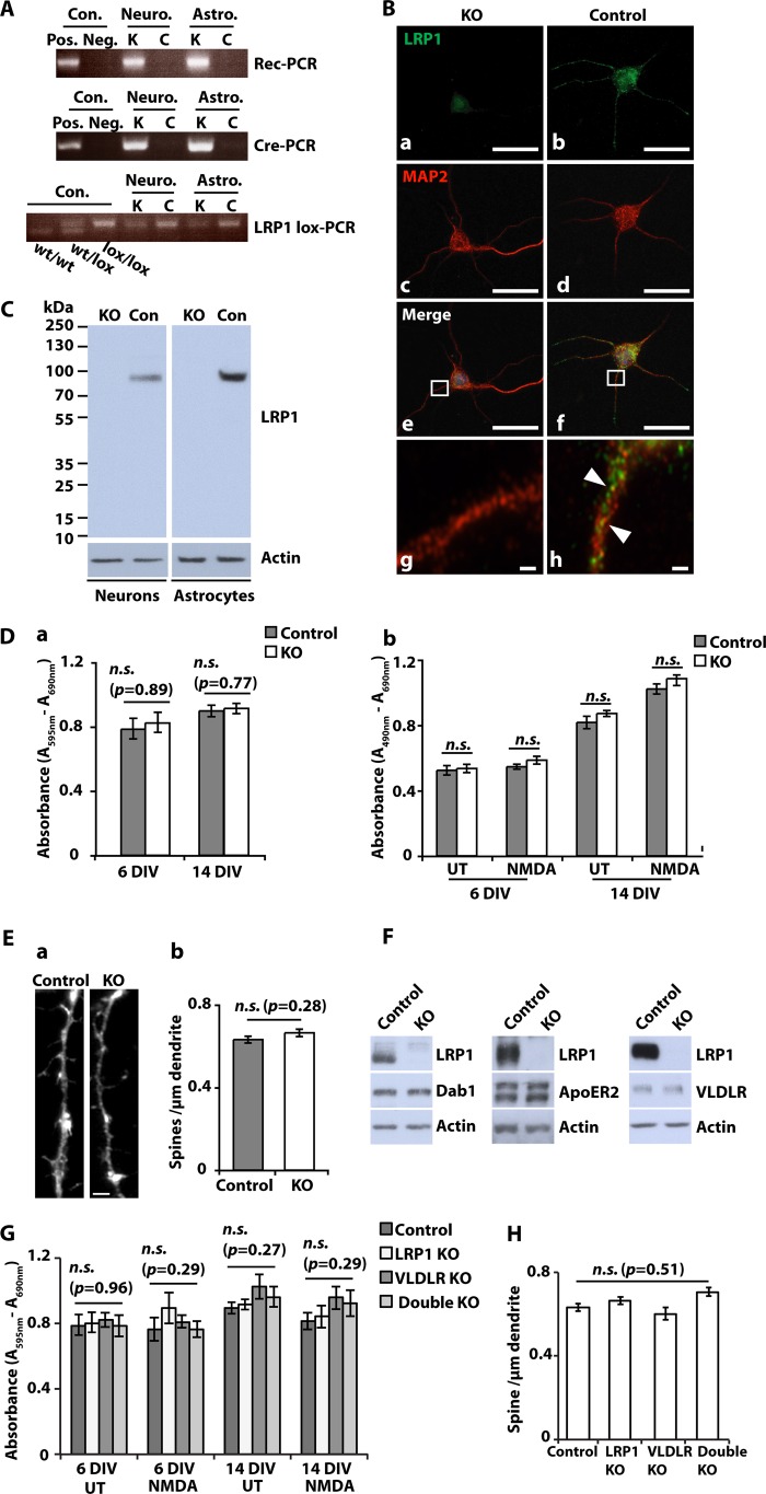
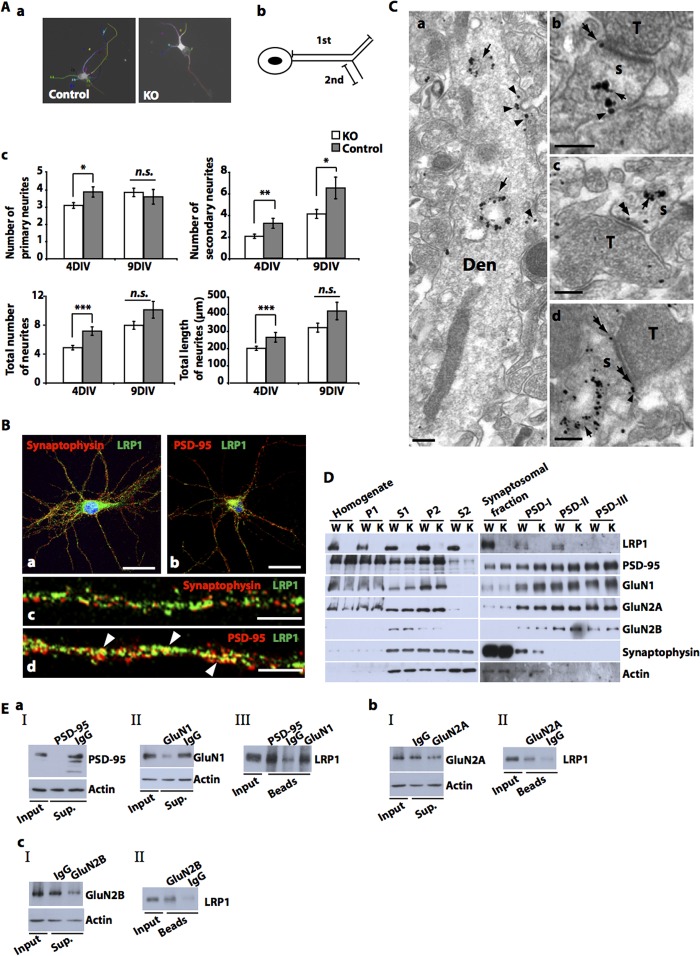
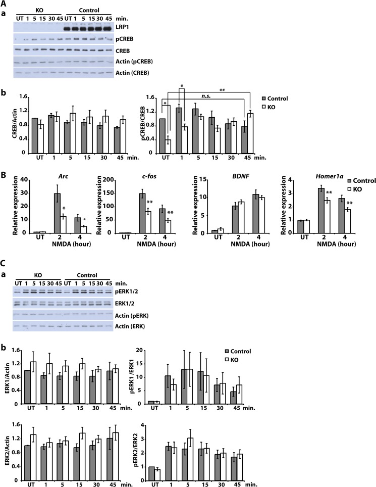
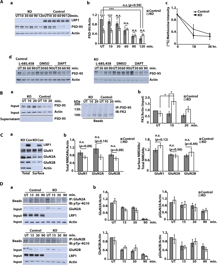
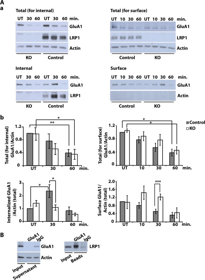
The lipoprotein receptor LRP1 is essential in neurons of the central nervous system, as was revealed by the analysis of conditional Lrp1-deficient mouse models. The molecular basis of its neuronal functions, however, is still incompletely understood. Here we show by immunocytochemistry, electron microscopy, and postsynaptic density preparation that LRP1 is located postsynaptically. Basal and NMDA-induced phosphorylation of the transcription factor cAMP-response element-binding protein (CREB) as well as NMDA target gene transcription are reduced in LRP1-deficient neurons. In control neurons, NMDA promotes γ-secretase-dependent release of the LRP1 intracellular domain (LRP1-ICD). However, pull-down and chromatin immunoprecipitation (ChIP) assays showed no direct interaction between the LRP1-ICD and either CREB or target gene promoters. On the other hand, NMDA-induced degradation of the postsynaptic scaffold protein PSD-95 was impaired in the absence of LRP1, whereas its ubiquitination was increased, indicating that LRP1 influences the composition of postsynaptic protein complexes. Accordingly, NMDA-induced internalization of the AMPA receptor subunit GluA1 was impaired in LRP1-deficient neurons. These results show a role of LRP1 in the regulation and turnover of synaptic proteins, which may contribute to the reduced dendritic branching and to the neurological phenotype observed in the absence of LRP1.

Keywords: Glutamate Receptors; Lipoprotein Receptor; Secretases; Signaling; Synapses.


Figures

FIGURE 1.
FIGURE 1.
Complete loss of LRP1 in NesCreLRPlox/lox neurons and astrocytes. A, primary cultured cortical neurons (Neuro.) and astrocytes (Astro.) were prepared from E15–E16 NesCreLRP1lox/lox (K) and E15–E16 LRP1lox/lox (C) mouse embryos. Genomic DNA was prepared from the cultures and examined by PCRs specific for the recombined LRP1 allele (Rec-PCR), the CRE transgene (Cre-PCR), and the non-recombined loxP-marked Lrp1 allele (LRP1lox-PCR), respectively. B, primary cultured cortical neurons were prepared from E15 NesCreLRP1lox/lox (KO) and E15 LRP1lox/lox (Control) mouse embryos and were seeded on glass coverslips. On DIV 5, immunocytochemical staining with antibodies directed against LRP1 and MAP-2 neuronal protein was performed. Scale bar, 25 μm. Inset, partial overlap of the LRP1 signal (green) with the MAP-2 signal (red) on dendrites of control neurons (arrowheads in h). Scale bars, 25 μm (a–f) and 1 μm (g and h). C, primary cultured cortical neurons and astrocytes were prepared from E15–E16 NesCreLRP1lox/lox (KO) and LRP1lox/lox (control (Con)) mouse embryos. Whole cell lysates were prepared on DIV 10 and analyzed by SDS-PAGE and immunoblotting with an antiserum directed against LRP1. Actin served as a loading control. D, primary cultured cortical neurons were prepared from E15–E16 NesCreLRP1lox/lox (KO) and E15–E16 LRP1lox/lox (control) mouse embryos. On DIV 6 and 14, MTT cell viability assays and LDH release cytotoxicity assays were performed. a, the results of MTT cell viability assays are shown as mean values. Error bars, S.E. Statistical analysis was done by Student's t test. n.s., not significant (p > 0.05, n = 4). b, LDH release was measured without (UT) and after 2 h of treatment with 50 μm NMDA. The results are shown as mean values ± S.E. Statistical analysis was done by Student's t test. n.s., not significant (p > 0.05, n = 4). E, primary cultured cortical neurons were prepared from E15–E16 NesCreLRP1lox/lox (KO) and E15-E16 LRP1lox/lox (Control) mouse embryos. On DIV 24, neurons were stained with phalloidin, and spine density was assessed on 20 dendrites/culture. a, representative spines and dendrite from KO and control. Scale bar, 5 μm. b, bars represent mean values ± S.E. from five independent cultures. Spine density did not differ significantly between KO and control neurons (statistical analysis was done by Student's t test). F, primary cultured cortical neurons from NesCreLRP1lox/lox (KO) and LRP1lox/lox (Control) embryos were prepared and lysed on 10–16 DIV with RIPA buffer. Whole cell lysates were subjected to SDS-PAGE, and expression levels of Dab1, ApoER2, and VLDLR were examined by Western blotting. Actin served as a loading control. Representative results of at least three independent experiments are shown. G, primary cultured cortical neurons were prepared from NesCreLRP1lox/lox (LRP1 KO), LRP1lox/lox (Control), LRP1lox/lox/VLDLR−/− (VLDLR KO), and NesCreLRP1lox/lox/VLDLR−/− (Double KO) mouse embryos. On DIV 6 and 14, MTT cell viability assays were performed. The cells were kept untreated or treated with 50 μm NMDA for 2 h before assays. Bars, means ± S.E. (Student's t test; n.s., not significant (p > 0.05), n = 4). H, primary cultured cortical neurons were prepared from NesCreLRP1lox/lox (LRP1 KO), LRP1lox/lox (Control), LRP1lox/lox/VLDLR−/− (VLDLR KO), and NesCreLRP1lox/lox/VLDLR−/− (Double KO) mouse embryos. Spine density on 24 DIV neurons was assessed as described above. Bars, means ± S.E. (error bars) (ANOVA; n.s., not significant (p > 0.05), n = 5).
FIGURE 2.
FIGURE 2.
Impaired neurite outgrowth of LRP1-deficient neurons and postsynaptic localization of LRP1. A, primary cultured cortical neurons were prepared from E15–E16 NesCreLRP1lox/lox (KO) and E15–E16 LRP1lox/lox (Control) mouse embryos and were seeded on glass coverslips. The total number of primary neurites, the total number of branches (secondary neurites), and the total number and length of neurites were assessed on DIV 4 and 9 after immunocytochemical staining with a MAP-2 antibody. a, representative micrographs of control and KO neurons. b, schematic representation of primary and secondary neurites. c, quantitative representation of the number of primary and secondary neurites per cell, average total number of neurites (primary neurites plus branches) per cell, and average total length of neurites. The average number of neurites/cell was obtained by dividing the total number of neurites by the number of analyzed cells (120 randomly chosen neurons from four independent experiments). Bars, means ± S.E. (error bars). Statistical analysis was done by Student's t test (*, p < 0.05; **, p < 0.01; ***, p < 0.001). B, primary cultured cortical neurons were prepared from E15–E16 wild type mouse embryos and were seeded on glass coverslips. On DIV 21, immunocytochemical analysis was performed with antibodies directed against LRP1 and synaptophysin (a and c) or PSD-95 (b and d), respectively, and documented by confocal microscopy. Co-localization of LRP1 and PSD-95 was observed (arrowheads in d). Scale bar, 25 μm (a and b) and 10 μm (c and d). C, electron microscopic analysis of the CA1 region (stratum lacunosum-moleculare) of 2–5-month-old LRP1lox/lox mice was performed by the pre-embedding immunogold method with an LRP1 antibody. Clusters of immunogold particles were found on intracellular membranes, such as lysosomes and endosomes (arrows in a–d) of dendritic compartments (Den, s) of presumed pyramidal cells. Immunoparticles were also found on the extrasynaptic (arrowheads in a, b, and d) and perisynaptic membrane (double arrows in b and d) of dendritic shafts (Den) and dendritic spines (s) and also appeared occasionally over the postsynaptic specialization at synapses between spines and axon terminals (T) (double arrowheads in c). No membrane-bound particles were detected in tissues obtained from NesCreLRP1lox/lox animals (not shown). Scale bars, 0.2 μm. D, synaptosomes were prepared from the brains of 3-month-old LRP1wt/wt (WT, W) and NesCreLRP1lox/lox (KO, K) mice, and postsynaptic proteins were extracted. Subsequent immunoblot analysis was done with antibodies against the proteins indicated. A representative photograph of three independent preparations is shown. P1, first pellet (nuclei and large debris); S1, supernatant 1 (cortex homogenate); P2, pellet 2 (crude synaptosomal membranes); S2, supernatant 2 (cytosol and light membranes); PSDI-III, postsynaptic fractions I–III (I, extracted with Triton X-100; II, additional extraction with SDS; III, additional extraction with Sarkosyl). E, primary cultured cortical neurons were prepared from E15 wild type mouse embryos. Detergent-soluble synaptosomal membrane fractions were prepared on DIV 10. Co-immunoprecipitation was performed using primary antibodies against PSD-95 (a), GluN1 (a), GluN2A (b), or GluN2B (c). After separation of precipitates by SDS-PAGE, immunoblotting with an antibody against LRP1 was performed. IgG was used as a negative control. Blots of IP supernatants (Sup.) (a, I and II; b, I; c, I) and of proteins extracted from beads (a, III; b, II; c, II) are represented. Representative blots of two independent experiments are shown.
FIGURE 3.
FIGURE 3.
NMDA-induced intracellular signaling and gene transcription are altered in LRP1-deficient neurons. A, primary cultured cortical neurons were prepared from E15–E16 NesCreLRP1lox/lox (KO) and E15 LRP1wt/lox (Control) mouse embryos. On DIV 7–8, neurons were treated with 50 μm NMDA for the time indicated or left untreated (UT). Then whole cell lysates were prepared and analyzed by immunoblotting with antibodies directed against LRP1, CREB, pCREB, and actin, which served as a loading control. Representative blots (a) and quantitative analysis (b) of four independent experiments are shown. Error bars, S.E. Statistical analysis was done by Student's t test (*, p < 0.05; **, p < 0.01, n = 4). B, primary cultured cortical neurons were prepared from E15–E16 NesCreLRP1lox/lox (KO) and E15 LRP1lox/lox (Control) mouse embryos. On DIV 5, neurons were treated with 50 μm NMDA for the time indicated. Then total RNA was isolated and analyzed by quantitative real-time PCR for the expression of NMDA target genes Arc, BDNF, c-fos, and Homer 1a. Expression levels were normalized to GAPDH and are represented as -fold induction over untreated control. Bars, means ± S.E. from at least seven independent experiments. Statistical analysis was done by Student's t test (*, p < 0.05; **, p < 0.01, n ≥ 7). C, primary cultured cortical neurons were prepared from E15–E16 NesCreLRP1lox/lox (KO) and E15 LRP1wt/lox (Control) mouse embryos. On DIV 7–8, neurons were treated with 50 μm NMDA for the time indicated or left untreated (UT). Then whole cell lysates were prepared and analyzed by immunoblotting with antibodies directed against LRP1, ERK1/2, phospho-ERK1/2 (pErk 1/2), and actin, which served as a loading control. Representative blots (a) and quantitative analysis (b) of four independent experiments are shown. Error bars, S.E. Statistical analysis was done by Student's t test (*, p < 0.05; **, p < 0.01, n = 4).
FIGURE 4.
FIGURE 4.
NMDA induces proteolytical processing of LRP1 in primary cultured cortical neurons. A (a), primary cultured cortical neurons were prepared from E15–E16 wild type mouse embryos. On DIV 5, neurons were pretreated with 1 μm DAPT for 2 h, and then 50 μm NMDA was added for the time indicated. DMSO was used as control (vehicle treatment). Whole cell lysates were prepared and analyzed by immunoblotting with antiserum directed against LRP1. Actin served as a loading control. b, primary cultured cortical neurons were prepared from E15–E16 wild type mouse embryos. On DIV 5, neurons were pretreated with 1 μm DAPT for 2 h, and then 50 μm NMDA, 50 μm AP5, 50 μm NMDA plus 50 μm AP5, or 50 μm N-methyl-l-aspartate was added for 12 h (UT, untreated control). Whole cell lysates were prepared and analyzed by immunoblotting with α-LRP1 antiserum. Actin served as a loading control. c, densitometric quantification of LRP1-CTF from b. Data are means ± S.E. (error bars) of five independent experiments. Statistical analysis was done by ANOVA (*, p < 0.05; **, p < 0.01; n.s., not significant). d, primary cultured cortical neurons were prepared from E15–E16 NesCreLRP1lox/lox (KO) and E15-E16 LRP1lox/lox (Control) mouse embryos. On DIV 5, control neurons were pretreated with 1 μm DAPT for 2 h, and then 50 μm NMDA was added for 12 h. KO neurons were pretreated with 1 μm DAPT for 2 h, and then 50 μm NMDA, 50 μm AP5, or 50 μm NMDA plus 50 μm AP5 was added for 12 h (UT, untreated control). Whole cell lysates were prepared and analyzed by immunoblotting with α-LRP1 antiserum. Actin served as a loading control. e, primary cultured cortical neurons were prepared from E15–E16 wild type mouse embryos. On DIV 5, neurons were pretreated with 1 μm DAPT for 2 h, and then 50 μm NMDA, 50 μm AP5, or 50 μm NMDA plus 50 μm AP5, 50 μm NMDA plus 1 μm ifenprodil, or 50 μm NMDA plus 10 μm ifenprodil was added for 12 h. DMSO was used as control (vehicle treatment). Whole cell lysates were prepared and analyzed by immunoblotting with α-LRP1 antiserum. Actin served as a loading control. B, primary cultured cortical neurons were prepared from E15 wild type mouse embryos. On DIV 5, neurons were pretreated with 10 μg/ml GST or 10 μg/ml GST-RAP for 30 min, and then 1 μm DAPT was added, followed by 50 μm NMDA for 12 h. Whole cell lysates were prepared and analyzed by immunoblotting with α-LRP1 antiserum. Actin served as a loading control. A representative blot (a) and quantitative analysis of four independent experiments (b) are shown. Error bars in b, S.E. Statistical analysis was done by ANOVA (*, p < 0.05). C, primary cultured cortical neurons were prepared from E15 wild type mouse embryos. On DIV 7, neurons were treated with 50 μm NMDA (N) for 4 h or left untreated (UT). After cross-linking with 1% paraformaldehyde, cell lysates were prepared, and DNA was sheared by sonification. Subsequently, chromatin immunoprecipitation was performed with an α-LRP1 antibody, rabbit IgG as negative control, or α-CREB as positive control. PCR amplification of c-fos promoter fragments was done with different primer sets to include transcription factor binding sites CRE −64, CRE −294, and CRE −342, and the serum-response element (SRE). The numbering refers to the fos gene in the Ensembl database (ENSMUSG00000021250).
FIGURE 5.
FIGURE 5.
Subcellular localization of NMDA receptor subunits and NMDA-dependent PSD-95 degradation in LRP1-deficient neurons. A (a and b), primary cultured cortical neurons were prepared from E15–E16 NesCreLRP1lox/lox (KO) and E15 LRP1wt/lox (Control) mouse embryos. On DIV 10–12, neurons were treated with NMDA (20 μm, 3 min), and whole cell lysates were prepared after the time indicated. Immunoblot analysis was performed with α-LRP1 and α-PSD-95 antibodies. Actin served as a loading control. Representative blot (a) and quantitative analysis (b) of six independent experiments are shown. Error bars, S.E. Statistical analysis was done by Student's t test (***, p < 0.001). c, cortical neurons were cultured from NesCreLRP1lox/lox (KO) and LRP1wt/lox (Control) mouse embryos and pulse-labeled with [35S]methionine on DIV 14–16. The cultures were chased with unlabeled methionine for 0, 18, or 36 h. PSD-95 was immunoprecipitated from the lysates, and samples were analyzed by SDS-PAGE and subsequent autoradiography. Densitometric analysis of the results is shown. Error bars, S.E. No statistical significance between KO and control was found by Student's t test (n = 3). d, cortical neurons from E15 LRP1lox/lox (Control) and NesCre/LRP1lox/lox (KO) animals were prepared. On DIV 12, neurons were pretreated with 1 μm DAPT, 10 μm L-685,458, or DMSO (1:1000; vehicle) for 2 h and were then further treated with NMDA (20 μm, 3 min). After the NMDA treatment, the cells were briefly washed with fresh medium and then cultured in fresh medium for the time indicated. Whole cell lysates were subjected to Western blotting to detect PSD-95 and actin. Actin served as a loading control. A representative blot of two independent experiments is shown. B, primary cultured cortical neurons were prepared from E15–E16 NesCreLRP1lox/lox (KO) and E15 LRP1wt/lox (Control) mouse embryos. On DIV 10–12, neurons were treated with NMDA (20 μm, 3 min). After further incubation for 10 or 20 min, whole cell lysates were prepared and subjected to immunoprecipitation (IP) with a PSD-95 antibody. Lysates, supernatants, and beads were analyzed by immunoblotting (IB) with the antibodies indicated. FK2, an antibody against mono- and polyubiquitin. Representative blots (a) and quantitative analysis (b) of five independent experiments are shown. Statistical analysis was done by Student's t test (*, p < 0.05). C, primary cultured cortical neurons were prepared from E15–E16 NesCreLRP1lox/lox (KO) and E15 LRP1wt/lox (Con) mouse embryos. On DIV 10–12, cell surface proteins were biotinylated. Subsequently, whole cell lysates were prepared, and biotinylated proteins were precipitated with Neutravidin. Aliquots from the lysates and precipitated proteins were analyzed by immunoblotting with anti-LRP1, -GluN1, -GluN2A, and -GluN2B antibodies. Actin served as a loading control. Representative blot (a) and quantitative analysis (b) of at least seven independent experiments are shown. Proteins in the lysate (total) were normalized to actin, and cell surface proteins were measured as biotinylated protein/total protein. Statistical analysis was done by Student's t test. n.s., not significant (p ≥ 0.05). D, primary cultured cortical neurons were prepared from E15–E16 NesCreLRP1lox/lox (KO) and E15 LRP1wt/lox (Control) mouse embryos. On DIV 10–12, cultures were treated with 50 μm NMDA for the time indicated or left untreated (UT). Whole cell lysates were prepared and subjected to immunoprecipitation with either α-GluN2A or α-GluN2B antibody. Input, supernatants, and precipitated proteins (beads) were analyzed by immunoblotting with the antibodies indicated. Actin served as a loading control. Representative blots (a) and quantitative analysis (b) of three independent experiments are shown. Statistical analysis was done by Student's t test (p ≥ 0.05).
FIGURE 6.
FIGURE 6.
NMDA-induced GluA1 turnover is altered in LRP1-deficient neurons. A, primary cultured cortical neurons were prepared from E15 NesCreLRP1lox/lox (KO) and E15 LRP1wt/lox (Control) mouse embryos. Internal and surface GluA1 were analyzed under NMDA treatment. To measure internal GluA1, on DIV 10–12, cell surface proteins were biotinylated, and then neurons were treated with NMDA (20 μm, 3 min). Neurons were washed and incubated in fresh medium for the time indicated to allow endocytosis of GluA1. After stripping off cell surface biotin, whole cell lysates were prepared, and biotinylated proteins were precipitated with Neutravidin. To measure surface GluA1, on DIV 10–12, neurons were first treated with NMDA (20 μm, 3 min). Then neurons were washed and incubated in fresh medium for the time indicated. Subsequently, cell surface proteins were biotinylated. Whole cell lysates were prepared, and biotinylated proteins were precipitated with Neutravidin. Aliquots from the lysates and supernatants were analyzed by immunoblotting with anti-LRP1 and anti-GluA1 antibodies. Actin served as a loading control and as an indicator of purity for surface samples. Shown are representative Western blots of total and internalized or surface GluA1, respectively (a), and quantitative analysis of total (for internalized analysis) GluA1 (b), internalized GluA1 (b), total (for surface analysis) GluA1 (b), and surface GluA1 (b). All values are normalized to actin from total lysate. Bars, means ± S.E. (error bars) (Student's t test; *, p < 0.05; **, p < 0.01; ***, p < 0.001, n = 4 for internal GluA1, n = 3 for surface GluA1). B, detergent-soluble synaptosomal membrane fractions from DIV 10–12 wild type cortical neurons were prepared and subjected to immunoprecipitation with an anti-GluA1 antibody or rabbit-IgG as a negative control. Precipitated proteins were examined with an antibody against LRP1 by immunoblotting. Representative blots of five independent experiments are shown.

References

    1. Lillis A. P., Van Duyn L. B., Murphy-Ullrich J. E., Strickland D. K. (2008) LDL receptor-related protein 1. Unique tissue-specific functions revealed by selective gene knockout studies. Physiol. Rev. 88, 887–918 - PMC - PubMed
    1. May P., Rohlmann A., Bock H. H., Zurhove K., Marth J. D., Schomburg E. D., Noebels J. L., Beffert U., Sweatt J. D., Weeber E. J., Herz J. (2004) Neuronal LRP1 functionally associates with postsynaptic proteins and is required for normal motor function in mice. Mol. Cell. Biol. 24, 8872–8883 - PMC - PubMed
    1. Liu Q., Trotter J., Zhang J., Peters M. M., Cheng H., Bao J., Han X., Weeber E. J., Bu G. (2010) Neuronal LRP1 knockout in adult mice leads to impaired brain lipid metabolism and progressive, age-dependent synapse loss and neurodegeneration. J. Neurosci. 30, 17068–17078 - PMC - PubMed
    1. Pietrzik C. U., Busse T., Merriam D. E., Weggen S., Koo E. H. (2002) The cytoplasmic domain of the LDL receptor-related protein regulates multiple steps in APP processing. EMBO J. 21, 5691–5700 - PMC - PubMed
    1. Kang D. E., Pietrzik C. U., Baum L., Chevallier N., Merriam D. E., Kounnas M. Z., Wagner S. L., Troncoso J. C., Kawas C. H., Katzman R., Koo E. H. (2000) Modulation of amyloid β-protein clearance and Alzheimer's disease susceptibility by the LDL receptor-related protein pathway. J. Clin. Invest. 106, 1159–1166 - PMC - PubMed

Publication types

MeSH terms

LinkOut - more resources