. 2013 Jul 26;288(30):21824-35.
doi: 10.1074/jbc.M113.472654. Epub 2013 Jun 12.

An out-of-frame overlapping reading frame in the ataxin-1 coding sequence encodes a novel ataxin-1 interacting protein

Affiliations

An out-of-frame overlapping reading frame in the ataxin-1 coding sequence encodes a novel ataxin-1 interacting protein

Danny Bergeron et al. J Biol Chem. .

Abstract

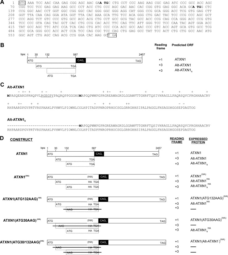
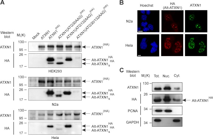
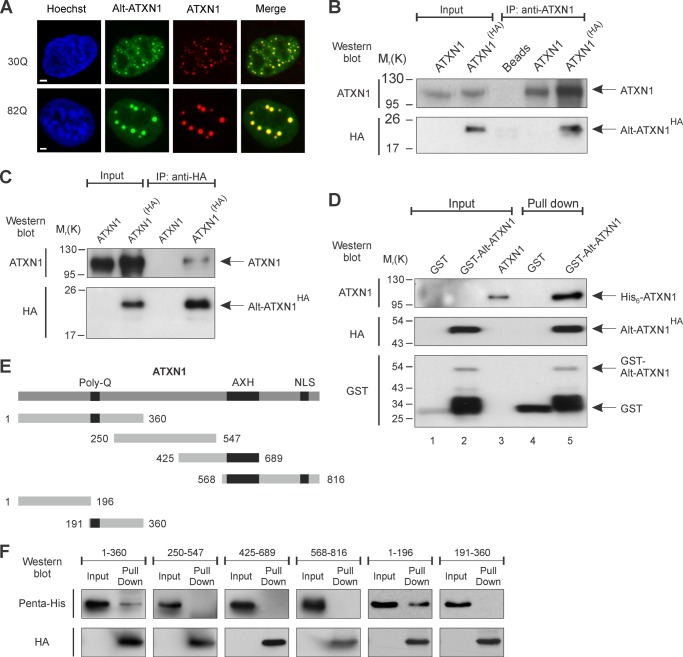
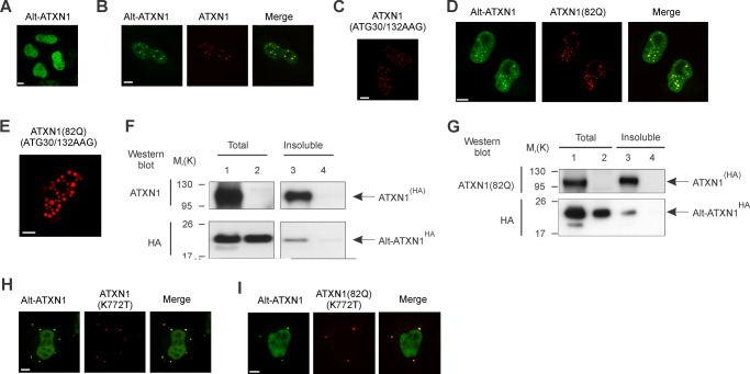
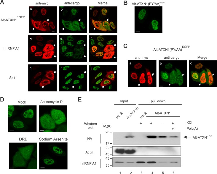
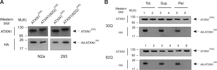
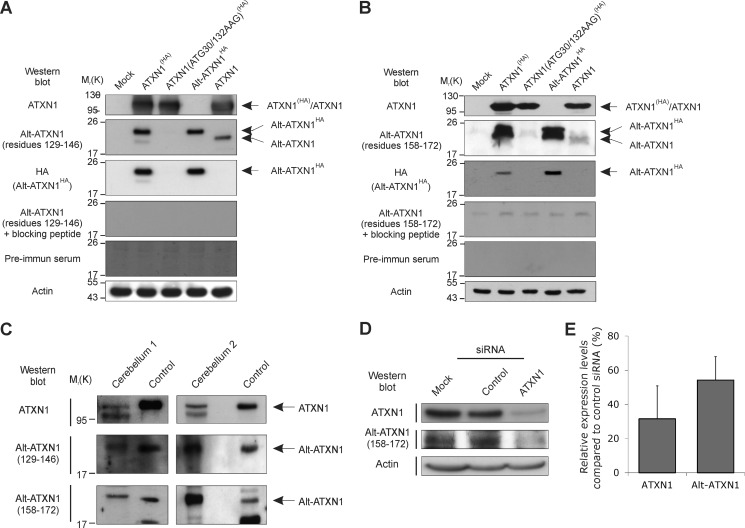
Spinocerebellar ataxia type 1 is an autosomal dominant cerebellar ataxia associated with the expansion of a polyglutamine tract within the ataxin-1 (ATXN1) protein. Recent studies suggest that understanding the normal function of ATXN1 in cellular processes is essential to decipher the pathogenesis mechanisms in spinocerebellar ataxia type 1. We found an alternative translation initiation ATG codon in the +3 reading frame of human ATXN1 starting 30 nucleotides downstream of the initiation codon for ATXN1 and ending at nucleotide 587. This novel overlapping open reading frame (ORF) encodes a 21-kDa polypeptide termed Alt-ATXN1 (Alternative ATXN1) with a completely different amino acid sequence from ATXN1. We introduced a hemagglutinin tag in-frame with Alt-ATXN1 in ATXN1 cDNA and showed in cell culture the co-expression of both ATXN1 and Alt-ATXN1. Remarkably, Alt-ATXN1 colocalized and interacted with ATXN1 in nuclear inclusions. In contrast, in the absence of ATXN1 expression, Alt-ATXN1 displays a homogenous nucleoplasmic distribution. Alt-ATXN1 interacts with poly(A)(+) RNA, and its nuclear localization is dependent on RNA transcription. Polyclonal antibodies raised against Alt-ATXN1 confirmed the expression of Alt-ATXN1 in human cerebellum expressing ATXN1. These results demonstrate that human ATXN1 gene is a dual coding sequence and that ATXN1 interacts with and controls the subcellular distribution of Alt-ATXN1.

Keywords: Alternative Translation Initiation; Ataxia; Dual Coding Gene; Gene Expression; Nucleus; Overlapping Reading Frame; RNA-Binding Protein; Spinocerebellar Ataxia Type 1; Translation.


Figures

FIGURE 1.
FIGURE 1.
An alternative ORF overlaps ATXN1 coding sequence in the +3 reading frame. A, shown is the human ATXN1 DNA sequence starting from the ATXN1 start codon (boxed) up to the Alt-ATXN1 stop codon between bp 585–587 (boxed). For clarity purposes, the remaining ATXN1 CDS (up to bp 2448) is not shown. Initiation codons for the long and short Alt-ATXN1 isoforms are shown in bold. B, a diagram of ATXN1 CDS shows different features of ATXN1, including the localization of the long (Alt-ATXN1) and short (Alt-ATXN1S) alternative ATXN1 protein isoforms. Note that Alt-ATXN1 stop codon is located just upstream of the (CAG)n repeats domain. C, shown is the amino acid sequence of Alt-ATXN1. The N-terminal methionine residues of Alt-ATXN1 and Alt-ATXN1S isoforms are labeled in bold. A putative PY-NLS with the sequence RGEGPY is underlined. D, shown is the strategy used to detect Alt-ATXN1 by introducing an HA tag at the C terminus of Alt-ATXN1. In ATXN1(HA), ATXN1(ATG132AAG)(HA), ATXN1(ATG30AAG)(HA) , and ATXN1(ATG30/132AAG)(HA), an HA tag (gray box), was inserted at the C terminus of Alt-ATXN1. The parentheses surrounding the HA in the ATXN1 reading frame represent the fact that the HA epitope sequence is encoded in the Alt-ATXN1 reading frame and is, therefore, undetected if expressed from the ATG codon at bp 1 of the ATXN1 CDS. ATXN1(ATG132AAG)(HA) and ATXN1(ATG30AAG)(HA) are identical to ATXN1(HA) except that the ATG codons at bp 132 and 30 have been mutated to AAG, respectively. ATXN1(ATG30/132AAG)(HA) is identical to ATXN1(HA) except that both ATG codons at bp 30 and 132 have been mutated to AAG.
FIGURE 2.
FIGURE 2.
Expression of Alt-ATXN1. A, shown is a Western blot against ATXN1 and Alt-ATXN1 (HA epitope) in HEK293, N2a cells, and HeLa cells mock-transfected or transfected with ATXN1, ATXN1(HA), ATXN1(ATG132AAG)(HA), ATXN1(ATG30AAG)(HA), or ATXN1(ATG30/132AAG)(HA) constructs. Molecular mass markers in kDa are indicated on the left. This experiment is representative of three independent experiments in each cell line. B, shown is confocal microscopy of cells transfected with ATXN1(HA) and immunostained with anti-HA (red channel) and anti-ATXN1 (green channel) antibodies. Nuclei were stained with Hoechst (blue channel). Scale bar, 5 μm. C, total extracts (Tot.), nucleus (Nuc.), and cytoplasmic (Cyt.) fractions from HEK293 cells expressing ATXN1(HA) were immunoblotted for Alt-ATXN1HA, the nuclear marker proliferating cell nuclear antigen, and the cytosolic marker GAPDH. Molecular mass markers in kDa are indicated on the left. This experiment is representative of two independent experiments.
FIGURE 3.
FIGURE 3.
Alt-ATXN1 and ATXN1 colocalize in nuclear inclusions and interact. A, HeLa cells were co-transfected with Alt-ATXN1EGFP (green channel) and ATXN1DsRed2 (red channel). Nuclei were stained with Hoechst (blue channel). ATXN1 contained 30 Gln (Q) repetitions or 82 Gln repetitions as indicated. Scale bar, 2 μm. B and C, co-immunoprecipitation (IP) experiments with anti-ATXN1 (B) or anti-HA (C) antibodies were performed using lysates from N2a cells expressing ATXN1 or ATXN1(HA). This experiment is representative of four independent experiments. D, binding assays were carried out with purified glutathione-Sepharose-bound GST (Input, lane 1) or GST-Alt-ATXN1HA (Input, lane 2) incubated with purified recombinant His6-ATXN1 (Input, lane 3). GST-Alt-ATXN1HA present in the binding reaction was detected using antibodies against HA or GST, and binding of His6-ATXN1 was detected with antibodies against ATXN1 (lane 5). GST was detected with antibodies against GST. Molecular mass markers in kDa are indicated on the left. E, shown is a schematic representation of the different ATXN1 fragments used to determine the region responsible for its interaction with Alt-ATXN1 (48). Residues 1–360 contain the polyglutamine repetitions (Poly-Q), 250–547 contain the SAD (self-associating domain), 425–689 contain the SAD and the AXH (ATXN1/HMG-box protein 1) domain, and 568–816 contain the AXH domain and the NLS region of ATXN1. Fragments 1–360 were subsequently divided into two subdomains, residues 1–196 and 191–360). F, binding assays with purified glutathione-Sepharose-bound GST-Alt-ATXN1HA incubated with purified recombinant His6-ATXN1 fragments are shown. GST-Alt-ATXN1HA present in the binding reaction was detected using antibodies against HA, and fragments of ATXN1 were detected with antibodies against pentahistidine.
FIGURE 4.
FIGURE 4.
ATXN1 and ATXN1(82Q) recruit Alt-ATXN1 inside inclusions. A–E, HeLa cells were transfected with Alt-ATXN1EGFP (A), Alt-ATXN1EGFP and ATXN1DsRed2 (B), ATXN1(ATG30/132AAG)DsRed2 (C), Alt-ATXN1EGFP and ATXN1(82Q)DsRed2 (D), or ATXN1(82Q)(ATG30/132AAG)DsRed2 (E) and observed by confocal microscopy. F and G, a Western blot with anti-ATXN1 and anti-HA antibodies shows the proportion of ATXN1(HA) and Alt-ATXN1HA in a total cell lysate (lanes 1 and 2) and in the insoluble fraction (lanes 3 and 4) from HEK293 cells expressing ATXN1(HA) (F, lanes 1 and 3), ATXN1(82Q)(HA) (G, lanes 1 and 3), or Alt-ATXN1 (F and G, lanes 2 and 4). Molecular mass markers in kDa are indicated on the left. H and I, HeLa cells were co-transfected with Alt-ATXN1EGFP and ATXN1(K772T)DsRed2 or Alt-ATXN1EGFP and ATXN1(82Q)(K772T)DsRed2, fixed, and observed by confocal microscopy. Scale bar, 10 μm.
FIGURE 5.
FIGURE 5.
Alt-ATXN1 is imported into the nucleus by Kapβ2 by a transcription-dependent mechanism and binds RNA. A, HeLa cells were co-transfected with Alt-ATXN1EGFP and myc-MBP-M9M (a–c) or transfected with myc-MBP-M9M only (d–i). Cells were processed for immunofluorescence with antibodies against myc-MBP-M9M (a, d, and g), endogenous hnRNP A1 (e), and endogenous Sp1 (h). Arrows indicate individual cells overexpressing myc-MBP-M9M at high levels. Scale bar, 10 μm. B, HeLa cells were transfected with Alt-ATXN1(PY/AA)EGFP for 24 h and visualized by confocal microscopy. C, HeLa cells were co-transfected with Alt-ATXN1(PY/AA)EGFP and myc-MBP-M9M. Cells were fixed 24 h post-transfection and processed for immunofluorescence with antibodies against myc. D, shown is confocal microscopy of Alt-ATXN1EGFP in mock-treated HeLa cells (Mock) and treated with actinomycin D, 5,6-dichlorobenzimidazole riboside (DRB), or with sodium arsenite, as indicated. Scale bar, 5 μm. E, lysates from mock-transfected cells or cells expressing Alt-ATXN1HA (lanes 1 and 2, respectively) were incubated with oligo(dT)-cellulose beads. After extensive washing, proteins were eluted (lanes 3–6). In control experiments, cells were lysed in the absence of KCl (lane 5), or poly(A) was bound to the beads before pulldown (lane 6). Alt-ATXN1HA, hnRNP A1 and actin were detected by Western blotting. Molecular mass markers in kDa are indicated on the left.
FIGURE 6.
FIGURE 6.
CAG repeats do not modify Alt-ATXN1 expression, and Alt-ATXN1 expression does not modify ATXN1 solubility. A, shown is a Western blot against ATXN1 and Alt-ATXN1 in lysates from N2a or HEK 293 cells transfected with ATXN1(HA) (30Q) or ATXN1(82Q)(HA) constructs. Molecular mass markers in kDa are indicated on the left. B, a Western blot with anti-ATXN1 and anti-HA antibodies shows the distribution of ATXN1(HA) and Alt-ATXN1HA in a total cell lysate (Tot., lanes 1 and 2), in the soluble fraction (Sup., lanes 3 and 4), and in the insoluble fraction (Pel., lanes 5 and 6) from HEK293 cells expressing ATXN1(HA) (upper panels; lanes 1, 3, and 5), ATXN1(ATG30/132AAG)(HA) (upper panels; lanes 2, 4, and 6), ATXN1(82Q)(HA) (lower panels; lanes 1, 3, and 5), or ATXN1(82Q)(ATG30/132AAG)(HA) (lower panels; lanes 2, 4, and 6).
FIGURE 7.
FIGURE 7.
Alt-ATXN1 is endogenously expressed. A and B, validation of polyclonal antibodies against Alt-ATXN1 is shown. N2a cells were either untransfected (Mock) or transfected with ATXN1(HA), ATXN1(ATG30/132AAG)(HA), Alt-ATXN1HA, or ATXN1. Lysates were probed for Alt-ATXN1 and Alt-ATXN1HA using polyclonal antibodies raised against residues 129CQLHPITADPPNRQPRHQ146 (A) or 158HSIPALPAGGLFHSAG172 (B) of Alt-ATXN1 or anti-HA antibodies. The same lysates were also probed with the anti-Alt-ATXN1 antibodies blocked with the immunogenic peptides or with the animal's pre-immune serum to demonstrate the specificity of the antibodies. Equal loading was assessed with an anti-β-actin antibody. C, two human cerebellum homogenates, cerebellum 1 (200 μg) and cerebellum 2 (100 μg), were probed for Alt-ATXN1 using two polyclonal antibodies raised against residues 129CQLHPITADPPNRQPRHQ146 or 158HSIPALPAGGLFHSAG172. A band at the same molecular weight as the one seen in 7.5-μg lysates from cells transfected with human ATXN1 (control) was detected. Molecular mass markers in kDa are indicated on the left. D, endogenous Alt-ATXN1 was detected in glioblastoma cells, which express a high level of ATXN1. A siRNA against the 3′ of the coding sequence of ATXN1 greatly reduced the expression of ATXN1 and Alt-ATXN1, whereas the control siRNA had no effect on the expression of both proteins. E, densitometric analysis of siRNA treatment showed a decrease (45–70%) in expression of both ATXN1 and Alt-ATXN1 after siRNA treatment in glioblastoma cells. Value is expressed as the mean value (±S.D.) from three independent experiments.

References

    1. Orr H. T., Chung M. Y., Banfi S., Kwiatkowski T. J., Jr, Servadio A., Beaudet A. L., McCall A. E., Duvick L. A., Ranum L. P., Zoghbi H. Y. (1993) Expansion of an unstable trinucleotide CAG repeat in spinocerebellar ataxia type 1. Nat. Genet. 4, 221–226 - PubMed
    1. Chung M. Y., Ranum L. P., Duvick L. A., Servadio A., Zoghbi H. Y., Orr H. T. (1993) Evidence for a mechanism predisposing to intergenerational CAG repeat instability in spinocerebellar ataxia type I. Nat. Genet. 5, 254–258 - PubMed
    1. Cummings C. J., Mancini M. A., Antalffy B., DeFranco D. B., Orr H. T., Zoghbi H. Y. (1998) Chaperone suppression of aggregation and altered subcellular proteasome localization imply protein misfolding in SCA1. Nat. Genet. 19, 148–154 - PubMed
    1. Matilla A., Roberson E. D., Banfi S., Morales J., Armstrong D. L., Burright E. N., Orr H. T., Sweatt J. D., Zoghbi H. Y., Matzuk M. M. (1998) Mice lacking ataxin-1 display learning deficits and decreased hippocampal paired-pulse facilitation. J. Neurosci. 18, 5508–5516 - PMC - PubMed
    1. Crespo-Barreto J., Fryer J. D., Shaw C. A., Orr H. T., Zoghbi H. Y. (2010) Partial loss of ataxin-1 function contributes to transcriptional dysregulation in spinocerebellar ataxia type 1 pathogenesis. PLoS Genet. 6, e1001021. - PMC - PubMed

Publication types

MeSH terms

Associated data