. 2013 Sep;51(9):2822-9.
doi: 10.1128/JCM.01397-13. Epub 2013 Jun 12.

Multiplex real-time PCR for detection of Campylobacter, Salmonella, and Shigella

Affiliations

Multiplex real-time PCR for detection of Campylobacter, Salmonella, and Shigella

F Barletta et al. J Clin Microbiol. 2013 Sep.

Abstract

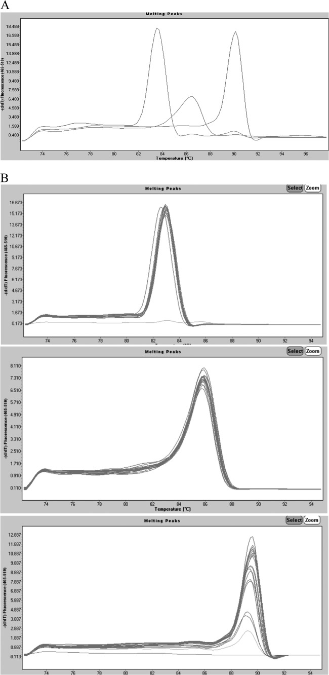
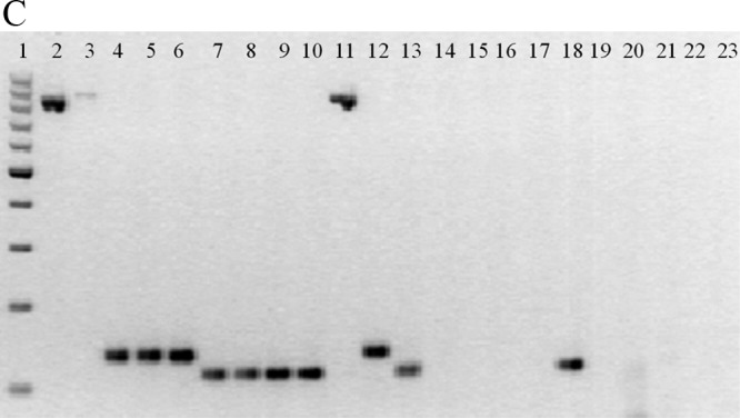
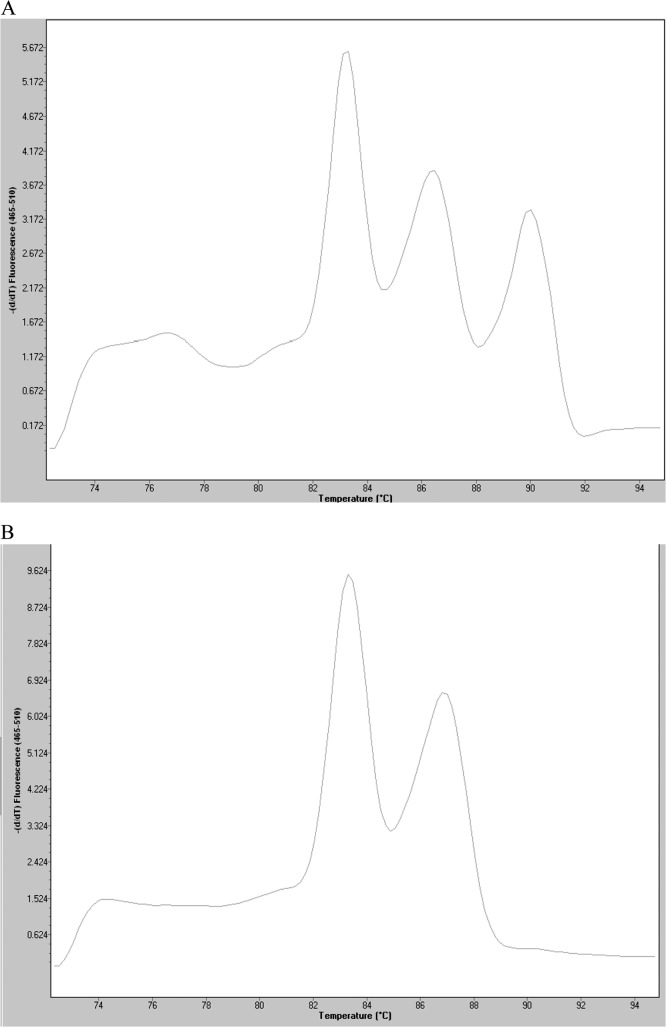
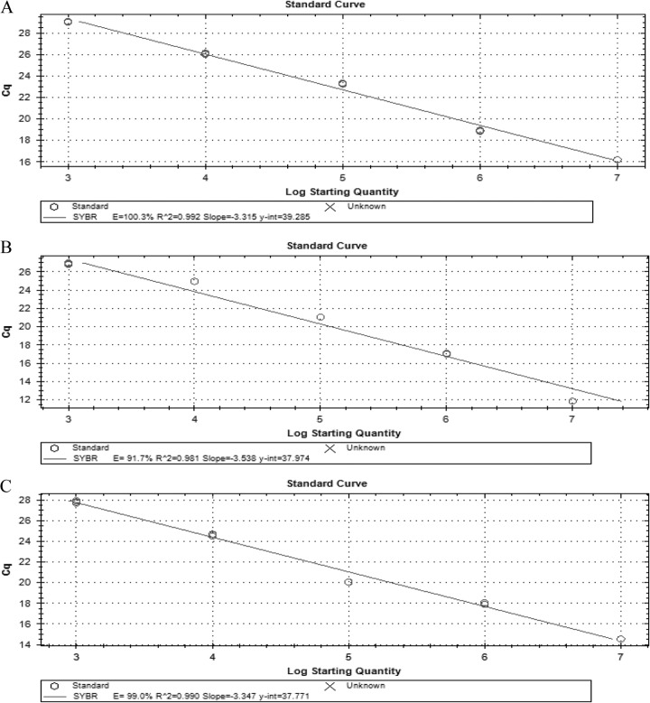
Infectious diarrhea can be classified based on its clinical presentation as noninflammatory or inflammatory disease. In developing countries, among inflammatory diarrhea cases, Shigella is the most common cause, followed by Campylobacter and Salmonella. Because the time frame in which treatment choices must be made is short and conventional stool cultures lack good sensitivity, there is a need for a rapid, sensitive, and inexpensive detection technique. The purpose of our study was to develop a multiplex real-time PCR procedure to simultaneously identify Campylobacter spp., Salmonella spp., and Shigella spp. Primers were designed to amplify the invA, ipaH, and 16S rRNA genes simultaneously in a single reaction to detect Salmonella, Shigella, and Campylobacter, respectively. Using this approach, we correctly identified 102 of 103 strains of the targeted enteropathogens and 34 of 34 other pathogens. The melting temperatures were 82.96 ± 0.05 °C for invA, 85.56 ± 0.28 °C for ipaH, and 89.21 ± 0.24 °C for 16S rRNA. The limit of accurate quantification for the assay in stool samples was 10(4) CFU g(-1); however, the limit of detection was 10(3) CFU g(-1). This assay is a simple, rapid, inexpensive, and reliable system for the practical detection of these three enteropathogens in clinical specimens.


Figures

Fig 1
Fig 1
(A) Real-time PCR simultaneously detects three different genes. Data from individual tubes, each containing the ATCC strains S. enteritidis 13076, S. flexneri 12022, and C. jejuni subsp. jejuni 33560, are shown in a single graph so that the separation between individual amplicon melting curves is illustrated (from left to right, invA, ipaH, and 16S rRNA). The y axis (fluorescence) represents the negative derivative of fluorescence over temperature versus temperature. (B) Melting curves of DNA isolated from pure cultures of (top) Salmonella (n = 26), (center) Shigella (n = 49), and (bottom) Campylobacter (n = 41). Curves are superimposed to show the reproducibility within species. The y axis (fluorescence) represents the negative derivative of fluorescence over temperature versus temperature. In the top and bottom panels, the horizontal lines at the bottom represent negative controls. In the bottom panel, the lowest peak represents a positive strain. (C) Agarose gel (2%) of amplicons of representative strains from the multiplex real-time PCR. From left to right, 1, 100-bp molecular weight ladder; 2, C. coli; 3, C. jejuni; 4, S. enteritidis; 5, S. infantis; 6, Salmonella spp.; 7, S. boydii; 8, S. dysenteriae; 9, S. flexneri; 10, S. sonnei; 11, C. jejuni subsp. jejuni ATCC 33560; 12, S. enteritidis ATCC 13076; 13, S. flexneri ATCC 12022; 14, diffusely adherent E. coli; 15, enteroaggregative E. coli; 16, enteropathogenic E. coli; 17, enterotoxigenic E. coli; 18, enteroinvasive E. coli; 19, Shiga-like toxin producer E. coli; 20, E. coli K-12; 21, Pseudomonas aeruginosa; 22, Klebsiella pneumoniae; 23, Proteus mirabilis.
Fig 1
Fig 1
(A) Real-time PCR simultaneously detects three different genes. Data from individual tubes, each containing the ATCC strains S. enteritidis 13076, S. flexneri 12022, and C. jejuni subsp. jejuni 33560, are shown in a single graph so that the separation between individual amplicon melting curves is illustrated (from left to right, invA, ipaH, and 16S rRNA). The y axis (fluorescence) represents the negative derivative of fluorescence over temperature versus temperature. (B) Melting curves of DNA isolated from pure cultures of (top) Salmonella (n = 26), (center) Shigella (n = 49), and (bottom) Campylobacter (n = 41). Curves are superimposed to show the reproducibility within species. The y axis (fluorescence) represents the negative derivative of fluorescence over temperature versus temperature. In the top and bottom panels, the horizontal lines at the bottom represent negative controls. In the bottom panel, the lowest peak represents a positive strain. (C) Agarose gel (2%) of amplicons of representative strains from the multiplex real-time PCR. From left to right, 1, 100-bp molecular weight ladder; 2, C. coli; 3, C. jejuni; 4, S. enteritidis; 5, S. infantis; 6, Salmonella spp.; 7, S. boydii; 8, S. dysenteriae; 9, S. flexneri; 10, S. sonnei; 11, C. jejuni subsp. jejuni ATCC 33560; 12, S. enteritidis ATCC 13076; 13, S. flexneri ATCC 12022; 14, diffusely adherent E. coli; 15, enteroaggregative E. coli; 16, enteropathogenic E. coli; 17, enterotoxigenic E. coli; 18, enteroinvasive E. coli; 19, Shiga-like toxin producer E. coli; 20, E. coli K-12; 21, Pseudomonas aeruginosa; 22, Klebsiella pneumoniae; 23, Proteus mirabilis.
Fig 2
Fig 2
Mixed infection detected in a pool of colonies corresponding to S. enteritidis ATCC 13076 (invA), S. flexneri ATCC 12022 (ipaH), and C. jejuni subsp. jejuni ATCC 33560 (16S rRNA) (A) and another corresponding to S. enteritidis ATCC 13076 (invA) and S. flexneri ATCC 12022 (ipaH) (B).
Fig 3
Fig 3
In stool samples, the qPCR detected serial dilutions of 107 to 103 CFU g−1 of Salmonella (A), Shigella (B), and Campylobacter (C).
Fig 4
Fig 4
In stool samples, the qPCR showed an efficiency of 100.3%, 91.7%, and 99.0% for the invA (A), ipaH (B), and 16S rRNA (C) genes, respectively, with correlation coefficients of 0.99, 0.98, and 0.99, respectively.

References

    1. Boschi-Pinto C, Velebit L, Shibuya K. 2008. Estimating child mortality due to diarrhoea in developing countries. Bull. World Health 86:710–717 - PMC - PubMed
    1. Navaneethan U, Giannella RA. 2008. Mechanisms of infectious diarrhea. Nat. Clin. Pract. Gastroenterol. Hepatol. 5:637–647 - PubMed
    1. Pfeiffer ML, DuPont HL, Ochoa TJ. 2012. The patient presenting with acute dysentery—a systematic review. J. Infect. 64:374–386 - PubMed
    1. Pawlowski SW, Warren CA, Guerrant R. 2009. Diagnosis and treatment of acute or persistent diarrhea. Gastroenterology 136:1874–1886 - PMC - PubMed
    1. Guion CE, Ochoa TJ, Walker CM, Barletta F, Cleary TG. 2008. Detection of diarrheagenic Escherichia coli by use of melting-curve analysis and real-time multiplex PCR. J. Clin. Microbiol. 46:1752–1757 - PMC - PubMed

Publication types

MeSH terms

LinkOut - more resources