. 2013 Sep;11(9):1078-87.
doi: 10.1158/1541-7786.MCR-12-0637. Epub 2013 Jun 12.

Snail cooperates with KrasG12D to promote pancreatic fibrosis

Affiliations

Snail cooperates with KrasG12D to promote pancreatic fibrosis

Mario A Shields et al. Mol Cancer Res. 2013 Sep.

Abstract

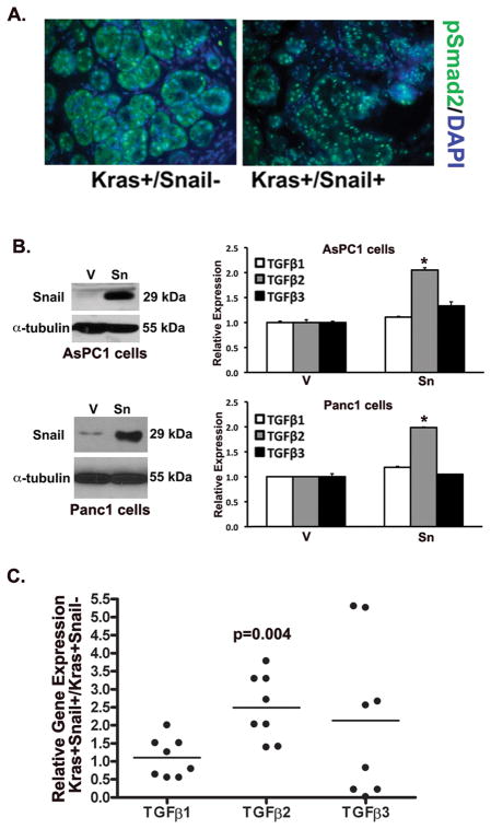
Patients with pancreatic cancer, which is characterized by an extensive collagen-rich fibrotic reaction, often present with metastases. A critical step in cancer metastasis is epithelial-to-mesenchymal transition (EMT), which can be orchestrated by the Snail family of transcription factors. To understand the role of Snail (SNAI1) in pancreatic cancer development, we generated transgenic mice expressing Snail in the pancreas. Because chronic pancreatitis can contribute to pancreatic cancer development, Snail-expressing mice were treated with cerulein to induce pancreatitis. Although significant tissue injury was observed, a minimal difference in pancreatitis was seen between control and Snail-expressing mice. However, because Kras mutation is necessary for tumor development in mouse models of pancreatic cancer, we generated mice expressing both mutant Kras(G12D) and Snail (Kras(+)/Snail(+)). Compared with control mice (Kras(+)/Snai(-)), Kras(+)/Snail(+) mice developed acinar ectasia and more advanced acinar-to-ductal metaplasia. The Kras(+)/Snail(+) mice exhibited increased fibrosis, increased phosphorylated Smad2, increased TGF-β2 expression, and activation of pancreatic stellate cells. To further understand the mechanism by which Snail promoted fibrosis, we established an in vitro model to examine the effect of Snail expression in pancreatic cancer cells on stellate cell collagen production. Snail expression in pancreatic cancer cells increased TGF-β2 levels, and conditioned media from Snail-expressing pancreatic cancer cells increased collagen production by stellate cells. Additionally, inhibiting TGF-β signaling in stellate cells attenuated the conditioned media-induced collagen production by stellate cells. Together, these results suggest that Snail contributes to pancreatic tumor development by promoting fibrotic reaction through increased TGF-β signaling.

Implications: Expression of the EMT regulator Snail in the context of mutant Kras provides new insight into pancreatic cancer progression.


Conflict of interest statement

Conflict of Interests: The authors have no conflict of interests.

Figures

Figure 1
Figure 1. Snail expression alone in the mouse pancreas does not cause any phenotypic changes
A. The TRE-Snail mice were generated as detailed in the Materials and Methods and crossed with EL-tTA mice to generate Snail− (EL-tTA+/TRE-Snail− or EL-tTA-/TRE-Snail−) and Snail+ (EL-tTA+/TRE-Snail+) mice. The mRNA samples from pancreas of control Snail− mice and Snail+ mice were analyzed for human (h) Snail, and mouse (m) GAPDH by RT-PCR (bottom). B. Pancreas from Snail− and Snail+ mice were examined for Snail expression by immunofluorescence using DAPI to counterstain the nuclei. C. The mRNA samples from pancreas of control Snail− and Snail+ mice were analyzed for mE-cadherin and mGAPDH by real time PCR (top). *, p<0.05. Tissue lysates from Snail− and Snail+ mice were analyzed for E-cadherin, hSnail and α-tubulin by Western blotting (bottom). E-cadherin and α-tubulin protein expression was quantified by densitometry and normalized to the relative expression in Snail− mice. D. Sections of mouse pancreas from Snail− and Snail+ mice were H&E stained and observed at low (left) and high (right) magnification by phase microscopy.
Figure 2
Figure 2. Snail expression in the mouse pancreas does not modulate pancreatitis-induced injury
A. Cerulein (100 μg/kg) or phosphate buffered saline (PBS) was i.p. injected 5 days per week for 3 weeks (left). Pancreatic tissue was then collected at 3 days after cessation of injections and analyzed for mouse (m) Snail and mGAPDH mRNA expression by real time PCR (right). B. Snail− and Snail+ mice were generated as detailed in Fig. 1 and i.p. injected with cerulein 5 days per week for 3 weeks. Pancreatic tissue from Snail− and Snail+ mice was collected at 3, 10 and 17 days after cessation of cerulein treatment and analyzed by H&E staining.
Figure 3
Figure 3. Snail expression in KrasG12D mice increases acinar to ductal metaplasia (ADM) and proliferation
A. Pancreas from EL-KrasG12D mice (Kras+) and control littermates (Kras-) were analyzed for mSnail and mGAPDH expression by real time PCR. B. The EL-tTA+/Snail+ (Snail+) mice were crossed with EL-KrasG12D mice to generate mice expressing both Snail and KrasG12D in the pancreas (EL-Kras+/EL-tTA+/TRE-Snail+ = Kras+/Snail+) or littermate control mice that expressed only KrasG12D and not Snail (EL-Kras+/EL-tTA+/TRE-Snail− or EL-Kras+/EL-tTA-/TRE-Snail− = Kras+/Snail−). The mRNA samples from pancreas of control Kras+/Snail− mice and Kras+/Snail+ mice were analyzed for human (h) Snail and mouse (m) GAPDH by RT-PCR. Pancreas from Kras+/Snail− and Kras+/Snail+ mice were analyzed for Snail and E-cadherin expression by immunofluorescence using DAPI to counterstain nuclei. C, D. H&E staining of sections of pancreas from littermates at 3 months of age demonstrating morphology (C) and the degree of ADM (D, top). Scale bar corresponds to 100 μm. The extent of ADM was quantified as described in the Materials and Methods. Number of Kras+/Snail− and Kras+/Snail+ mice with less than 25% (1+), 25%–75% (2+), or greater than 75% (3+) of their pancreas containing ADM was determined (n=28 Kras+/Snail− mice, n=41 Kras+/Snail+ mice, p<0.005) (D, bottom). E. Effect of Snail on CK-19 expression and proliferation (PCNA) was determined by immunofluorescence using DAPI to counterstain the nuclei. Relative PCNA(+) cells in paired Kras+/Snail− and Kras+/Snail+ littermates was quantified (n=7 pairs, p=0.014).
Figure 4
Figure 4. Snail expression in KrasG12D mice causes acinar ectasia and promotes pancreatic fibrosis
Kras+/Snail− and Kras+/Snail+ mice were generated as detailed in the Materials and Methods. A. Representative H&E comparison of pancreas from 3-month old mice showing the effect of Snail on the number of acinar ectasia/degenerative lesions. Scale bar corresponds to 100 μm. Number of degenerative lesions in Kras+/Snail− and Kras+/Snail+ mice was determined by microscopic examination of H&E stains (n=9 mice in each group, p<0.05). B. Representative H&E comparison of pancreas from 3 month old mice showing the effect of Snail on the number of cystic lesions. Scale bar corresponds to 100 μm. Average lesion number in 3-month old Kras+/Snail− and Kras+/Snail+ was determined by microscopic examination of H&E stains (n=10 mice in each group, ns). C. Representative trichrome staining (blue = fibrosis) of pancreas from 3-month old Kras+/Snail− and Kras+/Snail+ mice. Scale bar corresponds to 100 μm. The number of Kras+/Snail− and Kras+/Snail+ mice with less than 25% (1+), 25–50% (2+), or greater than 50% (3+) of the gland containing fibrosis was determined (n=14 Kras+/Snail− mice, 20 Kras+/Snail+ mice, p<0.05).
Figure 5
Figure 5. Snail expression in KrasG12D mice increases pSmad2 levels and TGF-β2 expression
A. Representative immunofluorescence staining of pSmad2 from 3-month old Kras+/Snail− and Kras+/Snail+ mice using DAPI to counterstain the nuclei. B. Western blot of AsPC1 and Panc1 pancreatic cancer cells expressing control vector (V) or Snail (Sn). The effect of Snail on human TGF-β1, TGF-β2 and TGF-β3 expression was determined by real time PCR and the mRNA levels normalized to the levels present in control vector-expressing cells (*, p<0.05). C. The mRNA samples from pancreas of control Kras+ mice (Kras+/Snail−) and mice expressing both Kras and Snail (Kras+/Snail+) mice were analyzed for mouse (m) TGF-β1, mTGF-β2, mTGF-β3 and mGAPDH by real time PCR and the relative expression normalized to the levels present in control Kras+/Snail− mice. The p-value was calculated using Wilcoxon Signed Rank test.
Figure 6
Figure 6. Snail expression in pancreatic cancer cells promotes collagen production by pancreatic stellate cells through increased TGF-β signaling
A. Representative immunofluorescence staining of α smooth muscle actin (α-SMA) from 3 month old Kras+/Snail− and Kras+/Snail+ mice using DAPI to counterstain the nuclei. B. Conditioned media from AsPC1-V (VCM) and AsPC1-Sn (SnCM) cells were added to human stellate cells grown on tissue culture plastic for 72 hours to examine the effect on collagen production by Western blot and real time PCR analysis (*, p<0.05). C. Stellate cells were grown in AsPC1-conditioned media, treated with either DMSO or TβRI inhibitor SB431542 (10 μM) for 48 hours, and the effect on collagen production was determined by Western blotting and by real time PCR. The mRNA levels were normalized to the levels present in DMSO-treated stellate cells grown in VCM (*, p<0.05). D. Stellate cells were transfected with control siRNA (Ctrlsi) or with TGF-β type I receptor siRNA (TβRIsi), allowed to recover for 24 hours and then treated with AsPC1 conditioned media for an additional 24 hours. Lysates were analyzed for TβRI and α-tubulin expression by Western blotting. The effect on collagen production was determined by Western blotting and by real time PCR and the mRNA levels normalized to the levels present in Ctrlsi-transfected stellate cells treated with VCM (*, p<0.05). The results are representative of at least 3 independent experiments.

References

    1. Shields MA, Dangi-Garimella S, Redig AJ, Munshi HG. Biochemical role of the collagen-rich tumour microenvironment in pancreatic cancer progression. The Biochemical journal. 2012;441:541–52. - PMC - PubMed
    1. Maitra A, Hruban RH. Pancreatic cancer. Annu Rev Pathol. 2008;3:157–88. - PMC - PubMed
    1. Bardeesy N, DePinho RA. Pancreatic cancer biology and genetics. Nat Rev Cancer. 2002;2:897–909. - PubMed
    1. Olive KP, Jacobetz MA, Davidson CJ, Gopinathan A, McIntyre D, Honess D, et al. Inhibition of Hedgehog signaling enhances delivery of chemotherapy in a mouse model of pancreatic cancer. Science. 2009;324:1457–61. - PMC - PubMed
    1. Provenzano PP, Cuevas C, Chang AE, Goel VK, Von Hoff DD, Hingorani SR. Enzymatic Targeting of the Stroma Ablates Physical Barriers to Treatment of Pancreatic Ductal Adenocarcinoma. Cancer Cell. 2012;21:418–29. - PMC - PubMed

Publication types

MeSH terms