

Dysapoptosis of osteoblasts and osteocytes increases cancellous bone formation but exaggerates cortical porosity with age
- PMID: 23761243
- PMCID: PMC3823639
- DOI: 10.1002/jbmr.2007
Dysapoptosis of osteoblasts and osteocytes increases cancellous bone formation but exaggerates cortical porosity with age
Abstract
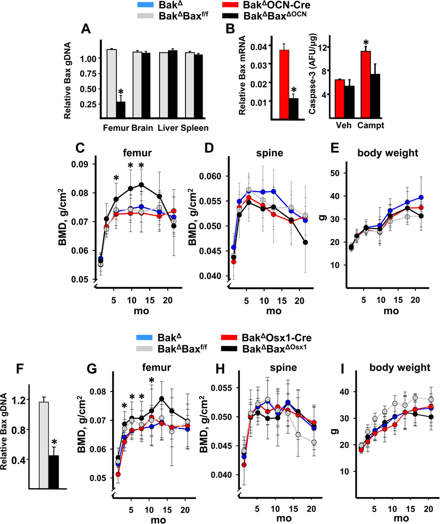
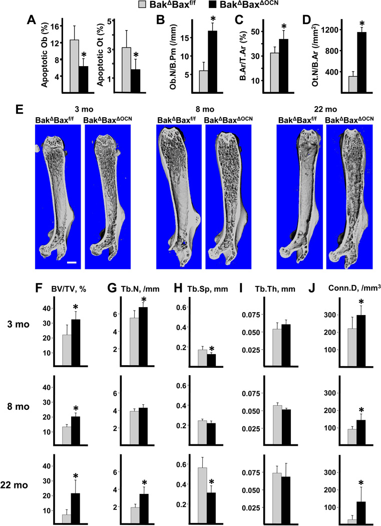
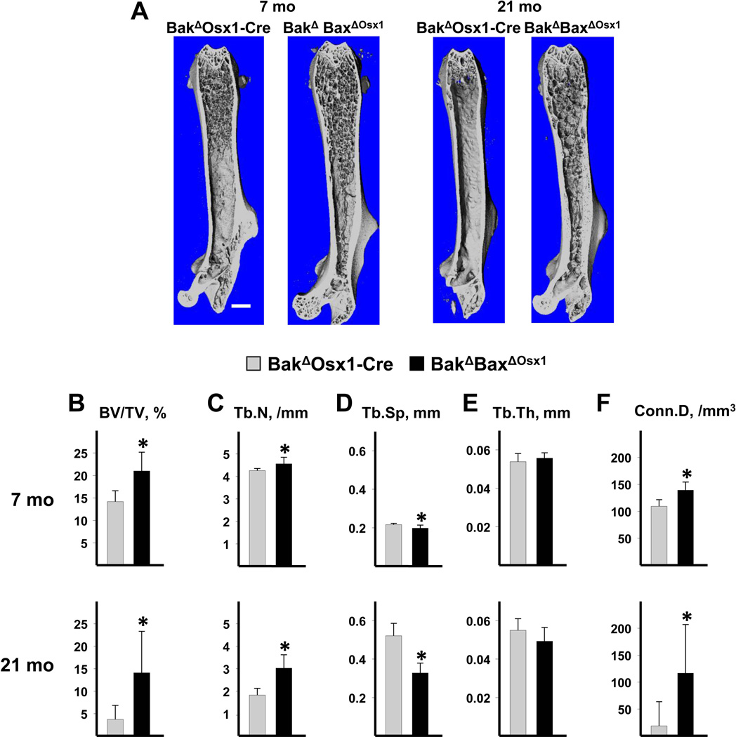
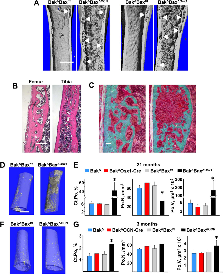
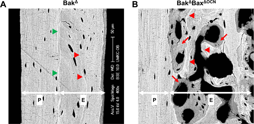
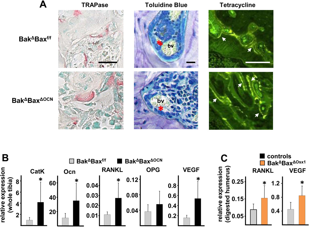
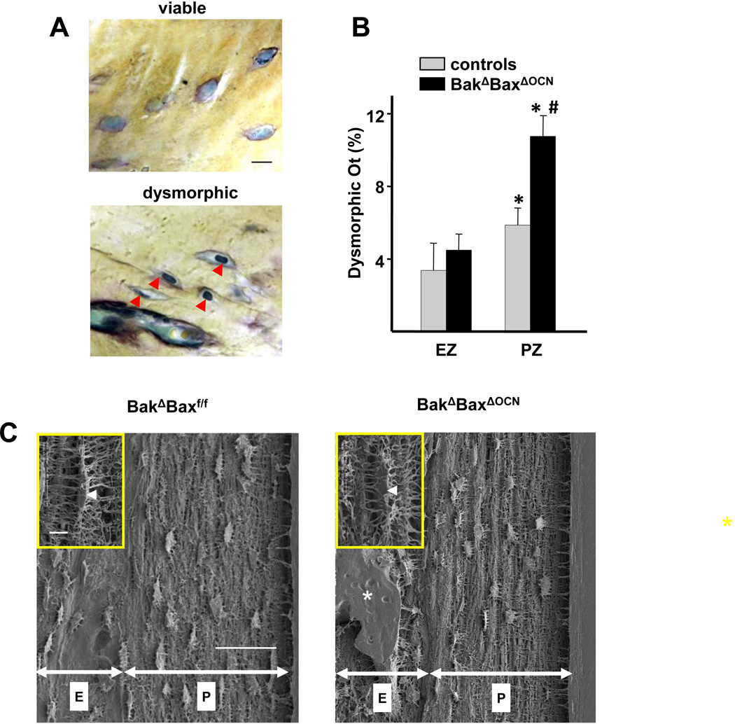
Skeletal aging is accompanied by decreased cancellous bone mass and increased formation of pores within cortical bone. The latter accounts for a large portion of the increase in nonvertebral fractures after age 65 years in humans. We selectively deleted Bak and Bax, two genes essential for apoptosis, in two types of terminally differentiated bone cells: the short-lived osteoblasts that elaborate the bone matrix, and the long-lived osteocytes that are immured within the mineralized matrix and choreograph the regeneration of bone. Attenuation of apoptosis in osteoblasts increased their working lifespan and thereby cancellous bone mass in the femur. In long-lived osteocytes, however, it caused dysfunction with advancing age and greatly magnified intracortical femoral porosity associated with increased production of receptor activator of nuclear factor-κB ligand and vascular endothelial growth factor. Increasing bone mass by artificial prolongation of the inherent lifespan of short-lived osteoblasts, while exaggerating the adverse effects of aging on long-lived osteocytes, highlights the seminal role of cell age in bone homeostasis. In addition, our findings suggest that distress signals produced by old and/or dysfunctional osteocytes are the culprits of the increased intracortical porosity in old age.
Keywords: AGING; APOPTOSIS; BONE FORMATION; CORTICAL POROSITY; OSTEOBLASTS; OSTEOCYTES; RANKL.
© 2014 American Society for Bone and Mineral Research.
Conflict of interest statement
The authors state that they have no conflicts of interest.
Figures
Similar articles
-
Osteocyte- and late osteoblast-derived NOTUM reduces cortical bone mass in mice.Am J Physiol Endocrinol Metab. 2021 May 1;320(5):E967-E975. doi: 10.1152/ajpendo.00565.2020. Epub 2021 Mar 22. Am J Physiol Endocrinol Metab. 2021. PMID: 33749332
-
Suppression of autophagy in osteocytes mimics skeletal aging.J Biol Chem. 2013 Jun 14;288(24):17432-40. doi: 10.1074/jbc.M112.444190. Epub 2013 May 3. J Biol Chem. 2013. PMID: 23645674 Free PMC article.
-
For whom the bell tolls: distress signals from long-lived osteocytes and the pathogenesis of metabolic bone diseases.Bone. 2013 Jun;54(2):272-8. doi: 10.1016/j.bone.2012.09.017. Epub 2012 Sep 23. Bone. 2013. PMID: 23010104 Free PMC article. Review.
-
Osteocytes, not Osteoblasts or Lining Cells, are the Main Source of the RANKL Required for Osteoclast Formation in Remodeling Bone.PLoS One. 2015 Sep 22;10(9):e0138189. doi: 10.1371/journal.pone.0138189. eCollection 2015. PLoS One. 2015. PMID: 26393791 Free PMC article.
-
Cortical bone development, maintenance and porosity: genetic alterations in humans and mice influencing chondrocytes, osteoclasts, osteoblasts and osteocytes.Cell Mol Life Sci. 2021 Aug;78(15):5755-5773. doi: 10.1007/s00018-021-03884-w. Epub 2021 Jul 1. Cell Mol Life Sci. 2021. PMID: 34196732 Free PMC article. Review.
Cited by
-
The Quest for Osteoporosis Mechanisms and Rational Therapies: How Far We've Come, How Much Further We Need to Go.J Bone Miner Res. 2018 Mar;33(3):371-385. doi: 10.1002/jbmr.3400. Epub 2018 Feb 22. J Bone Miner Res. 2018. PMID: 29405383 Free PMC article.
-
The mechanism of oxytocin and its receptors in regulating cells in bone metabolism.Front Pharmacol. 2023 May 9;14:1171732. doi: 10.3389/fphar.2023.1171732. eCollection 2023. Front Pharmacol. 2023. PMID: 37229246 Free PMC article. Review.
-
The Effects of Aging and Sex Steroid Deficiency on the Murine Skeleton Are Independent and Mechanistically Distinct.J Bone Miner Res. 2017 Mar;32(3):560-574. doi: 10.1002/jbmr.3014. Epub 2016 Nov 3. J Bone Miner Res. 2017. PMID: 27714847 Free PMC article.
-
Fenofibrate decreases the bone quality by down regulating Runx2 in high-fat-diet induced Type 2 diabetes mellitus mouse model.Lipids Health Dis. 2017 Oct 13;16(1):201. doi: 10.1186/s12944-017-0592-5. Lipids Health Dis. 2017. PMID: 29029615 Free PMC article.
-
Estrogens decrease osteoclast number by attenuating mitochondria oxidative phosphorylation and ATP production in early osteoclast precursors.Sci Rep. 2020 Jul 20;10(1):11933. doi: 10.1038/s41598-020-68890-7. Sci Rep. 2020. PMID: 32686739 Free PMC article.
References
-
- Manolagas SC. Birth and death of bone cells: basic regulatory mechanisms and implications for the pathogenesis and treatment of osteoporosis. Endocr Rev. 2000;21:115–137. - PubMed
-
- Almeida M, Han L, Martin-Millan M, Plotkin LI, Stewart SA, Roberson PK, Kousteni S, O'Brien CA, Bellido T, Parfitt AM, Weinstein RS, Jilka RL, Manolagas SC. Skeletal involution by age-associated oxidative stress and its acceleration by loss of sex steroids. J Biol Chem. 2007;282:27285–27297. - PMC - PubMed
Publication types
MeSH terms
Substances
Grants and funding
LinkOut - more resources
Full Text Sources
Other Literature Sources
Medical
Research Materials
