. 2013 Aug;41(15):7231-9.
doi: 10.1093/nar/gkt528. Epub 2013 Jun 12.

Visualization of multivalent histone modification in a single cell reveals highly concerted epigenetic changes on differentiation of embryonic stem cells

Affiliations

Visualization of multivalent histone modification in a single cell reveals highly concerted epigenetic changes on differentiation of embryonic stem cells

Naoko Hattori et al. Nucleic Acids Res. 2013 Aug.

Abstract

Combinations of histone modifications have significant biological roles, such as maintenance of pluripotency and cancer development, but cannot be analyzed at the single cell level. Here, we visualized a combination of histone modifications by applying the in situ proximity ligation assay, which detects two proteins in close vicinity (∼30 nm). The specificity of the method [designated as imaging of a combination of histone modifications (iChmo)] was confirmed by positive signals from H3K4me3/acetylated H3K9, H3K4me3/RNA polymerase II and H3K9me3/H4K20me3, and negative signals from H3K4me3/H3K9me3. Bivalent modification was clearly visualized by iChmo in wild-type embryonic stem cells (ESCs) known to have it, whereas rarely in Suz12 knockout ESCs and mouse embryonic fibroblasts known to have little of it. iChmo was applied to analysis of epigenetic and phenotypic changes of heterogeneous cell population, namely, ESCs at an early stage of differentiation, and this revealed that the bivalent modification disappeared in a highly concerted manner, whereas phenotypic differentiation proceeded with large variations among cells. Also, using this method, we were able to visualize a combination of repressive histone marks in tissue samples. The application of iChmo to samples with heterogeneous cell population and tissue samples is expected to clarify unknown biological and pathological significance of various combinations of epigenetic modifications.


Figures

Figure 1.
Figure 1.
Visualization of combinations of epigenetic modifications in a single cell. Immunofluorescence staining was performed using mouse ESCs and antibodies against H3K4me3 and H3K9ac (A), H3K4me3 and RNAPII (B), H4K20me3 and H3K9me3 (C) and H3K4me3 and H3K9me3 (D). Colocalizations of H3K4me3/H3K9Ac, of H3K4me3/RNAP2 and of H4K20me3/H3K9me3 were observed, whereas that of H3K4me3/H3K9me3 was not. iChmo was performed using mouse ESCs and antibodies against H3K4me3 and H3K9ac (E), H3K4me3 and RNAPII (F), H4K20me3 and H3K9me3 (G) and H3K4me3 and H3K9me3 (H). Coexistence of H3K4me3/H3K9ac, of H3K4me3/RNAPII and of H4K20me3/H3K9me3, but not of H3K4me3/H3K9me3, was observed. Scale bar represents 10 µm. (I) The number of iChmo spots was counted for individual combinations in the nuclei of ESCs (H3K4me3 and H3K9ac, n = 71; H3K4me3 and RNAPII, n = 70; H4K20me3 and H3K9me3, n = 60; and H3K4me3 and H3K9me3, n = 80).
Figure 2.
Figure 2.
Application of iChmo to visualization of bivalent modification. (A) Mouse ESCs were stained by immunofluorescence with antibodies against H3K4me3 and H3K27me3 (scale bar: 10 µm). (B) Coexistence of H3K4me3 and H3K27me3 in WT ESCs was detected by iChmo, but hardly in Suz12 KO ESCs and MEFs (scale bar: 10 µm). (C) The mean number of fluorescence spots was significantly larger in WT ESCs (15.2; n = 58) than in Suz12 KO ESCs (0.9; n = 69) and MEFs (4.1; n = 60) (Mann–Whitney U-test; *P < 0.001).
Figure 3.
Figure 3.
Visualization of epigenetic and phenotypic layers of differentiation in an early stage of ESC differentiation. (A) Differentiation of ESCs was induced by treatment of all-trans RA, and Oct-4 mRNA expression was measured before, and 24 and 48 h after the RA treatment. The expression levels in the mouse liver and brain are shown as those in Oct-4-negative tissues. Values show mean + SD of three experiments. (B) Expression of Oct-4 and ßIII-tubulin proteins was analyzed in ESCs before, and 24 and 48 h after the RA treatment by immunofluorescence (scale bar: 20 µm). (C) Images of phase contrast and iChmo for the bivalent modification in ESC colonies before, and 24 and 48 h after the RA treatment (upper panel), and differentiated neuron-like cells at 24 and 48 h (lower panel). Regardless of the phenotypic differentiation statuses, the bivalent modification was absent both at 24 and 48 h, supporting highly concerted regulation of epigenetic changes. Scale bar represents 10 µm. (D) The number of fluorescence spots was counted in ESCs before (n = 33), and 24 (n = 37) and 48 (n = 38) h after the RA treatment (Mann–Whitney U-test; *P < 0.001). Although the decrease of Oct-4 expression and increase of ßIII-tubulin were highly variable among the ESCs treated with RA, the decrease of the bivalent modification was highly coordinated.
Figure 4.
Figure 4.
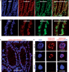
Application of iChmo to the analysis of human colonic tissue. (A) Human colonic tissues were stained by immunofluorescence with antibodies against H3K9me3 and H4K20me3 (scale bar: 50 µm). Colocalization of H3K9me3/ H4K20me3 was observed in the cells of colonic tissue. (B) High-magnification images of (A) (scale bar: 10 µm). (C) Coexistence of H3K9me3 and H4K20me3 was visualized in the nuclei of cells as fluorescence spots (scale bar: 50 µm). (D) High-magnification images of (C) (scale bar: 10 µm). The cells with iChmo spots (Cell #1 and #2) coincided with the cells having weak DAPI intensity, and the cells without iChmo spots (Cell #3) showed strong DAPI intensity.

References

    1. Meissner A. Epigenetic modifications in pluripotent and differentiated cells. Nat. Biotechnol. 2010;28:1079–1088. - PubMed
    1. Kondo Y. Epigenetic cross-talk between DNA methylation and histone modifications in human cancers. Yonsei Med. J. 2009;50:455–463. - PMC - PubMed
    1. Bernstein BE, Mikkelsen TS, Xie X, Kamal M, Huebert DJ, Cuff J, Fry B, Meissner A, Wernig M, Plath K, et al. A bivalent chromatin structure marks key developmental genes in embryonic stem cells. Cell. 2006;125:315–326. - PubMed
    1. Lien WH, Guo X, Polak L, Lawton LN, Young RA, Zheng D, Fuchs E. Genome-wide maps of histone modifications unwind in vivo chromatin states of the hair follicle lineage. Cell Stem Cell. 2011;9:219–232. - PMC - PubMed
    1. Mikkelsen TS, Ku M, Jaffe DB, Issac B, Lieberman E, Giannoukos G, Alvarez P, Brockman W, Kim TK, Koche RP, et al. Genome-wide maps of chromatin state in pluripotent and lineage-committed cells. Nature. 2007;448:553–560. - PMC - PubMed

Publication types

MeSH terms