. 2013 Jul 1;216(Pt 13):2469-77.
doi: 10.1242/jeb.082404.

Mechanisms of muscle gene regulation in the electric organ of Sternopygus macrurus

Affiliations

Mechanisms of muscle gene regulation in the electric organ of Sternopygus macrurus

Robert Güth et al. J Exp Biol. .

Abstract

Animals perform a remarkable diversity of movements through the coordinated mechanical contraction of skeletal muscle. This capacity for a wide range of movements is due to the presence of muscle cells with a very plastic phenotype that display many different biochemical, physiological and morphological properties. What factors influence the maintenance and plasticity of differentiated muscle fibers is a fundamental question in muscle biology. We have exploited the remarkable potential of skeletal muscle cells of the gymnotiform electric fish Sternopygus macrurus to trans-differentiate into electrocytes, the non-contractile electrogenic cells of the electric organ (EO), to investigate the mechanisms that regulate the skeletal muscle phenotype. In S. macrurus, mature electrocytes possess a phenotype that is intermediate between muscle and non-muscle cells. How some genes coding for muscle-specific proteins are downregulated while others are maintained, and novel genes are upregulated, is an intriguing problem in the control of skeletal muscle and EO phenotype. To date, the intracellular and extracellular factors that generate and maintain distinct patterns of gene expression in muscle and EO have not been defined. Expression studies in S. macrurus have started to shed light on the role that transcriptional and post-transcriptional events play in regulating specific muscle protein systems and the muscle phenotype of the EO. In addition, these findings also represent an important step toward identifying mechanisms that affect the maintenance and plasticity of the muscle cell phenotype for the evolution of highly specialized non-contractile tissues.

Keywords: electrocyte; muscle regulatory factors; muscle-derived cells; post-transcriptional regulation.


Figures

Fig. 1.
Fig. 1.
Mature electrocytes of Sternopygus macrurus lack sarcomeric structures. Electron micrograph of a longitudinal section taken from a control tail showing part of a skeletal muscle fiber (SM) and an electrocyte (EC). The muscle fiber contains a regular array of sarcomeres. These structures are absent in the electrocyte. Mitochondria and vacuole-like structures that resemble sarcoplasmic reticula are found peripherally in electrocytes (arrows), as are nuclei (n). Note the presence of satellite cells (sc) associated with both the muscle fiber and electrocyte. Scale bar, 10 μm.
Fig. 2.
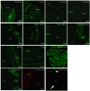
Fig. 2.
Muscle protein expression in S. macrurus electric organ (EO). Immunolabeling of tail cryosections for various muscle proteins (green) reveals staining of electrocytes (EC, arrow in α-actinin panel) for desmin, α-actinin, α-acetylcholine receptor (AChR) and titin but not for myosin heavy chain (MHC), tropomyosin or troponin-T. Electrocytes were immunolabeled with anti-α-actin but not phalloidin, which stains filamentous actin (red). In contrast to electrocytes, skeletal muscle fibers (mm; * in the slow MHC panel; arrowhead in the α-actinin panel) stained for all muscle markers. The α-actinin image is reproduced from Kim et al. (Kim et al., 2004) (see their fig. 3) with permission from Springer Science+Business Media; all other images are reproduced from Cuellar et al. (Cuellar et al., 2006) with permission from FASEB Journal.
Fig. 3.
Fig. 3.
Muscle regulatory factors (MRFs) are expressed in S. macrurus EO. (A) Transcripts of all four MRFs are detected in EO and skeletal muscle of S. macrurus. Quantitative RT-PCR shows significantly higher levels of myogenin, Myf5 and MRF4 transcripts (*P≤0.05) in EO than in skeletal muscle. Bars represent means + s.e.m. Reproduced with permission from Kim et al. (Kim et al., 2008). (B) MyoD and myogenin proteins are detected in the nuclei of muscle fibers and electrocytes (EC) by immunolabeling of adult tail cryosections with S. macrurus-specific antibodies (green). Nuclei were counterstained with Hoechst 33342 (blue). White arrowheads point to nuclei in electrocytes and muscle fibers that were double-labeled with antibody and Hoechst. epi, epidermis. Scale bars, 100 μm. Reproduced from Kim et al. (Kim et al., 2009) with permission from International Journal of Developmental Biology.
Fig. 4.
Fig. 4.
Some muscle genes exhibit post-transcriptional regulation in S. macrurus myogenic tissues. (A) The abundance of contraction-associated and metabolic transcripts was determined in EO relative to skeletal muscle (SM) by quantitative RT-PCR. The expression of metabolic proteins was determined in EO relative to SM using western blotting. Coloration indicates a higher abundance in EO (>2.5 times, red) or in muscle (>2.5 times, green), or a similar abundance in the two tissues (<2.5 times difference between EO and SM, yellow). *Lactate dehydrogenase protein data represent an indirect measure of LDH enzymatic activity determined by assaying lactate concentration per gram of tissue. Data from R.G. and G.A.U. (unpublished). (B) Detection of muscle gene transcripts and corresponding proteins in S. macrurus SM and EO using RT-PCR and western blotting. ‘No RT’ lanes (−) demonstrate the absence of DNA contamination in samples without reverse transcriptase treatment. Adapted from Cuellar et al. (Cuellar et al., 2006) with permission from FASEB Journal. Muscle creatine kinase protein expression was undetectable in EO in B but detectable with increased protein loading in A. AChR, acetylcholine receptor; MHC, myosin heavy chain; MCK, muscle creatine kinase; Trop-T, troponin-T; TPM, tropomyosin; GAPDH, glyceraldehyde 3-phosphate dehydrogenase; SDH, succinate dehydrogenase; LDH, lactate dehydrogenase; SERCA, sarco/endoplasmic reticulum Ca2+-ATPase.
Fig. 5.
Fig. 5.
Schematic diagram of regulation of the muscle program in S. macrurus electrocytes. Gene expression in S. macrurus electrocytes is regulated by a range of mechanisms as determined by quantitative analyses at the transcript and protein levels. Possible mechanisms include translational silencing of mRNAs via microRNA–nucleoprotein complexes (miRNP) and P body pathways, as well as post-translational regulation of protein stability and assembly into myofibrillar structures. The identity of upstream signals regulating these processes is currently unknown, but likely includes neural factors. EMN, electromotor neuron; MRFs, myogenic regulatory factors; all other abbreviations as given in Fig. 4.

Similar articles

Cited by

References

    1. Ai J., Zhang R., Gao X., Niu H.-F., Wang N., Xu Y., Li Y., Ma N., Sun L.-H., Pan Z.-W., et al. (2012). Overexpression of microRNA-1 impairs cardiac contractile function by damaging sarcomere assembly. Cardiovasc. Res. 95, 385-393 - PubMed
    1. Allen D. L., Sartorius C. A., Sycuro L. K., Leinwand L. A. (2001). Different pathways regulate expression of the skeletal myosin heavy chain genes. J. Biol. Chem. 276, 43524-43533 - PubMed
    1. Ambros V. (2004). The functions of animal microRNAs. Nature 431, 350-355 - PubMed
    1. Anderson C., Catoe H., Werner R. (2006). MIR-206 regulates connexin43 expression during skeletal muscle development. Nucleic Acids Res. 34, 5863-5871 - PMC - PubMed
    1. Arnold H. H., Braun T. (1996). Targeted inactivation of myogenic factor genes reveals their role during mouse myogenesis: a review. Int. J. Dev. Biol. 40, 345-353 - PubMed

Publication types