Review
. 2013 Jul 1;216(Pt 13):2515-22.
doi: 10.1242/jeb.082362.

The evolution and development of vertebrate lateral line electroreceptors

Affiliations
Review

The evolution and development of vertebrate lateral line electroreceptors

Clare V H Baker et al. J Exp Biol. .

Abstract

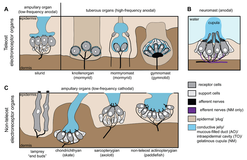
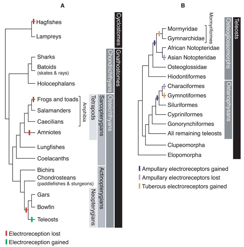
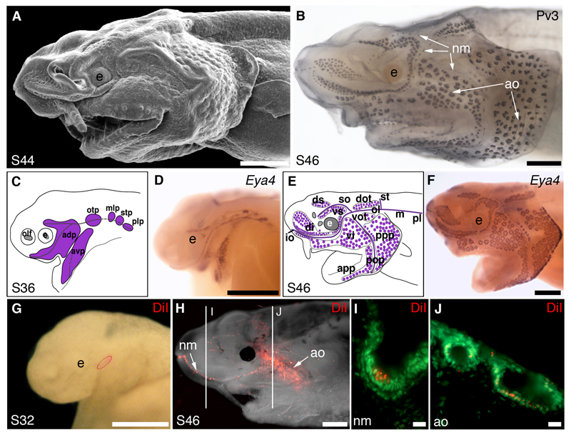
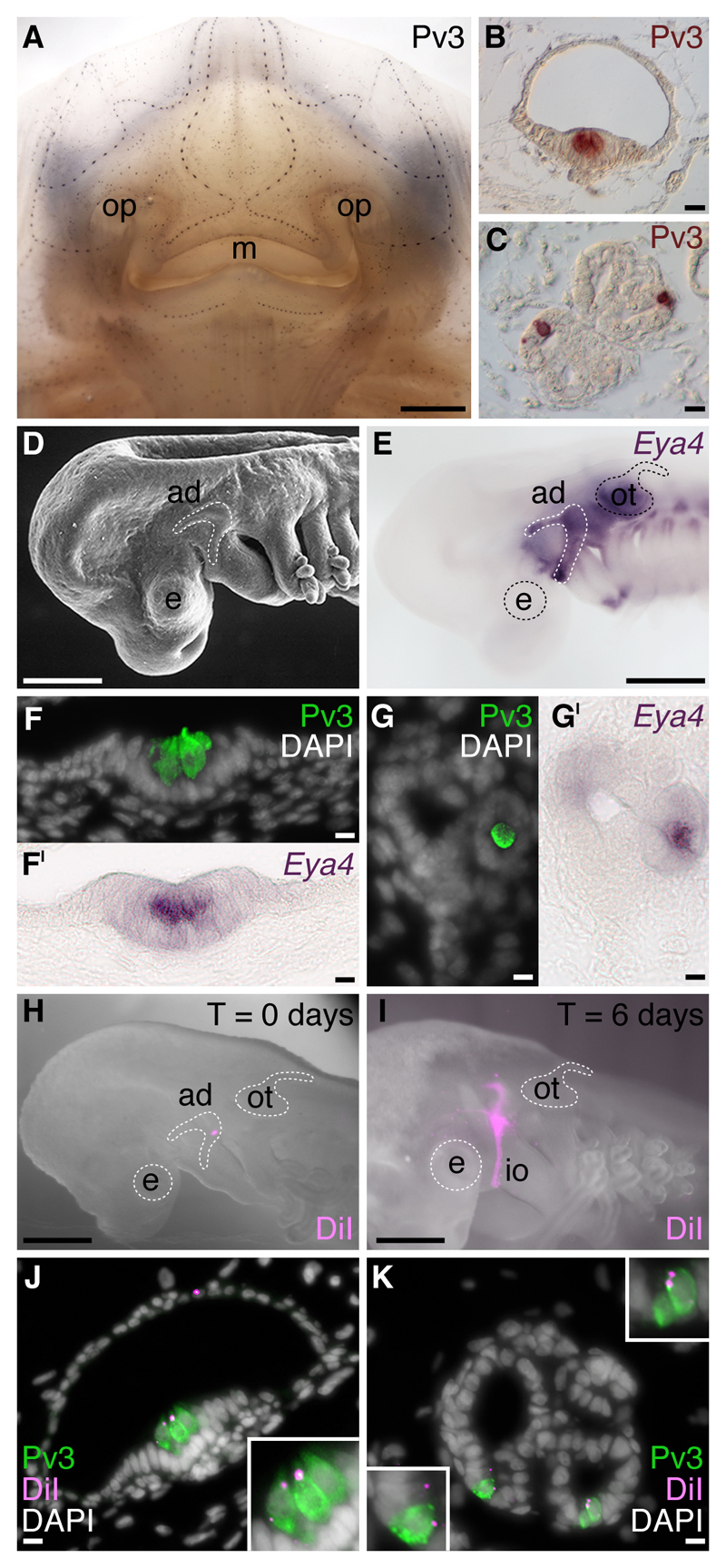
Electroreception is an ancient vertebrate sense with a fascinating evolutionary history involving multiple losses as well as independent evolution at least twice within teleosts. We review the phylogenetic distribution of electroreception and the morphology and innervation of electroreceptors in different vertebrate groups. We summarise recent work from our laboratory that has confirmed the homology of ampullary electroreceptors in non-teleost jawed vertebrates by showing, in conjunction with previously published work, that these are derived embryonically from lateral line placodes. Finally, we review hypotheses to explain the distribution of electroreception within teleosts, including the hypothesis that teleost ampullary and tuberous electroreceptors evolved via the modification of mechanosensory hair cells in lateral line neuromasts. We conclude that further experimental work on teleost electroreceptor development is needed to test such hypotheses.

Keywords: ampullary; electroreception; electroreceptors; hair cell; lateral line; neuromast; tuberous.


Figures

Fig. 1
Fig. 1
Schematics illustrating the range of lateral line organ morphologies (not to scale). (A) Teleost ampullary organs (e.g. silurid, based on Northcutt et al., 2000), which respond to low-frequency anodal stimuli, contain electroreceptor cells with short, sparse microvilli, located at the base of mucous-filled ducts that open to the surface. Tuberous organs, which respond to high-frequency anodal stimuli, are morphologically varied but the electroreceptor cells (which have many microvilli) are generally located within an intraepidermal cavity plugged by epidermal cells. Both types of mormyrid tuberous organs (knollenorgan and mormyromast; adapted from Jørgensen, 2005) and a gymnotid tuberous organ (gymnomast; adapted from Cernuda-Cernuda and García-Fernández, 1996) are shown. (B) Neuromast receptor cells, which are mechanosensory but can also respond to large anodal stimuli, have a single cilium flanked by a stepped array of microvilli (the “hair bundle”). The cilia and hair bundles of all the receptor cells in the neuromast are encased together in a gelatinous cupula in contact with water. Unlike electroreceptors, which only receive afferent innervation, neuromast hair cells receive both afferent and efferent innervation. (C) Examples of non-teleost electroreceptor organs, which all respond to low-frequency cathodal stimuli: lamprey "end buds" containing multiple electroreceptor cells, each with multiple microvilli but no cilia (adapted from Jørgensen, 2005), and chondrichthyan (e.g. skate), sarcopterygian (e.g. axolotl) and non-teleost actinopterygian (e.g. paddlefish) ampullary organs, whose electroreceptor cells generally have a single cilium and variable numbers of microvilli. AO, ampullary organ; NM, neuromast; TO, tuberous organ.
Fig. 2
Fig. 2
The phylogenetic distribution of electroreception among (A) vertebrates and (B) teleost fishes. Neopterygian and teleost phylogenies drawn after Near et al. (2012). (A) The distribution of electroreception among the vertebrates reveals it to be an ancient sense that was lost independently (red bar) in various lineages, including the lineage leading to neopterygian fishes (gars, bowfin and teleosts). Electroreception subsequently evolved independently within the teleosts (green bar). (B) The distribution of electroreception among teleost fishes suggests that ampullary electroreceptors (blue bar) evolved independently twice: once in the Osteoglossomorpha, along the lineage leading to the notopterids and mormyriforms (with subsequent loss in Asian notopterids); and once in the Ostariophysi, along the lineage leading to the siluriforms, gymnotiforms and characiforms (Near et al., 2012) (with subsequent loss in characiforms). Electric organs and tuberous electroreceptors (brown bar) subsequently evolved independently in the mormyriforms within the Osteoglossomorpha, and in the gymnotiforms within the Ostariophysi. Alternative hypotheses are discussed in the text.
Fig. 3
Fig. 3
Lateral line placodes give rise to ampullary organs and neuromasts in a basal ray-finned bony fish, the North American (Mississippi) paddlefish, Polyodon spathula. Lateral views, anterior to the left, unless otherwise noted; staging according to Bemis and Grande (1992). All panels were previously published in Modrell et al. (2011a) and are reproduced here in accordance with the terms of the authors’ Licence to Publish agreement with Nature Publishing Group. (A) Scanning electron micrograph of a stage 44 embryo showing differentiated ampullary organ fields, particularly on the operculum. (B) Stage 46 embryo immunostained for the Ca2+-binding protein parvalbumin-3 (Pv3), which is strongly expressed in the sensory receptor cells of both neuromasts and ampullary organs (also see Modrell et al., 2011a). (C-F) Schematic diagrams and whole-mount in situ hybridisation for the transcription co-factor gene Eya4 at (C,D) stage 36, when Eya4 is expressed in developing neuromast canal lines and the ampullary organ fields flanking those lines (purple in C) and (E,F) stage 46, when Eya4 expression is maintained in both neuromasts and ampullary organs (purple in E). (G) Stage 32 embryo immediately following a focal DiI injection into the anterodorsal lateral line placode (injection site outlined in red). (H) The same embryo as in G, at stage 46. DiI-labelled cells are visible both in a neuromast canal line and ampullary organ fields. Lines indicate the plane of transverse sections showing DiI-labelled cells (red) in (I) a neuromast and (J) ampullary organs, both counterstained with the nuclear marker Sytox Green (green). Abbreviations: adp, anterodorsal placode; ao, ampullary organ; app, anterior preopercular ampullary field; avp, anteroventral placode; dot, dorsal otic ampullary field; di, dorsal infraorbital ampullary field; ds, dorsal supraorbital ampullary field; e, eye; epi, epibranchial placode region; io, infraorbital lateral line; m, middle lateral line; mlp, middle lateral line placode; ol; otic lateral line; otp, otic lateral line placode; plp, posterior lateral line placode; pll, posterior lateral line; pop, preopercular lateral line; ppp, posterior preopercular field; S, stage; stp, supratemporal placode; so, supraorbital lateral line; st, supratemporal lateral line; vi, ventral infraorbital field; vot, ventral otic field; vs, ventral supraorbital field. Scale bars: (A,B,D,G) 0.5mm, (F,H) 1mm, (I,J) 10μm.
Fig. 4
Fig. 4
Lateral line placodes give rise to ampullary organs and neuromasts in a cartilaginous fish, the little skate, Leucoraja erinacea. All panels except E, H and I were previously published in Gillis et al. (2012) and are reproduced here in accordance with the terms of the authors’ Licence Agreement with the Company of Biologists. (A) Whole-mount immunostaining for the Ca2+-binding protein parvalbumin-3 (Pv3) in an L. erinacea embryo at stage 33 (Maxwell et al., 2008) reveals superficial lines of cephalic mechanosensory neuromasts, as well as clusters of ampullary organs located deeper within the dermis. Immunohistochemical localisation of Pv3 in (B) neuromasts and (C) ampullary organs reveals small clusters of Pv3-positive sensory receptor cells nested among Pv3-negative supporting cells. To test the hypothesis that lateral line placodes give rise to neuromasts and ampullary organs, we fate-mapped the anterodorsal lateral line placode in L. erinacea, which is recognisable (D) as a horseshoe-shaped thickening of cranial ectoderm caudal to the eye and dorsal to the mandibular arch, and (E) by its expression of the transcription co-factor gene Eya4. Eya4 expression is maintained at later stages in the Pv3-positive sensory receptor cells of (F,FI) neuromasts and (G,GI) ampullary organs. (H) Example of an embryo immediately after focal labelling of the anterodorsal lateral line placode with the lipophilic vital dye DiI. (I) After 6 days of incubation, DiI-positive cells were observed migrating away from the placode, in the infraorbital sensory primordium. In embryos with DiI-labelled anterodorsal lateral line placodes, sensory receptor cells, support cells and canal cells of (J) neuromasts and (K) ampullary organs were DiI-positive, indicating their lateral line placodal origin. Abbreviations: ad, anterodorsal lateral line placode; ad, anterodorsal lateral line placode; e, eye; io, infraorbital sensory primordium; m, mouth; op, olfactory pit; ot, otic vesicle. Scale bars: (A) 2.5mm, (B-C) 10μm, (D,E,H) 0.5mm, (I) 0.4mm, (F-G’,J,K) 10μm.

References

    1. Alves-Gomes JA. The evolution of electroreception and bioelectrogenesis in teleost fish: a phylogenetic perspective. J Fish Biol. 2001;58:1489–1511.
    1. Aman A, Piotrowski T. Cell-cell signaling interactions coordinate multiple cell behaviors that drive morphogenesis of the lateral line. Cell Adh Migr. 2011;5:499–508. - PMC - PubMed
    1. Barry MA, White RL, Bennett MV. The elasmobranch spiracular organ. II. Physiological studies. J Comp Physiol A. 1988;163:93–98. - PubMed
    1. Baumann M, Roth A. The Ca++ permeability of the apical membrane of neuromast hair cells. J Comp Physiol A. 1986;158:681–688. - PubMed
    1. Bell CC, Maler L. Central neuroanatomy of electrosensory systems in fish. In: Bullock TH, Hopkins CD, Popper AN, Fay RR, editors. Electroreception. New York: Springer; 2005. pp. 68–111.

Publication types

LinkOut - more resources