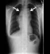
Resolution of an intracardiac mass with chemotherapy
- PMID: 23761492
- PMCID: PMC3702828
- DOI: 10.1136/bcr-2012-008371
Resolution of an intracardiac mass with chemotherapy
Abstract
Right atrial intracardiac tumours are uncommonly seen during echocardiography. Our patient presented with primary mediastinal large B-cell lymphoma with intracardiac involvement. The tumour was seen by echocardiography and the extent of the tumour was defined by CT scan of the chest. Following chemotherapy directed to her specific tumour cell type, there was complete resolution of the intracardiac mass.
Figures


Similar articles
-
Intracardiac diffuse large B-cell lymphoma: an unexpected diagnosis.BMJ Case Rep. 2024 Jun 24;17(6):e259242. doi: 10.1136/bcr-2023-259242. BMJ Case Rep. 2024. PMID: 38914528
-
Intracardiac tumor regression documented by two-dimensional echocardiography.Am J Cardiol. 1986 Jul 1;58(1):186-7. doi: 10.1016/0002-9149(86)90274-2. Am J Cardiol. 1986. PMID: 3487969 No abstract available.
-
[Primary cardiac lymphoma in an immunocompetent young adult: outcome with chemotherapy].G Ital Cardiol (Rome). 2017 Jan;18(1):7-10. doi: 10.1714/2628.27021. G Ital Cardiol (Rome). 2017. PMID: 28287209 Italian.
-
[Primary cardiac lymphoma: cytological diagnosis and treatment with response to polychemotherapy and hematopoietic precursor autotransplant. Presentation of a case a review of the literature].An Med Interna. 2002 Jun;19(6):305-9. An Med Interna. 2002. PMID: 12152391 Review. Spanish.
-
18F-FDG PET/CT in a cardiac metastasis in a patient with history of malignant neuroectodermal tumour of the chest wall: Case report and review of the literature.Rev Esp Med Nucl Imagen Mol (Engl Ed). 2018 Mar-Apr;37(2):110-113. doi: 10.1016/j.remn.2017.04.008. Epub 2017 Jun 23. Rev Esp Med Nucl Imagen Mol (Engl Ed). 2018. PMID: 28648525 Review. English, Spanish.
References
-
- Van besien K, Kelta M, Bahaguna P. Primary mediastinal B-cell lymphoma: a review of pathology and management. J Clin Oncol 2001;2013:1855–64 - PubMed
-
- Cazals-Hatem D, Lepage E, Brice P, et al. Primary mediastinal large B-cell lymphoma. A clinicopathologic study of 141 cases compared with 916 nonmediastinal large B-cell lymphomas, a GELA (“Groupe d'Etude des Lymphomes de l'Adulte”) study. Am J Surg Pathol 1996;2013:877–88 - PubMed
-
- McAllister HA, Hall RJ, Cooley DA. Tumors of the heart and pericardium. Curr Probl Cardiol 1999;2013:57–116 - PubMed
-
- Tanaka T, Sato T, Akifuji Y, et al. Aggressive non-Hodgkin's lymphoma with massive involvement of the right ventricle. Intern Med 1996;2013:826–30 - PubMed
-
- McDonnell PJ, Mann RB, Bulkley BH. Involvement of the heart by malignant lymphoma: a clinicopathological study. Cancer 1982;2013:944–51 - PubMed
Publication types
MeSH terms
LinkOut - more resources
Full Text Sources
Other Literature Sources
