Review
. 2013 Aug 1;305(3):C241-52.
doi: 10.1152/ajpcell.00173.2013. Epub 2013 Jun 12.

Cellular mechanisms of tissue fibrosis. 4. Structural and functional consequences of skeletal muscle fibrosis

Affiliations
Review

Cellular mechanisms of tissue fibrosis. 4. Structural and functional consequences of skeletal muscle fibrosis

Richard L Lieber et al. Am J Physiol Cell Physiol. .

Abstract

Skeletal muscle fibrosis can be a devastating clinical problem that arises from many causes, including primary skeletal muscle tissue diseases, as seen in the muscular dystrophies, or it can be secondary to events that include trauma to muscle or brain injury. The cellular source of activated fibroblasts (myofibroblasts) may include resident fibroblasts, adult muscle stem cells, or inflammatory or perivascular cells, depending on the model studied. Even though it is likely that there is no single source for all myofibroblasts, a common mechanism for the production of fibrosis is via the transforming growth factor-β/phosphorylated Smad3 pathway. This pathway and its downstream targets thus provide loci for antifibrotic therapies, as do methods for blocking the transdifferentiation of progenitors into activated fibroblasts. A structural model for the extracellular collagen network of skeletal muscle is needed so that measurements of collagen content, morphology, and gene expression can be related to mechanical properties. Approaches used to study fibrosis in tissues, such as lung, kidney, and liver, need to be applied to studies of skeletal muscle to identify ways to prevent or even cure the devastating maladies of skeletal muscle.

Keywords: muscle mechanics; myofibroblast; passive mechanics; stiffness.


Figures

Fig. 1.
Fig. 1.
A: micrograph of a cross section of healthy skeletal (rat tibialis anterior) muscle demonstrating normal morphology that consists of tightly packed polygonal fibers with a small amount (∼5%) of extracellular material. Traditional location of endomysial connective tissue is outlined with a solid line; perimysial tissue is outlined with a dashed line. However, as can be seen elsewhere in the micrograph, this distinction can be arbitrary. B: micrograph of a cross section of skeletal muscle demonstrating fibrotic morphology in which extracellular material is increased to ∼20% of the cross section, fibers are loosely packed, extracellular space is hypercellular, and fiber sizes are highly variable. This muscle was injected twice with botulinum toxin type A (Botox, Allergan; 6 U/kg in 100 μl) at a 3-mo interval and tested after 6 mo.
Fig. 2.
Fig. 2.
A: scanning electron micrograph of perimysial cables in a stretched mouse extensor digitorum longus (EDL) muscle. These cables run longitudinally along the length of the muscle fiber without noticeable connections to the fiber itself over ∼100-μm distances (arrowheads). Cables consist of bundles of collagen fibrils, as can be seen in the cable that has frayed (arrows). (Micrograph modified from Ref. .) B: transmission electron micrograph of wild-type mouse EDL muscle in the extracellular space region. A perimysial cable is demarcated with the box, and a wide ribbonlike perimysial cable is marked with arrowheads. C: higher-magnification view of the region in B indicates that the perimysial cable is composed of collagen fibrils. (This is most easily seen on longitudinal sections that demonstrate the characteristic striation pattern of type I collagen fibrils.) Also shown are superficial aspects of the muscle cell and a section of an extracellular-resident fibroblast (F). (Micrographs courtesy of Allison Gillies.)
Fig. 3.
Fig. 3.
Correlation between collagen content as measured by hydroxyproline assay and mechanical tangent stiffness measured biomechanically in human muscle fiber bundles, as described in Ref. . Note that collagen content alone is a very poor predictor of bundle stiffness.
Fig. 4.
Fig. 4.
A: schematic illustration of the arrangement of 3 specimen types. Single fibers (curved pink lines) were isolated from the muscle and tested individually or secured in groups. Bundles of a similar number of fibers embedded in ECM (light pink) were isolated and similarly secured. (Modified from Ref. with permission from Elsevier). B: normalized stiffness of fibers, fiber groups, and fiber bundles. Fiber bundles have a significantly higher modulus than either individual fibers or fiber groups, demonstrating that the ECM provides a large fraction of the load bearing. Fiber and fiber group moduli were not significantly different from each other. Connecting bars represent significant differences (P < 0.05). (Data replotted from Ref. with permission from Elsevier.)
Fig. 5.
Fig. 5.
Micrograph of a cross section of a regenerated skeletal muscle from a control mouse (A) and a mouse model of Duchenne muscular dystrophy, the mdx mouse (B). In contrast with the control muscle and the muscle shown in Fig. 1, the mdx muscle has more loosely packed fibers of highly varying size with a large amount (>25%) of hypercellular extracellular material. Scale bars, 60 μm. (From Ref. .)
Fig. 6.
Fig. 6.
Active (dotted lines) and passive (red and blue symbols) length-tension curves of a control rabbit muscle and a rabbit muscle subjected to chronic stretch at a fixed length (Fibrotic). Chronic surgical stretch results in a leftward shift in passive mechanical properties, demonstrating the functional mechanical effects of fibrosis. (Data replotted from Ref. .)
Fig. 7.
Fig. 7.
Data from a transcriptional profile of control muscles [Young wild-type (wt), n = 4] and muscles with a desmin intermediate filament deletion [desmin-deficient (Des−/−), n = 4]. Des−/− muscle demonstrates increased ECM-specific gene expression, as shown in the normalized gene expression pattern for 42 genes involved in ECM structure and maintenance. Expression levels are shown on a color scale, with green and red representing low and high expression, respectively. Hierarchical clustering is represented by connecting lines at the top of the grid, with lines closest to the grid denoting the most similar samples. Des−/− muscle samples have a distinct expression pattern of ECM genes compared with wild-type muscles, showing a higher expression of the majority of the listed genes indicated by the red color scheme. ECM-related genes are subdivided into 4 categories: basement membrane, ECM constituents, proteases, and protease inhibitors. Inf, inflammatory; Reg, regulatory. *Significantly higher expression values, as determined by 2-way ANOVA. (Data from Ref. .)
Fig. 8.
Fig. 8.
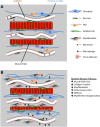
A: schematic illustration of the location of cells involved in skeletal muscle fibrosis. Skeletal muscle contains numerous sources of mononuclear cells that can ultimately become activated fibroblasts (myofibroblasts; multicolored). These sources include inflammatory cells (black), resident fibroblasts (blue), fibroadipogenic progenitor (FAP) cells (orange), and pericytes (black). There is no evidence that satellite cells (green) become myofibroblasts. B: after an insult that results in fibrosis (cf. Table 2), muscle tissue is characterized by muscle fiber atrophy, increased collagen content, presence of myofibroblasts and inflammatory cells, increased muscle mechanical stiffness, and myofibrillar disorganization that presents primarily as disrupted muscle cell z disks.
Fig. 9.
Fig. 9.
Two different progenitor cells differentiate into either muscle or fat. A: myogenic precursor cells (identified as CD34+/Sca1; see Ref. 63) that express green fluorescent protein (GFP) were injected into muscle and engrafted to existing muscle fibers that were damaged along the needle track. Red outline of the fiber indicates laminin immunostaining. B: adipogenic and fibrogenic precursor cells (identified as PDGCα+/CD34+/Sca1+; see Ref. 63) that express GFP were injected subcutaneously and differentiated into adipocytes, as evidenced by the positive perlipin stain. Scale bars, 50 μm. (Images from Ref. .)

References

    1. Abrams RA, Tsai AM, Watson B, Jamali A, Lieber RL. Skeletal muscle recovery after tenotomy and 7-day delayed muscle length restoration. Muscle Nerve 23: 707–714, 2000 - PubMed
    1. Ahsan T, Sah RL. Biomechanics of integrative cartilage repair. Osteoarthritis Cartilage 7: 29–40, 1999 - PubMed
    1. Alnaqeeb MA, Al Zaid NS, Goldspink G. Connective tissue changes and physical properties of developing and ageing skeletal muscle. J Anat 139: 677–689, 1984 - PMC - PubMed
    1. Bahi L, Koulmann N, Sanchez H, Momken I, Veksler V, Bigard AX, Ventura-Clapier R. Does ACE inhibition enhance endurance performance and muscle energy metabolism in rats? J Appl Physiol 96: 59–64, 2004 - PubMed
    1. Barton ER, Gimbel JA, Williams GR, Soslowsky LJ. Rat supraspinatus muscle atrophy after tendon detachment. J Orthop Res 23: 259–265, 2005 - PubMed

Publication types

MeSH terms

LinkOut - more resources