. 2011 Jun;26(2):159-162.
doi: 10.1154/1.3583156.

Binding of Mn-deoxyribonucleoside triphosphates to the active site of the DNA polymerase of bacteriophage T7

Affiliations

Binding of Mn-deoxyribonucleoside triphosphates to the active site of the DNA polymerase of bacteriophage T7

Barak Akabayov et al. Powder Diffr. 2011 Jun.

Abstract

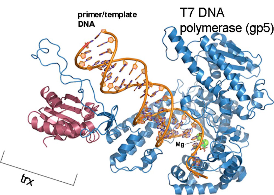
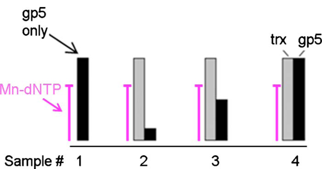
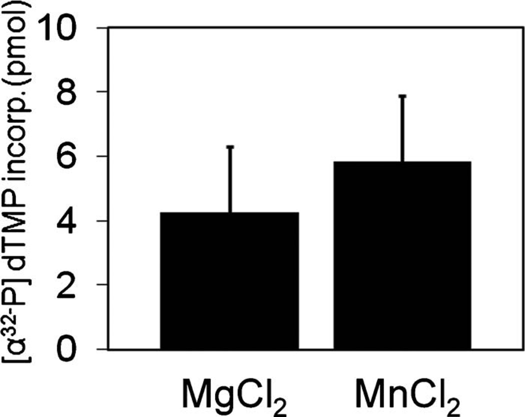
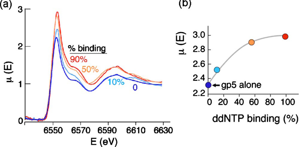
Divalent metal ions are crucial as cofactors for a variety of intracellular enzymatic activities. Mg2+, as an example, mediates binding of deoxyribonucleoside 5'-triphosphates followed by their hydrolysis in the active site of DNA polymerase. It is difficult to study the binding of Mg2+ to an active site because Mg2+ is spectroscopically silent and Mg2+ binds with low affinity to the active site of an enzyme. Therefore, we substituted Mg2+ with Mn2+:Mn2+ that is not only visible spectroscopically but also provides full activity of the DNA polymerase of bacteriophage T7. In order to demonstrate that the majority of Mn2+ is bound to the enzyme, we have applied site-directed titration analysis of T7 DNA polymerase using X-ray near edge spectroscopy. Here we show how X-ray near edge spectroscopy can be used to distinguish between signal originating from Mn2+ that is free in solution and Mn2+ bound to the active site of T7 DNA polymerase. This method can be applied to other enzymes that use divalent metal ions as a cofactor.

Keywords: DNA polymerase; XAFS; manganese; metal cofactor; metalloenzyme.


Figures

Figure 1
Figure 1
(Color online) Crystal structure of the bacteriophage T7 DNA polymerase (PDB entry 1t8e). The DNA polymerase (gp5) and the host encoded processivity factor trx are indicated. The figure was created using PyMOL (http://www.pymol.org).
Figure 2
Figure 2
(Color online) Schematic representation of sample preparation. Bound nucleotide obtained upon titration of T7 DNA polymerase (gp5) above and below its binding constant to dATP (20 µM) using the binding equation (1). The four samples are depicted schematically in columns where black is the amount of DNA polymerase used, gray is the amount of trx, and the amount of Mn-ddATP is fixed as indicated (300 µM).
Figure 3
Figure 3
Effect of substitution of Mg with Mn on T7 DNA polymerase activity. DNA polymerase activity was measured in a reaction containing 40 mM tris-HCl, pH 7.5, 2.75 mM MgCl2, 10 mM DTT, 50 mM potassium glutamate, 0.25 mM dTTP, dCTP, dGTP, and [α-32P] dTTP, 20 nM primed M13 DNA, and 10 nM T7 gp5/trx. After incubation at 37 °C for 10 min, the amount of [α32-P] dTMP incorporated into DNA was measured.
Figure 4
Figure 4
(Color online) (a) Site directed titration XANES analysis of T7 DNA polymerase above and below its binding constant to Mn-dNTP [according to Eq. (1), experimental]. Each sample contained the same concentration of Mn-dNTP (300 µM) and increasing concentrations of T7 DNA polymerase (blue: 0 µM; light blue: 32-µM; orange; 170-µM; red: 450-µM) mixed with E. coli thioredoxin (450-µM) (see Figure 2). (b) A typical binding curve when each point in this curve represents the maximum amplitude value of each XANES spectrum vs. dNTP binding [color coding as in (a)].

Similar articles

Cited by

References

    1. Akabayov B, Doonan CJ, Pickering IJ, George GN, Sagi I. Using softer X-ray absorption spectroscopy to probe biological systems. J. Synchrotron Radiat. 2005;12:392–401. - PubMed
    1. Akabayov B, Lee SJ, Akabayov SR, Rekhi S, Zhu B, Richardson CC. DNA recognition by the DNA primase of bacteriophage T7: A structure-function study of the zinc-binding domain. Biochemistry. 2009;48:1763–1773. - PMC - PubMed
    1. Bedford E, Tabor S, Richardson CC. The thioredoxin binding domain of bacteriophage T7 DNA polymerase confers processivity on Escherichia coli DNA polymerase I. Proc. Natl. Acad. Sci. U.S.A. 1997;94:479–484. - PMC - PubMed
    1. Blumberg WE, Eisenberger P, Peisach J, Shulman RG. X-ray absorption spectroscopy: Probing the chemical and electronic structure of metalloproteins. Adv. Exp. Med. Biol. 1976;74:389–399. - PubMed
    1. Hamdan SM, Johnson DE, Tanner NA, Lee JB, Qimron U, Tabor S, van Oijen AM, Richardson CC. Dynamic DNA helicase-DNA polymerase interactions assure processive replication fork movement. Mol. Cell. 2007;27:539–549. - PubMed

LinkOut - more resources