. 2013 Jun 5:19:1222-30.
Print 2013.

A mouse model of corneal endothelial decompensation using cryoinjury

Affiliations

A mouse model of corneal endothelial decompensation using cryoinjury

Sang Beom Han et al. Mol Vis. .

Abstract

Purpose: To develop a mouse model of bullous keratoplasty and evaluate the safety and efficacy of cryoinjury-induced corneal endothelial decompensation.

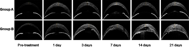
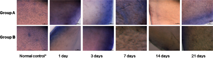
Methods: Transcorneal freezing was performed on the right eye of each mouse. One cycle of cryoinjury was performed in 18 eyes (group A), and three cycles were performed in 17 eyes (group B). Pachymetry and intraocular pressure (IOP) measurements were done preoperatively, as well as at 1, 3, 7, 14, and 21 days after cryoinjury. At each post-cryoinjury time point, three mice from each group were euthanized, and the corneas underwent histology and electron microscopy.

Results: In both groups, significant corneal edema was noted at post-cryoinjury day 1, which was maintained throughout the study period. IOP remained within normal range in group A, but increased significantly with time in group B (p=0.011 at day 1, 0.038 at day 3, 0.026 at day 14, and 0.008 at day 21). In group B, serious complications including hyphema (one case), severe iridocorneal adhesion (15 cases), and total cataract (three cases) were detected, while only mild iridocorneal adhesion (four cases) and cataract (three cases) were noted in group A. Live/dead cell assay, hematoxylin and eosin staining, and scanning electron microscopy revealed successful ablation of corneal endothelial cells and absence of regeneration in both groups. Hematoxylin and eosin staining and terminal deoxynucleotidyl transferase-mediated nick end labeling assay showed that apoptosis was mainly confined to the posterior stroma and endothelium in group A, while severe apoptosis was observed throughout all layers of the cornea in group B.

Conclusions: One cycle of cryoinjury was safer than three, while both were equally effective in inducing bullous keratopathy. This cryoinjury mouse model of bullous keratopathy was a consistently reproducible model that can be used for further studies on endothelial cell damage and rescue therapy.


Figures

Figure 1
Figure 1
Transcorneal freezing using a cryoprobe with a 2.5 mm diameter on the right eye of a mouse.
Figure 2
Figure 2
Anterior segment optical coherence tomography. Significant corneal edema developed in groups A and B, and the increased corneal thickness was maintained during the study period. Although the anterior segment appears normal except the corneal edema and mild cataract in group A, severe anterior chamber inflammation and iridocorneal adhesion are remarkable in group B.
Figure 3
Figure 3
Representative photos showing complications following the cryoinjury. In group A, mild peripheral iridocorneal adhesion (left) and mild cataract (middle and right) were found. In group B, serious complications including severe anterior chamber inflammation and cataract (left), total iridocorneal adhesion (middle), and hyphema (right) were detected.
Figure 4
Figure 4
Live/dead cell assay using alizarin red S and Trypan Blue staining of the corneal endothelial cells. Successful ablation of the corneal endothelial cells was observed in samples from day 1, and no evidence of cell regrowth was detected during the study period. Descemet’s membrane was preserved over most of the area. Scale bar=50 μm. *taken from the opposite eye that did not undergo cryoinjury.
Figure 5
Figure 5
Hematoxylin and eosin staining. Ablation of endothelial cells was observed at all time points in both groups. At 1 day after cryoinjury, more severe infiltration of inflammatory cells throughout all layers of the cornea in group B compared to that in group A was observed. Although Descemet’s membrane (DM) was mostly preserved, detachment of DM was detected in some areas (black arrow). Scale bar=50 μm. *taken from the opposite eye that did not undergo cryoinjury.
Figure 6
Figure 6
Results of the terminal deoxynucleotidyl transferase-mediated nick end labeling assay. A: Apoptosis (red color) was observed only in a few cells in the normal cornea. B: In a cornea from group A, apoptosis was detected mainly in the posterior stroma and endothelial layer. C: In a cornea from group B, severe apoptosis throughout all layers of the cornea occurred. Scale bar=50 μm. *taken from the opposite eye that did not undergo cryoinjury.
Figure 7
Figure 7
Scanning electron microscopy. At day 1, some corneal endothelial cells (CECs) are still attached to Descemet’s membrane (DM) in both groups, although most of the cells were ablated in both groups. Most of the remnant cells have detached between day 3 and day 14 in both groups. At day 21, denuded DM with only scant distribution of remnant CECs was noted in both groups. Scale bar=20 μm. *taken from the opposite eye that did not undergo cryoinjury.

References

    1. Khodadoust AA, Green K. Physiological function of regenerating endothelium. Invest Ophthalmol. 1976;15:96–101. - PubMed
    1. Maurice DM, Giardini AA. Swelling of the cornea in vivo after the destruction of its limiting layers. Br J Ophthalmol. 1951;35:791–7. - PMC - PubMed
    1. Stocker FW. The endothelium of the cornea and its clinical implications. Trans Am Ophthalmol Soc. 1953;51:669–786. - PMC - PubMed
    1. Morton PL, Ormsby HL, Basu PK. Healing of endothelium and Descemet's membrane of rabbit cornea. Am J Ophthalmol. 1958;46:62–7. - PubMed
    1. McDonald JE. The effect of endothelial curettement on corneal wound healing: experimental study. Am J Ophthalmol. 1961;51:930–41. - PubMed

Publication types

LinkOut - more resources