

Using μPIXE for quantitative mapping of metal concentration in Arabidopsis thaliana seeds
- PMID: 23761799
- PMCID: PMC3669754
- DOI: 10.3389/fpls.2013.00168
Using μPIXE for quantitative mapping of metal concentration in Arabidopsis thaliana seeds
Abstract
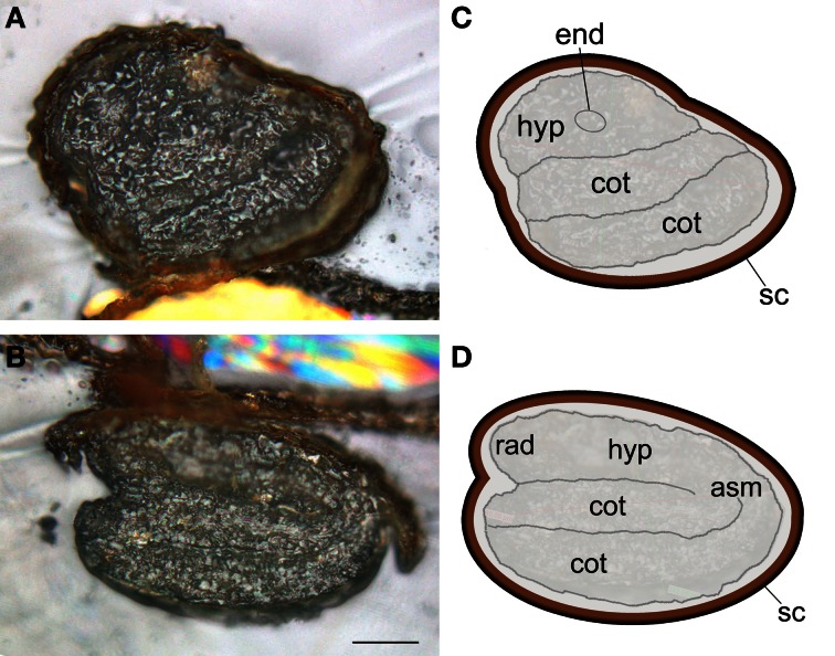
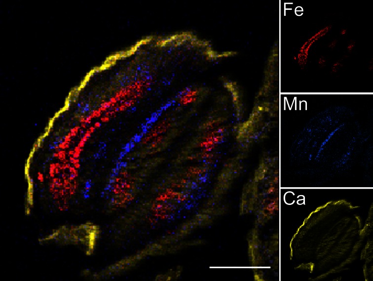
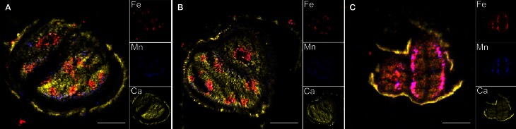
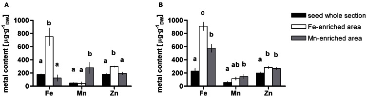
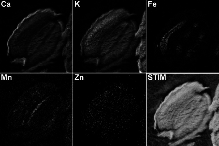
Seeds are a crucial stage in plant life. They contain the nutrients necessary to initiate the development of a new organism. Seeds also represent an important source of nutrient for human beings. Iron (Fe) and zinc (Zn) deficiencies affect over a billion people worldwide. It is therefore important to understand how these essential metals are stored in seeds. In this work, Particle-Induced X-ray Emission with the use of a focused ion beam (μPIXE) has been used to map and quantify essential metals in Arabidopsis seeds. In agreement with Synchrotron radiation X-ray fluorescence (SXRF) imaging and Perls/DAB staining, μPIXE maps confirmed the specific pattern of Fe and Mn localization in the endodermal and subepidermal cell layers in dry seeds, respectively. Moreover, μPIXE allows absolute quantification revealing that the Fe concentration in the endodermal cell layer reaches ~800 μg·g(-1) dry weight. Nevertheless, this cell layer accounts only for about half of Fe stores in dry seeds. Comparison between Arabidopsis wild type (WT) and mutant seeds impaired in Fe vacuolar storage (vit1-1) or release (nramp3nramp4) confirmed the strongly altered Fe localization pattern in vit1-1, whereas no alteration could be detected in nramp3nramp4 dry seeds. Imaging of imbibed seeds indicates a dynamic localization of metals as Fe and Zn concentrations increase in the subepidermal cell layer of cotyledons after imbibition. The complementarities between μPIXE and other approaches as well as the importance of being able to quantify the patterns for the interpretation of mutant phenotypes are discussed.
Keywords: Arabidopsis; elemental mapping; iron; quantitative; seed; μPIXE.
Figures

References
-
- Briat J. F., Lobreaux S. (1997). Iron transport and storage in plants. Trends Plant Sci. 2, 187–193 10.1016/S1360-1385(97)85225-9 - DOI
-
- Budka D., Mesjasz-Przybylowicz J., Tylko G., Przybylowicz W. J. (2005). Freeze-substitution methods for Ni localization and quantitative analysis in Berkheya coddii leaves by means of PIXE. Nucl. Instrum. Methods Phys. Res. B 231, 338–344 10.1016/j.nimb.2005.01.080 - DOI
-
- Campbell J. L., Hopman T. L., Maxwell J. A., Nejedly Z. (2000). Guelph PIXE software package III: alternative proton database Nucl. Instrum. Methods Phys. Res. B 170, 193–204 10.1016/S0168-583X(00)00156-7 - DOI
LinkOut - more resources
Full Text Sources
Other Literature Sources