. 2013 Jun 12;33(24):10110-22.
doi: 10.1523/JNEUROSCI.5606-12.2013.

Conditional deletion of Atoh1 reveals distinct critical periods for survival and function of hair cells in the organ of Corti

Affiliations

Conditional deletion of Atoh1 reveals distinct critical periods for survival and function of hair cells in the organ of Corti

Tiantian Cai et al. J Neurosci. .

Abstract

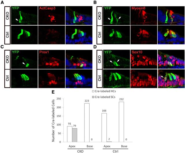
Atonal homolog1 (Atoh1) encodes a basic helix-loop-helix protein that is the first transcription factor to be expressed in differentiating hair cells. Previous work suggests that expression of Atoh1 in prosensory precursors is necessary for the differentiation and survival of hair cells, but it is not clear whether Atoh1 is required exclusively for these processes, or whether it regulates other functions later during hair cell maturation. We used EGFP-tagged Atoh1 knock-in mice to demonstrate for the first time that Atoh1 protein is expressed in hair cell precursors several days before the appearance of differentiated markers, but not in the broad pattern expected of a proneural gene. We conditionally deleted Atoh1 at different points in hair cell development and observe a rapid onset of hair cell defects, suggesting that the Atoh1 protein is unstable in differentiating hair cells and is necessary through an extended phase of their differentiation. Conditional deletion of Atoh1 reveals multiple functions in hair cell survival, maturation of stereociliary bundles, and auditory function. We show the presence of distinct critical periods for Atoh1 in each of these functions, suggesting that Atoh1 may be directly regulating many aspects of hair cell function. Finally, we show that the supporting cell death that accompanies loss of Atoh1 in hair cells is likely caused by the abortive trans-differentiation of supporting cells into hair cells. Together our data suggest that Atoh1 regulates multiple aspects of hair cell development and function.


Figures

Figure 1.
Figure 1.
Atoh1 protein expression in the mouse cochlea. A, Schematic diagram of the Atoh1A1GFP/A1GFP knock-in mouse line, referred to in the figure as Atoh1-EGFP. An Atoh1-EGFP fusion gene was targeted to the Atoh1 genomic locus. B, Whole-mount Atoh1A1GFP/A1GFP cochleas from E13.5 to P0. The right two rows show higher magnification of the base and apex regions marked by box at the left. Hey2 (red) was used to mark the prosensory domain at E13.5 and E14.5. Scale bars, 200 μm. C, Sections of Atoh1A1GFP/A1GFP cochlea at E13.5. Scattered EGFP-labeled cells (arrows) were localized within the prosensory domain labeled by p27kip1 and Hey2. D, Sections through different regions of E16.5 Atoh1A1GFP/A1GFP cochlea. Hair cells were labeled by Myosin6 (red). Hair cell differentiation begins near the base of the cochlea and spreads down to the apex over a period of days. Sections through the base, middle turn, and apex of the cochlea at E16.5 therefore reveal different stages in the differentiation of hair cell precursors. In the base, most Atoh1-EGFP protein is localized in the nucleus of Myosin6-expressing hair cells. However, in the apex, much cytoplasmic Atoh1-EGFP protein can be observed in precursors that have not yet expressed Myosin6. E, An Atoh1 antibody also reveals nuclear staining in hair cells in the base of the cochlea, more cytoplasmic staining in the middle turn of the cochlea, but no detectable staining in the apex. Arrowhead, inner hair cells; bracket, outer hair cells.
Figure 2.
Figure 2.
Precursor cells in the sensory epithelia start to undergo apoptosis at E15.5 in the Atoh1-null (−/−) mice. A–C, E–G, Whole-mount cochleas from E15.5 Atoh1-null (A–C) and Atoh1 heterozygous (E–G) littermates. p27kip1 (green) marks the prosensory epithelial region. ActCasp3 (red) labels apoptotic cells in the prosensory epithelium of the Atoh1-null cochlea. C', G', Higher-power magnifications of the midbasal region of the cochlea marked by box in C and G, respectively. The dotted lines highlight the prosensory epithelium. D, H, Sections of the basal cochlea at E15.5. ActCasp3 (red) staining was only observed in the Atoh1 mutants (D) but not their heterozygous littermates (H).
Figure 3.
Figure 3.
A, Breeding scheme to generate Atoh1-CKO mice. Female Atoh1flox/flox; R26R-YFP mice were mated with Atoh1+/−; Atoh1-CreERT2 males to generate mice that carried the CreERT2 allele, one copy of the R26R-YFP Cre reporter, and either an Atoh1flox/+ (50%) or Atoh1flox/− (50%) allele. Use of the R26R-YFP Cre reporter provides a readout of the leakiness, speed, and efficiency of the system. B, The base and apex of a cochlea from a P3 Atoh1flox/−; Atoh1-CreERT2; R26R-YFP mouse whose mother did not receive tamoxifen during pregnancy. No YFP-labeled cells can be detected in either region of the cochlea, and we typically observed an average of 1 YFP+ cell in every five cochleas examined. C, The base and apex of cochleas from Atoh1flox/+; Atoh1-CreERT2; R26R-YFP mice whose mothers received a single dose of tamoxifen (TMX) at E17.5. The embryos were collected 8, 24, and 48 h after tamoxifen dosing. A small number of YFP cells can be observed in both base and apex after 8 h. After 24 h, significantly more labeled cells can be observed in the base, in which hair cells differentiate first, compared with the apex. After 48 h, similar numbers of YFP-labeled cells (80–85%) are present in both regions of the cochlea. D, The Atoh1-CreERT2 transgene remains active until at least 2 d after birth. Neonatal pups were injected with tamoxifen at either P0 or P2 and allowed to develop for 24 h. After birth, Atoh1 starts to be downregulated from the cochlea, starting at the base and proceeding down to the apex. Consequently, significantly fewer YFP-labeled cells can be seen in the base at both ages, but ∼50% YFP+ cells are seen in the apex.
Figure 4.
Figure 4.
Atoh1 is required for the survival of hair cells in a time-dependent manner. A, Diagram showing the experimental design for the analysis of Atoh1-CKO cochleas in B–D. Female Atoh1flox/flox; R26R-YFP mice were mated with Atoh1+/−; Atoh1-CreERT2 males and received a single dose of tamoxifen (TMX) at E15.5, E16.5, or E17.5. Mouse pups were analyzed on the day of birth (P0). B–D, Different regions of whole-mount cochleas from P0 Atoh1-CKO and control littermates in which one dose of tamoxifen was administered at E15.5 (B), E16.5 (C), and E17.5 (D). YFP (green) was used as a reporter to show Cre-mediated recombination. Red, Myosin6-labeled hair cells. The number of hair cells in both CKO and control cochleas is quantified and shown in the graphs at the right (n ≥ 3, error bars show SD).
Figure 5.
Figure 5.
Atoh1 is required for stereociliary bundle development in hair cells. A, Phalloidin staining of different regions of whole-mount cochlea preparations from P2 Atoh1-CKO and control littermates. One dose of tamoxifen administration at E17.5 resulted in disorganized hair bundles in the middle and apical turns of CKO cochleas, but no defects in the more mature hair cells in the basal region. However, only very subtle defects in hair bundles were observed in the apical cochlea of CKO animals that received one dose of tamoxifen at P0. This suggests that Atoh1 is initially required for hair cell survival, but later for other functions, including hair bundle morphology B, Whole-mount cochleas from Atoh1-CKO and control animals receiving one dose of tamoxifen at E17.5 and stained with anti-espin antibodies at P2. C, Whole-mount cochleas from Atoh1-CKO and control animals receiving one dose of tamoxifen at E17.5 and then dissected at P5 and exposed to 5 μm of the styryl dye FM1-43 for 15 s to detect dye uptake through mechanotransduction channels. Surviving hair cells from apical regions of the cochlea displaying abnormal bundle morphology (A, B) still take up FM1-43. Scale bars, 20 μm. TMX, tamoxifen.
Figure 6.
Figure 6.
Timing of Atoh1 deletion affects extent of hair cell survival and preservation of hearing function. A, B, Atoh1 CKO mice have profound hearing loss when tamoxifen is administered at E17.5 (ANOVA p = 7.96e–11) (A), but only mild hearing loss when tamoxifen administration is delayed until P2 (ANOVA p = 0.1849) (B). Error bars represent SD. The Student's t test with Benjamini–Hochberg adjustment of p values for multiple comparisons was used to estimate statistical significance between genotypes at individual frequencies. *p < 0.05, **p < 0.02, ***p < 0.001. C, When tamoxifen is administered at E17.5, hair cells, visualized with Myosin6 immunostaining (red), are only missing from the apex of the cochlea at P7. However, by P15, hair cell loss is seen in the middle turn of the cochlea and, by P42, at the base. Control animals show no hair cell loss. Regions lacking hair cells also have defects in afferent and efferent innervation, revealed by staining with the TuJ1 antibody (green). D, When tamoxifen is administered at P2, no significant defects are seen in either hair cell numbers or innervation.
Figure 7.
Figure 7.
Atoh1 is required for the survival of the surrounding supporting cells in a time-dependent manner. A, Sections of middle turn regions of the cochlea from Atoh1-null and heterozygous embryos at E19. The expression of supporting markers Prox1, Sox2, and Jag1 was abolished in the knock-out mutants. Similar, albeit slightly milder phenotypes were also seen in Atoh1-CKO mice that received three daily doses of tamoxifen between E13.5 and E15.5 and were killed at E19. B–D, Whole-mount cochleas from P0 Atoh1-CKO and control littermates receiving one dose of tamoxifen at E15.5 (B), E16.5 (C), and E17.5 (D). This is the same experimental regimen shown in Figure 4A. Supporting cells were visualized by staining with a Prox1 antibody (red). The number of Prox1-labeled cells in both CKO and control cochleas on each injection day is quantified and shown in the graph.
Figure 8.
Figure 8.
Atoh1 is indirectly required for differentiation and survival of cochlear supporting cells. A–D, Sections of apical regions of the cochlea from Atoh1-CKO and control animals harvested 24 h after tamoxifen treatment at E17.5. YFP shows cells that underwent recombination after tamoxifen administration. ActCasp3 (red) labels apoptotic cells in the apex of CKO organ (A). Myosin6 labels differentiated hair cells (B). Prox1 (C) and Sox10 (D) were used to examine supporting cell development. YFP+ cells (arrow) in the supporting cell layer were only observed in the apical region of CKO cochlea. This suggested that cells in the supporting cell layer of the CKO, but not wild-type cochlea, were activating the Atoh1-CreERT2 transgene. In the apex of control cochlea, we often saw inner hair cells labeled by Sox10 (D, arrowhead). E, Quantification of YFP+ cells in the hair cell and supporting cell layers of Atoh1-CKO and control mice harvested 24 h after tamoxifen treatment at E17.5. The total number of YFP+ cells was quantified in regions approximating to the basal and apical thirds of the cochlea from three different specimens of each genotype. Cells were assigned based on their position in the hair cell or supporting cell layers. Significant numbers of YFP-labeled cells (79) were observed in the supporting cell layer from the apical regions of three Atoh1-CKO cochleas. In contrast, only two YFP-labeled supporting cells from three cochleas could be observed in the apical turn of control cochleas. HC, hair cell; SC, supporting cell.

References

    1. Alagramam KN, Murcia CL, Kwon HY, Pawlowski KS, Wright CG, Woychik RP. The mouse Ames waltzer hearing-loss mutant is caused by mutation of Pcdh15, a novel protocadherin gene. Nat Genet. 2001;27:99–102. doi: 10.1038/83837. - DOI - PubMed
    1. Ben-Arie N, Bellen HJ, Armstrong DL, McCall AE, Gordadze PR, Guo Q, Matzuk MM, Zoghbi HY. Math1 is essential for genesis of cerebellar granule neurons. Nature. 1997;390:169–172. doi: 10.1038/36579. - DOI - PubMed
    1. Bermingham NA, Hassan BA, Price SD, Vollrath MA, Ben-Arie N, Eatock RA, Bellen HJ, Lysakowski A, Zoghbi HY. Math1: an essential gene for the generation of inner ear hair cells. Science. 1999;284:1837–1841. doi: 10.1126/science.284.5421.1837. - DOI - PubMed
    1. Burns JC, Cox BC, Thiede BR, Zuo J, Corwin JT. In vivo proliferative regeneration of balance hair cells in newborn mice. J Neurosci. 2012;32:6570–6577. doi: 10.1523/JNEUROSCI.6274-11.2012. - DOI - PMC - PubMed
    1. Chen P, Johnson JE, Zoghbi HY, Segil N. The role of Math1 in inner ear development: uncoupling the establishment of the sensory primordium from hair cell fate determination. Development. 2002;129:2495–2505. - PubMed

Publication types

MeSH terms

LinkOut - more resources