. 2013 Jun 12;33(24):10123-31.
doi: 10.1523/JNEUROSCI.4646-12.2013.

Temporal components in the parahippocampal place area revealed by human intracerebral recordings

Affiliations

Temporal components in the parahippocampal place area revealed by human intracerebral recordings

Julien Bastin et al. J Neurosci. .

Abstract

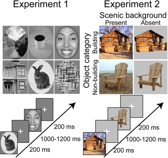
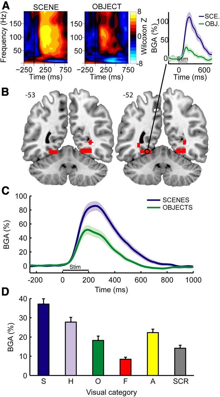
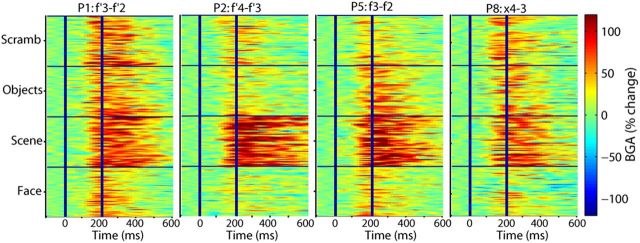
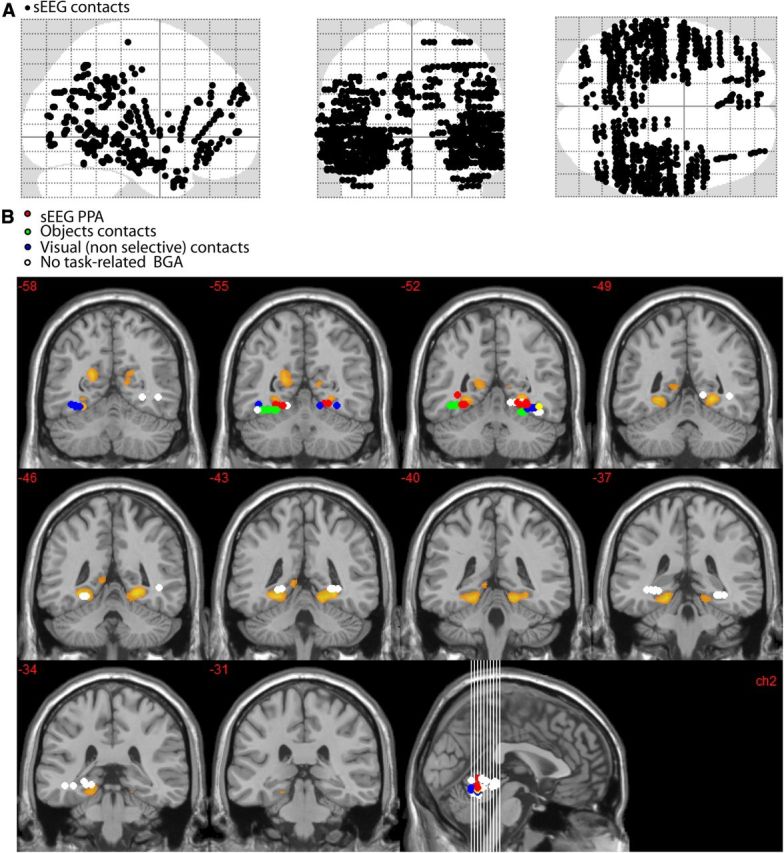
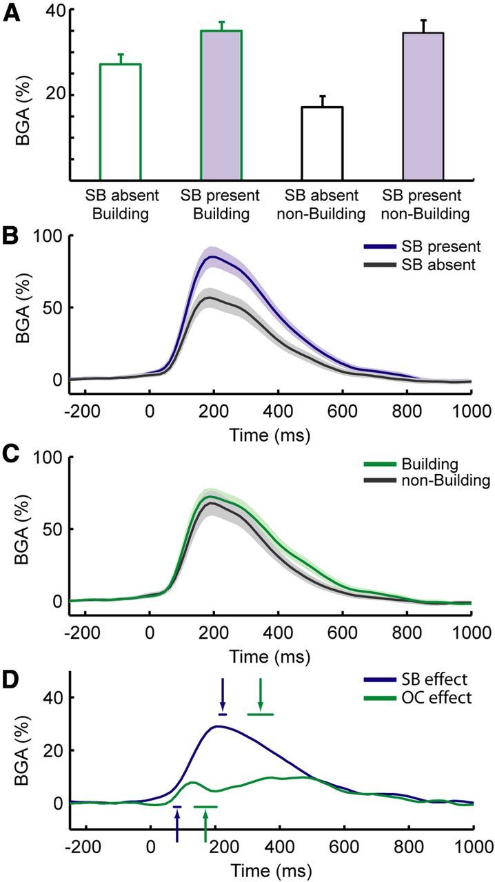
Many high-level visual regions exhibit complex patterns of stimulus selectivity that make their responses difficult to explain in terms of a single cognitive mechanism. For example, the parahippocampal place area (PPA) responds maximally to environmental scenes during fMRI studies but also responds strongly to nonscene landmark objects, such as buildings, which have a quite different geometric structure. We hypothesized that PPA responses to scenes and buildings might be driven by different underlying mechanisms with different temporal profiles. To test this, we examined broadband γ (50-150 Hz) responses from human intracerebral electroencephalography recordings, a measure that is closely related to population spiking activity. We found that the PPA distinguished scene from nonscene stimuli in ∼80 ms, suggesting the operation of a bottom-up process that encodes scene-specific visual or geometric features. In contrast, the differential PPA response to buildings versus nonbuildings occurred later (∼170 ms) and may reflect a delayed processing of spatial or semantic features definable for both scenes and objects, perhaps incorporating signals from other cortical regions. Although the response preferences of high-level visual regions are usually interpreted in terms of the operation of a single cognitive mechanism, these results suggest that a more complex picture emerges when the dynamics of recognition are considered.


Figures

Figure 1.
Figure 1.
Experimental design: examples of stimuli and presentation rates used in Experiments 1 and 2. Stimulus duration was set to 200 ms during both experiments.
Figure 2.
Figure 2.
TF response and broadband BGA in the PPA. A, Example of statistical TF maps (Wilcoxon z scores) and BGA response time course (right) during scene and object viewing from a single recording site (Patient 2; MNI coordinates: −23, −54, −12) located in the posterior part of the collateral sulcus. Shaded areas represent ±1 SEM across trials. B, Anatomical location of the contacts that respond preferentially to scenes. Two adjacent slices from MNI brain are shown. Numbers on the right of each image indicate MNI coordinates in the anterior–posterior (y) axis. C, Average BGA responses aligned to stimulus onset during scene and object encoding in scene-selective contacts (n = 14 contacts recorded from 6 patients). Shaded areas represent ±1 SEM across contacts. D, Average BGA responses during a 0–800 ms time interval to six different stimulus types in these contacts. S, Scenes; H, houses; O, objects; F, faces; A, animals; SCR, scrambled. Error bars represent SEM across contacts.
Figure 3.
Figure 3.
Reproducibility across trials and across patients of BGA responses recorded from the PPA during Experiment 1 (2 left PPA and 2 right PPA contacts recorded from four different patients are shown).
Figure 4.
Figure 4.
Anatomical locations of all intracerebral contact-pairs (A) and relationship between fMRI-defined PPA and contact-pairs that were within an ROI centered on the fMRI-defined PPA (MNI coordinates: abs(x) <45; −35<y<−60; −18<z<0) (B). Voxels that respond more to scenes than to objects are shown in orange (fMRI data from R.A.E. laboratory). sEEG contacts that were located within the ROI were categorized into four functionally distinct response types: red dots represent sEEG-defined PPA contacts (i.e., BGA for these contacts was selective to scene stimuli); green dots, contacts that were selective to objects; blue dots, contacts that showed a BGA increase that was similar across categories (visual nonselective); white dots, sEEG contacts that did not respond to visual stimulation (nonresponsive). The sEEG-defined PPA fits within the posterior tail of the fMRI-defined PPA. The center of fMRI-defined PPA was not sampled by intracerebral contacts. The more lateral location of intracerebral contacts selective to objects is consistent with the neuroimaging literature.
Figure 5.
Figure 5.
BGA at PPA contacts in Experiment 2. A, Grand-average BGA activity during the four conditions of Experiment 2 (average BGA during the 0–800 ms after stimulus interval): building with no background, building with background, nonbuilding object without background, and nonbuilding object with background. B, Grand-average BGA responses during viewing of stimuli that either had a scenic background (SB present) or not (SB absent). C, Grand-average BGA responses during viewing of stimuli that either contained buildings or nonbuilding objects. D, Time course of the scene (SB present − SB absent stimuli) versus object (building − nonbuilding stimuli) effects. Arrows indicate the average latencies of the onsets (arrows pointing upward) and peak times (arrows pointing downward) of experimental effects. Shaded areas and horizontal lines below or above arrows represent ±1 SEM. SB effect, Scenic background effect; OC effect, object category effect.

Similar articles

Cited by

References

    1. Amit E, Mehoudar E, Trope Y, Yovel G. Do object-category selective regions in the ventral visual stream represent perceived distance information? Brain Cogn. 2012;80:201–213. doi: 10.1016/j.bandc.2012.06.006. - DOI - PubMed
    1. Antes JR, Penland JG, Metzger RL. Processing global information in briefly presented pictures. Psychol Res. 1981;43:277–292. doi: 10.1007/BF00308452. - DOI - PubMed
    1. Bar M. Visual objects in context. Nat Rev Neurosci. 2004;5:617–629. doi: 10.1038/nrn1476. - DOI - PubMed
    1. Bar M, Aminoff E. Cortical analysis of visual context. Neuron. 2003;38:347–358. doi: 10.1016/S0896-6273(03)00167-3. - DOI - PubMed
    1. Bar M, Kassam KS, Ghuman AS, Boshyan J, Schmid AM, Dale AM, Hamalainen MS, Marinkovic K, Schacter DL, Rosen BR, Halgren E. Top-down facilitation of visual recognition. Proc Natl Acad Sci U S A. 2006;103:449–454. doi: 10.1073/pnas.0507062103. - DOI - PMC - PubMed

Publication types

LinkOut - more resources