. 2013 May 15:7:85-102.
doi: 10.4137/GRSB.S10751. Print 2013.

Investigation of dmyc Promoter and Regulatory Regions

Affiliations

Investigation of dmyc Promoter and Regulatory Regions

Jasmine Kharazmi et al. Gene Regul Syst Bio. .

Abstract

Products of the myc gene family integrate extracellular signals by modulating a wide range of their targets involved in cellular biogenesis and metabolism; the purpose of this integration is to regulate cell death, proliferation, and differentiation. However, understanding the regulation of myc at the transcription level remains a challenge. We performed rapid amplification of dmyc cDNA ends (5' RACE) and mapped the transcription start site at P1 promoter, 18 base pairs upstream of the start of the known EST GM01143 and within the 5' UTR. Our data show that the first TATA box, previously computationally predicted, is utilized to generate dmyc full length mRNA. The largest transcript contains all three exons, generated after the removal of the introns by constitutively regulated splicing events. Further investigation of Downstream Promoter Element (DPE) was achieved by studying lacZ reporter activity; investigation revealed that this element and its upstream cluster of binding sites are required for the dmyc intron 2 activity. These findings may provide valuable tools for further analysis of dmyc cis-elements.

Keywords: 5′ RACE; Downstream Promoter Element (DPE); Drosophila; RNA splicing; TATA-box; dmyc.


Figures

Figure 1
Figure 1
Extension of the dmyc cDNA at its 5′ end. Notes: Polymerase chain reaction amplification of the dmyc cDNA 5′ end was performed with the forward primer UPM and the reverse primer GSP-N01; two different settings of annealing and extension temperatures were used (see Materials and Methods). Sequences for the oligonucleotides are listed in Table S1. Abbreviations: TFR, Transferrin Receptor; UPM, Universal Primer Mix; GSP-N01, Gene Specific Primer N-term.
Figure 2
Figure 2
5′ RACE analysis of dmyc cDNA at its 5′ site detects TATA box1 as main promoter region. dmyc genomic organization is shown at the top (not drawn to scale). Notes: Gene specific primer GSP-N01 (blue arrow) was used to amplify the dmyc cDNA 5′ end and the sequencing primer (black arrow) was used to sequence the dmyc cDNA end. (The UPM forward amplification primer and the UPM-sh the forward sequencing primer are not plotted). The 5′ end sequence of the dmyc cDNA is shown at the bottom. The sequenced portion of the cDNA 5′ end is highlighted in magenta and the first transcribed nucleotide is in green. For the sequencing chromatogram, see Figure S1. Sequences for the oligonucleotides are listed in Table S1.
Figure 3
Figure 3
Insertion of the P-element P{lacW} at the TATA box1 region of the dmyc gene is lethal. Notes: Insertion site of the reporter lacZ P-element (10.3 kb in size) at the dmyc locus is shown. Breakpoints of the insertion are as follows: 3D2, X:3267141..3267197, which maps to the region 213 nucleotides upstream of dmyc exon1 start site. Insertion of the P-element P{lacW} causes deletion of 56 base pairs (red highlight), including GC box1 and TATA box1, at the breakpoint of insertion. A residue (green highlight adjacent to the breakpoint) was shown to be the first transcribed base (Fig. 2) that favors capping of the dmyc mRNA.
Figure 4
Figure 4
5′ RACE analysis of the dmyc cDNA ends detects splice junctions of the transcript. Notes:dmyc genomic organization (not drawn to scale) with the nucleotides at the exon/intron junctions are shown in green and red. Gel picture shows polymerase chain reaction amplification products of the dmyc cDNA with the gene specific reverse primers GSP-C05 and GSP-C06. For all of the amplification reactions, the Universal Primer Mix (UPM, Clontech) was used as forward primer.
Figure 5
Figure 5
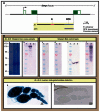
dmyc downstream promoter element (DPE) and its upstream binding sites cluster are required for the intron 2 activity. (A) The full length intron 2 in the J8 transgene and its truncation, J8.2, the enhancer region in J8.4 and the 3′ truncation J8.5 (containing the DPE element), and their relative location with respect to the dmyc locus and the genomic organization are shown. The insertion site of the wild type dmyc-lacZ P-element in the fly stock w67c23P{lacW}dmG0354/FM7c at the 5′ end of the dmyc gene is depicted. (BJ) In the wild type dmyc-lacZ enhancer trap line, lacZ patterning reflects the pattern reported for the dmyc endogenous mRNA distribution (B, brain; C, wing disc; D, eye disc; E, leg disc). (FI) In the embryos, lacZ is expressed with high similarities to dmyc mRNA distribution, predominantly in the midgut, hindgut, pharynx, anal pad, and partly in the mesodermal tissue (embryo stages are as follows: F, G, 5–9; H, I, 12–16). The staining in the ovary (J) reflects the dmyc mRNA localization, which has been reported in nurse cells and oocyte, but is weakly expressed at the tip of the germarium. (KS) The J8 and J8.2 transgenes retain normal patterning of the dmyc in the brain and discs (KN), throughout different stages of embryogenesis (OR embryo stages: O, P, 5–9; Q, R, 9–13), and in the ovary (S). (T–Z) J8.4 and the J8.5 transgenes fail to express lacZ in the larval brain and discs (TW), in the embryos (XY′ embryo stages: X, 2–6; X′, Y, 9–12; Y′, 13–16), and in the ovary (Z). (Z′) Reverse transcriptase polymerase chain reaction on J8.4 brain and ovary (shown in T, Z) and J8.5 disc (shown in U) detects no lacZ transcripts beyond background level. Controls used were as follows: -RT, negative control with no transcriptase (brain from K); P, K, positive control; N, y[1] w[1118] leg discs, negative control; actin, Drosophila actin as internal control. Notes: The yellow arrow indicates lacZ expression and the white arrow indicates lack of lacZ activity. Staining time for discs and ovary is indicated above the scale bar; embryos were stained overnight. Scale bar in (BZ) indicates 50 μm.
Figure 6
Figure 6
Detection of lacZ protein by Western Blot analysis of ovary extracts isolated from flies carrying J8 or J8.5 transgenes. (A) The intron 2 full length in the J8 transgene and its truncation, J8.5, and their relative location with respect to the dmyc locus and the genomic organization are shown. (B) Proteins extracted from the ovaries of the female flies carrying J8 or J8.5 transgenes were analyzed by SDS-PAGE and immunoblotting; lacZ protein was detectable in the J8 ovaries (panel b, lane 2), whereas J8.5 extract was negative (panel b, lane 3). Notes: (a): Gel SafeBlue staining of the protein extracts; 1: protein marker in kDa unit, 2: J8 lysate, 3: J8.5 lysate. (b): The extracts in (a) subjected to immunoblotting with antibody anti-beta Gal. (c): J8 extracts with no primary antibody added, (d): J8 extracts with no secondary antibody added, (e): The extracts in (a) subjected to immunoblotting with antibody anti-Drosophila actin. (f): positive Control: beta-Gal protein. (g): lacZ staining of J8 ovaries and (h): lacZ staining of J8.5 ovaries. The staining time for ovaries is indicated above the scale bar. Scale bar in (g, h) is 50 μm.
Figure 7
Figure 7
Combined action of the downstream promoter element and its upstream binding sites are required for the intron 2 activity. (A) The 40 kb dmyc locus with the predicted regulatory elements and the deletion constructs derived from the intron 2 region are shown. (BJ) Fusion of the enhancer region (J8.4) to the downstream promoter element (J8.5) in J8.3 construct recapitulates the J8/J8.2 transgenes activity in the larval brain and discs (BE), during embryogenesis (FI embryo stages: F, 2–5; G, 9–12; H, I, 12–15), and in the ovary (J). Notes: Staining time for discs and the ovary is indicated above the scale bar; embryos were stained overnight. Scale bar in (BJ) indicates 50 μm.
Figure 8
Figure 8
Major splice cis-elements and mechanism of the dmyc RNA splicing. (A) dmyc genomic organization (not drawn to scale) with the nucleotides at the 5′ splice donor and 3′ splice acceptor sites (GT/AG), are shown on top. Splicing cis-elements of the dmyc RNA are shown below and include the following: 5′ and 3′ splice signals (GU/AG), branchpoint consensus sequence, and a polypyrimidine tract located adjacent to the AG sequence at the 3′ end of each intron. (B) dmyc RNA splicing is regulated constitutively, following the strict GU/AG splicing rules and includes the ligation of each upstream exon to its adjacent downstream exon. The A residue (branchpoint) within the branchpoint consensus sequence participates in the formation of the ester bond between the 5′ phosphate of the intron and the 2′ oxygen of the branchpoint A residue. Notes: The second transesterification reaction between the free 3′ oxygen of the upstream exon and the 5′ phosphate of the downstream exon initiates the joining of the exons together and the excision of the intron as a lariat. The exon/exon splice junctions in the mRNA molecule are indicated at the bottom.

Similar articles

Cited by

References

    1. King MW, Roberts JM, Eisenman RN. Expression of the c-myc proto-oncogene during development of Xenopus laevis. Mol Cell Biol. 1986;6:4499–508. - PMC - PubMed
    1. Nishikura K. Expression of c-myc proto-oncogene during the early development of Xenopus laevis. Oncogene Res. 1987;1:179–91. - PubMed
    1. Gallant P, Shiio Y, Cheng PF, Parkhurst SM, Eisenman RN. Myc and Max homologs in Drosophila. Science. 1996;274:1523–7. - PubMed
    1. Chin L, et al. Contrasting roles for Myc and Mad proteins in cellular growth and differentiation. Proc Natl Acad Sci U S A. 1995;92:8488–92. - PMC - PubMed
    1. Sodir NM, Evan GI. Nursing some sense out of Myc. J Biol. 2009;8:77. - PMC - PubMed

LinkOut - more resources