. 2013;9(6):e1003394.
doi: 10.1371/journal.ppat.1003394. Epub 2013 Jun 6.

Streptolysin O and its co-toxin NAD-glycohydrolase protect group A Streptococcus from Xenophagic killing

Affiliations

Streptolysin O and its co-toxin NAD-glycohydrolase protect group A Streptococcus from Xenophagic killing

Maghnus O'Seaghdha et al. PLoS Pathog. 2013.

Abstract

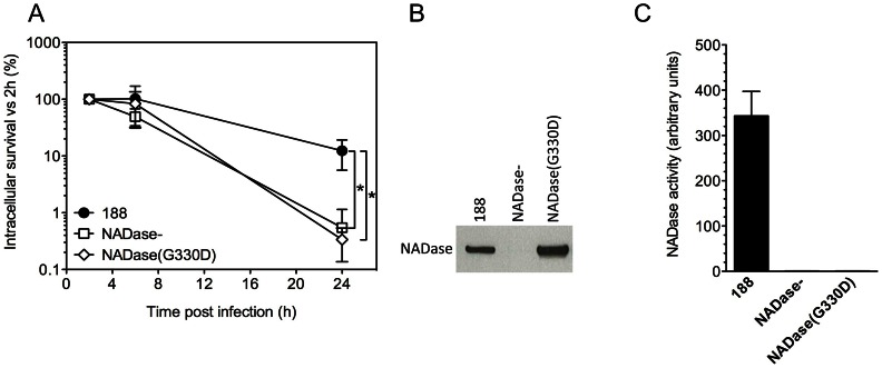
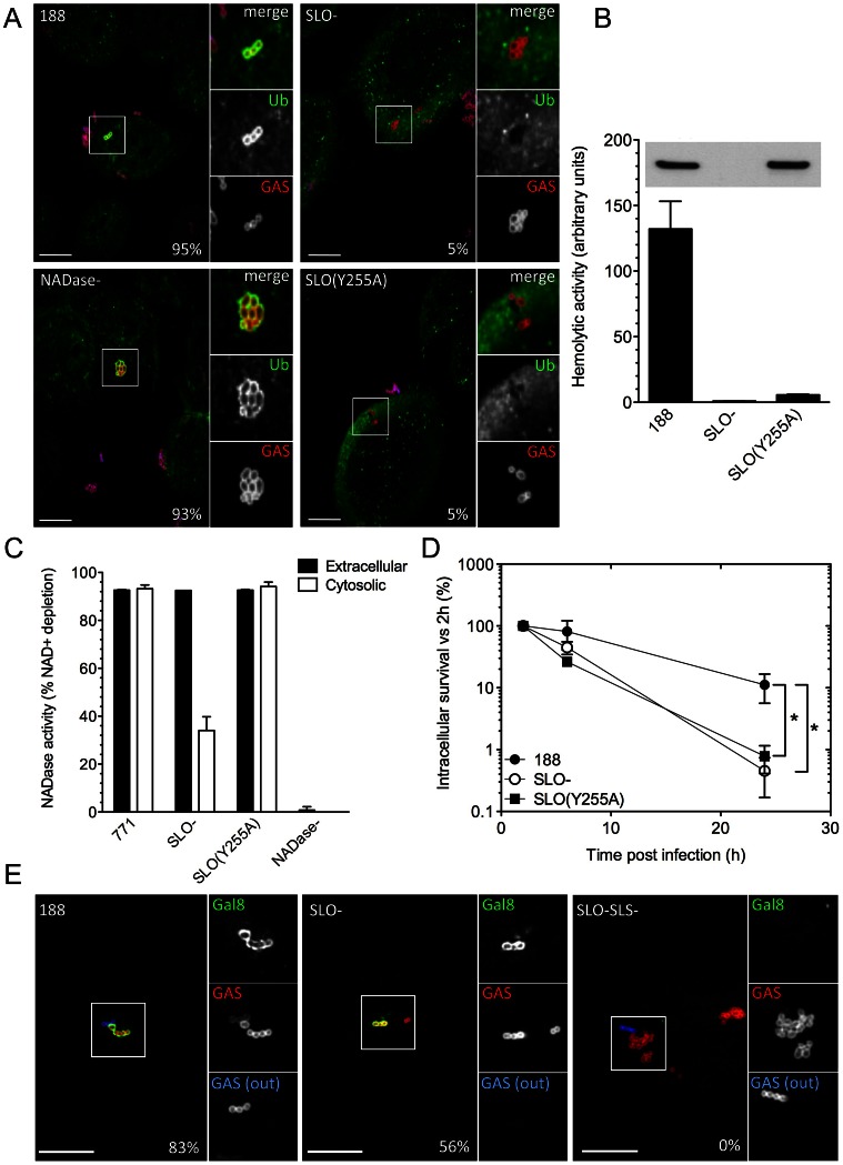
Group A Streptococcus (Streptococcus pyogenes or GAS) causes pharyngitis, severe invasive infections, and the post-infectious syndromes of glomerulonephritis and rheumatic fever. GAS can be internalized and killed by epithelial cells in vitro, a process that may contribute to local innate defense against pharyngeal infection. Secretion of the pore-forming toxin streptolysin O (SLO) by GAS has been reported to stimulate targeted autophagy (xenophagy) upon internalization of the bacteria by epithelial cells. Whereas this process was associated with killing of GAS in HeLa cells, studies in human keratinocytes found SLO production enhanced intracellular survival. To reconcile these conflicting observations, we now report in-depth investigation of xenophagy in response to GAS infection of human oropharyngeal keratinocytes, the predominant cell type of the pharyngeal epithelium. We found that SLO expression was associated with prolonged intracellular survival; unexpectedly, expression of the co-toxin NADase was required for this effect. Enhanced intracellular survival was lost upon deletion of NADase or inactivation of its enzymatic activity. Shortly after internalization of GAS by keratinocytes, SLO-mediated damage to the bacteria-containing vacuole resulted in exposure to the cytosol, ubiquitination of GAS and/or associated vacuolar membrane remnants, and engulfment of GAS in LC3-positive vacuoles. We also found that production of streptolysin S could mediate targeting of GAS to autophagosomes in the absence of SLO, a process accompanied by galectin 8 binding to damaged GAS-containing endosomes. Maturation of GAS-containing autophagosome-like vacuoles to degradative autolysosomes was prevented by SLO pore-formation and by SLO-mediated translocation of enzymatically active NADase into the keratinocyte cytosol. We conclude that SLO stimulates xenophagy in pharyngeal keratinocytes, but the coordinated action of SLO and NADase prevent maturation of GAS-containing autophagosomes, thereby prolonging GAS intracellular survival. This novel activity of NADase to block autophagic killing of GAS in pharyngeal cells may contribute to pharyngitis treatment failure, relapse, and chronic carriage.


Conflict of interest statement

The authors have declared that no competing interests exist.

Figures

Figure 1
Figure 1. SLO and NADase promote intracellular survival of GAS in oropharyngeal keratinocytes.
A. Intracellular survival of GAS strains 188, 188SLO- (SLO-), and 188NADase- (NADase-). Intracellular CFU were quantified at 2 h, 4 h, 6 h, 8 h, and 24 h, and intracellular survival was calculated as the percent viable CFU compared to 2 h. B. Intracellular survival of GAS strain 188SLO- that was partially complemented from plasmid piSLO (SLO-(piSLO)), strain 188 and 188SLO- that contained the empty complementation vector, 188(pSIV4) and SLO-(pSIV4), respectively. C. Intracellular survival of GAS strain JRS4, JRS4SLO- (SLO-), and JRS4NADase- (NADase-). Experiments were performed in triplicate and values represent the mean of three independent experiments ± SD. *, P<0.001.
Figure 2
Figure 2. Xenophagy in oropharyngeal keratinocytes can be stimulated by SLO or SLS and is not associated with killing of intracellular GAS.
A. Confocal microscopy of the association between EGFP-LC3 and GAS strain 188, 188SLO- (SLO-), 188SLS- (SLS-), or 188SLO-SLS- (SLO-SLS-). Immunofluorescent staining distinguished intracellular (Alexa-568, red) from extracellular (Alexa-568 and Alexa-660, red and blue, respectively) GAS. Scale bar = 10 µm. The percent of intracellular GAS that were associated with EGFP-LC3 at 3 h post-infection is shown for each strain. B. Electron microscopy of GAS strains 188, 188SLO-, and 188SLO-SLS- at 3 h post-infection. Arrowheads indicate the presence of double or multiple membranes (188 and 188SLO-) or single membranes (188SLO-SLS-) partially or completely surrounding bacteria. Vacuolar compartments associated with 188 and 188SLO- also contain cytosolic material. C. Intracellular survival of GAS strains 188, 188SLO-, 188SLS-, and 188SLO-SLS-. *, P<0.05. D. Intracellular survival of 188 or 188SLO- in the presence or absence of Beclin1 knockdown.
Figure 3
Figure 3. Expression of active NADase plays a central role in the intracellular survival of GAS in oropharyngeal keratinocytes.
A. Intracellular survival of GAS strains 188, 188NADase-, and 188NADase(G330D). Deletion of the gene encoding NADase or inactivation of NADase by the amino acid substitution G330D resulted in a 20- to 40-fold reduction in intracellular survival compared to parent strain 188. *, P<0.001. NADase western blot (B) and activity measurements (C) of culture supernatants from GAS strains 188, 188NADase-, and 188NADase(G330D).
Figure 4
Figure 4. SLO, SLS, and NADase affect xenophagy and GAS intracellular survival.
A. Confocal microscopy demonstrating the association between ubiquitin (green) and intracellular GAS (red) at 30 min post-infection in keratinocytes infected with GAS strain 188, 188SLO-, 188NADase-, or 188SLO(Y255A). Extracellular GAS were stained blue and red for identification as described in Figure 2. Scale bar = 10 µm. The percentage of intracellular GAS that were associated with ubiquitin is indicated for each strain, based on quantification of at least 100 bacteria in three independent experiments. B. Hemolytic activity of culture supernatants of GAS strain 188, 188SLO-, and 188SLO(Y255A). The amino acid substitution Y255A in SLO abrogates the ability of SLO to porate the host cell membrane, demonstrated by the failure of culture supernatants from 188SLO(Y255A) to lyse erythrocytes despite producing wild-type amounts of the variant SLO protein. Inset, corresponding Western blot for SLO in culture supernatants from these strains. C. NADase activity in cell culture supernatants and in the cytosol of keratinocytes infected with GAS strain 771, 771SLO-, 771SLO(Y255A), or 771NADase-. Intracellular NADase activity due to translocation by extracellular GAS was identical between 771 and 771SLO(Y255A), but reduced in 771SLO-. D. The intracellular survival of 188SLO(Y255A) was significantly lower than that of its parental strain, 188, and similar to that of 188SLO-. *, P<0.01. E. Confocal microscopy demonstrating the association between galectin 8 (green) and intracellular GAS (red) in keratinocytes infected for 30 min with GAS strain 188, 188SLO-, or 188 SLO-SLS-. Extracellular GAS (GAS(out)) were stained blue and red for identification as described in Figure 2. Scale bar = 10 µm. The percentage of intracellular GAS that were associated with galectin 8 is indicated for each strain, based on quantification of at least 100 bacteria in three independent experiments. 188SLO-SLS- was never associated with galectin 8 (0%).
Figure 5
Figure 5. SLO and NADase inhibit lysosomal fusion to GAS-containing autophagosomes in oropharyngeal keratinocytes.
A. Confocal microscopy of keratinocytes infected with GAS strain 188, 188SLO-, or 188NADase- demonstrating the association of the lysosomal marker LAMP-1 (red) with GAS (blue) contained within EGFP-LC3 (green)-positive compartments at 1 h, 3 h, and 6 h post-infection. Scale bar = 10 µm. B. Quantification of the percent of GAS within EGFP-LC3-positive compartments that are co-localized with LAMP-1 at 1 h, 3 h, and 6 h. Data represent mean values from three independent experiments in which at least 100 intracellular GAS were quantified for each time point in each experiment.
Figure 6
Figure 6. Model of the intracellular fate of GAS in oropharyngeal keratinocytes.
Shortly after internalization, GAS are associated with early endosomes. Within minutes, however, damage to the endosomal membrane by the action of SLO and SLS exposes glycans that bind the cytosolic lectin, galectin 8. In addition, SLO produces sufficient damage to the GAS-containing endosome that the bacteria become ubiquitinated upon exposure to the cytosol. Ubiquitin and/or galectin 8 target cytosolic bacteria and damaged endosomes for incorporation into autophagosome-like compartments, which eventually fuse with lysosomes. Lysosomal fusion is delayed or inhibited by SLO pore-formation and SLO-mediated delivery of NADase into the cell cytosol, thus prolonging GAS intracellular survival.

References

    1. Stevens DL (2000) Group A beta-hemolytic streptococci: virulence factors, pathogenesis, and spectrum of clinical infections. In: Stevens DL, Kaplan EL, editors. Streptococcal Infections: Clinical Aspects, Microbiology, and Molecular Pathogenesis. New York: Oxford University Press. pp. 19–36.
    1. Gerber MA, Tanz RR, Kabat W, Bell GL, Siddiqui B, et al. (1999) Potential mechanisms for failure to eradicate group A streptococci from the pharynx. Pediatrics 104: 911–917. - PubMed
    1. Pichichero ME, Casey JR, Mayes T, Francis AB, Marsocci SM, et al. (2000) Penicillin failure in streptococcal tonsillopharyngitis: causes and remedies. Pediatr Infect Dis J 19: 917–923. - PubMed
    1. Greco R, De Martino L, Donnarumma G, Conte MP, Seganti L, et al. (1995) Invasion of cultured human cells by Streptococcus pyogenes . Res Microbiol 146: 551–560. - PubMed
    1. Jadoun J, Ozeri V, Burstein E, Skutelsky E, Hanski E, et al. (1998) Protein F1 is required for efficient entry of Streptococcus pyogenes into epithelial cells. J Infect Dis 178: 147–158. - PubMed

Publication types

MeSH terms