. 2013 Jun 10;8(6):e56330.
doi: 10.1371/journal.pone.0056330. Print 2013.

Novel biodegradable porous scaffold applied to skin regeneration

Affiliations

Novel biodegradable porous scaffold applied to skin regeneration

Hui-Min Wang et al. PLoS One. .

Erratum in

  • PLoS One. 2013;8(11). doi:10.1371/annotation/4d5ef06d-b800-4d0c-b809-d3cb7a5d00c6. Wang, Zhao-Ren [corrected to Wang, Chau-Zen]

Abstract

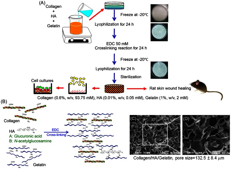
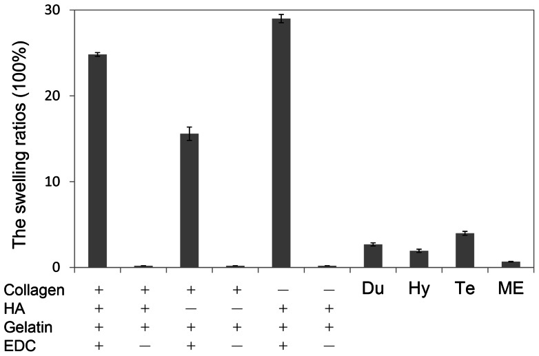
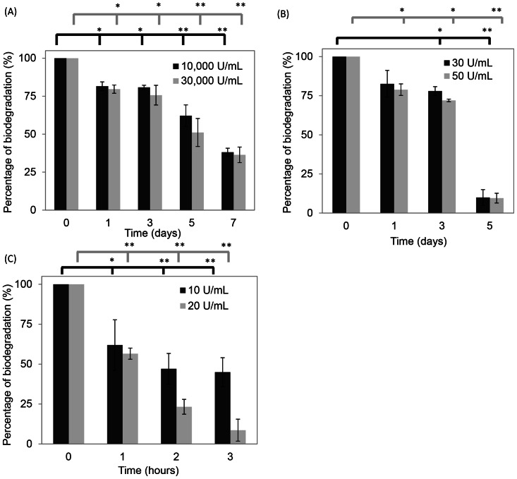
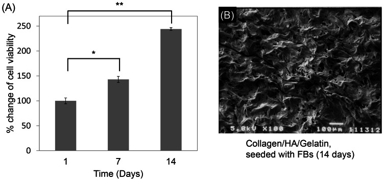
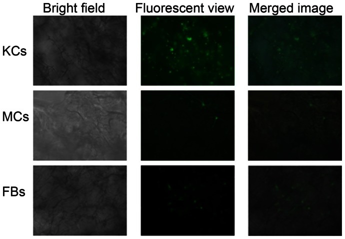
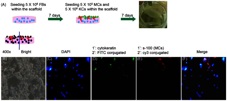
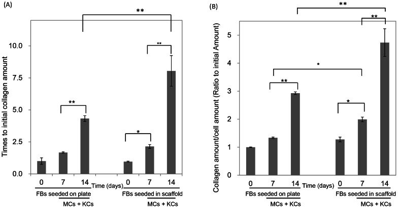
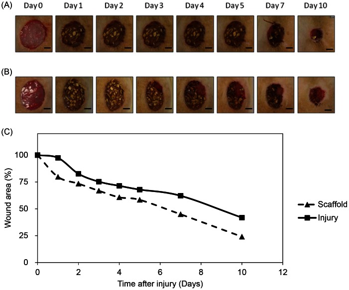
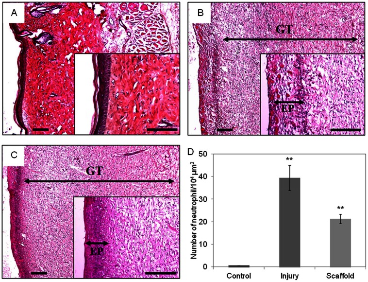
Skin wound healing is an important lifesaving issue for massive lesions. A novel porous scaffold with collagen, hyaluronic acid and gelatin was developed for skin wound repair. The swelling ratio of this developed scaffold was assayed by water absorption capacity and showed a value of over 20 g water/g dried scaffold. The scaffold was then degraded in time- and dose-dependent manners by three enzymes: lysozyme, hyaluronidase and collagenase I. The average pore diameter of the scaffold was 132.5±8.4 µm measured from SEM images. With human skin cells growing for 7 days, the SEM images showed surface fractures on the scaffold due to enzymatic digestion, indicating the biodegradable properties of this scaffold. To simulate skin distribution, the human epidermal keratinocytes, melanocytes and dermal fibroblasts were seeded on the porous scaffold and the cross-section immunofluorescent staining demonstrated normal human skin layer distributions. The collagen amount was also quantified after skin cells seeding and presented an amount 50% higher than those seeded on culture wells. The in vivo histological results showed that the scaffold ameliorated wound healing, including decreasing neutrophil infiltrates and thickening newly generated skin compared to the group without treatments.


Conflict of interest statement

Competing Interests: The authors have declared that no competing interests exist.

Figures

Figure 1
Figure 1. The manufacturing process and structural diagram.
(A) The biomaterial manufacture, skin culture and mouse skin wound healing model. (B) Proposed schematic presentation of collagen (0.6%, w/v, 93.75 mM), HA (0.01%, w/v, 0.05 mM), gelatin (1%, w/v, 2 mM) cross-linked by EDC. (C), (D) SEM of collagen/HA/gelatin scaffolds. The pore size was 132.5±8.4 μm.
Figure 2
Figure 2. The swelling studies of the scaffolds fabricated with collagen (0.6%, w/v), HA (0.01%, w/v), and gelatin (1.0%, w/v) and crosslinked with EDC (50 mM) (n = 3).
Without EDC crosslinking reactions, the scaffolds were dissoluble into water (symbol: ×). Compared with commercial materials include, Du (DuoDERM 9C52552), Hy (Hydro Coll), Te (Tegaderm M1635), and ME (MEDPOR®).
Figure 3
Figure 3. The degradation rates of the enzymes.
(A) Lysozyme, (B) hyaluronidase, and (C) collagenase. A significant difference compare to the control group was defined as *p<0.05 and **p<0.01.
Figure 4
Figure 4. Cell proliferation ratios of human skin FBs seeded in the scaffolds (n = 4). From 1 to 14 days, the proliferation rate was observed by MTT assay (A).
The SEM image of FBs seeding in scaffold for 14 days (B).
Figure 5
Figure 5. Photographed human KCs, MCs and FBs cultured in the scaffold on bright field, fluorescent and merged phase.
Fluorescent compound, PKH-67 (green), was used to stain cells.
Figure 6
Figure 6. The protocols and fluorescent photos of 3D human skin equivalent.
(A) The protocols of 3D human skin equivalent. (B) Paraffin section of the 3D human skin equivalent under microscope in bright view (400×). (C–E) Fluorescent images of KCs, MCs, and FBs cultured in scaffold for 14 days, and were stained with DAPI (blue); anti-cytokeratin to mark KCs (green); anti-s-100 for MCs (red). (F) The merged image was of KCs, MCs, and FBs together. Arrows pointed to KCs, MCs, and FBs with specific colors.
Figure 7
Figure 7. Collagen amount secreted from cells seeded on plate or in the scaffold.
On the plate or in the scaffold, FBs raised for the first 7 days, after that, KCs and MCs seeded in for another 7 days. (A), the total collagen amounts on the plate or in the scaffold were shown. (B), the specific collagen amount measured with dividing collagen amount by cell amount.
Figure 8
Figure 8. The healing pattern of the wounds in different conditions.
(A) Scaffold treated and (B) injury wound after 0, 1, 2, 3, 4, 5, 7 and 10 days after injury. The wound healing efficacy of the scaffold was evaluated in a full thickness wound model. Following anaesthetized a full thickness excisions of 2 cm in diameter were created by a surgical knife of male Wistar rats. For treatment group after excision was made, the scaffold was covered on the wound immediately. For injury group wounds were not covered for comparison. From the first day after injury, the healing of wound from injury group was slower than scaffold treated wound until 10 days after injury. Scale bar  = 0.5 cm. (C) Wound contraction ratios of scaffold and injury at different times. By examining the wound area at definite days, the reduction of wound area was calculated. The surface area of the excisional wounds was calculated as described in methods. The wound area decreased rapidly in the presence of scaffold when compared with the control since first day after injury. The wound area in control group was 60% of the original size on day 7. This percentage was reached almost 3 days earlier at scaffold group. The difference between wounds of injury and scaffold group were statistically significant at day 10. Data are presented as the mean ± standard error.
Figure 9
Figure 9. H&E stained sections for the morphological evaluation of skin wounds.
Ten days after injury, rats were scarified, wound skin was fixing in 4% of paraformaldehyde. The skin was stained with H&E for histological observation. Ten randomly selected areas of dermis from each sample were examined at a magnification of 400× for counting neutrophil. Scaffold group (A), injury group (B) and control (C) wounds at 10 days after injury. Both scaffold and injury group wounds have granulation tissue. The epidermis of treatment group was denser than injury epidermis. Wounds of treatment group were had less neutrophil infiltrated compare to injury group (D). Scale bar  = 200 μm. EP, epithelial layer, GT, granulation tissue.

Similar articles

Cited by

References

    1. Pomahac B, Svensjo T, Yao F, Brown H, Eriksson E (1998) Tissue engineering of skin. Crit Rev Oral Biol Med 9: 333–344. - PubMed
    1. Choi W, Wolber R, Gerwat W, Mann T, Batzer J, et al. (2010) The fibroblast-derived paracrine factor neuregulin-1 has a novel role in regulating the constitutive color and melanocyte function in human skin. J Cell Sci 123: 3102–3111. - PMC - PubMed
    1. Boyce ST (2001) Design principles for composition and performance of cultured skin substitutes. Burns 27: 523–533. - PubMed
    1. Ma L, Gao C, Mao Z, Zhou J, Shen J, et al. (2003) Collagen/chitosan porous scaffolds with improved biostability for skin tissue engineering. Biomaterials 24: 4833–4841. - PubMed
    1. Schulz JT 3rd, Tompkins RG, Burke JF (2000) Artificial skin. Annu Rev Med 51: 231–244. - PubMed

Publication types

MeSH terms